Introduction
Eosinophil cationic protein (ECP), found in the
secondary granules of human eosinophils, is a single-chain peptide
of 133 amino acids, with a molecular mass ranging between 15 and 22
kDa, encoded by the RNSE3 gene located on chromosome
14q11.2. Its amino acid sequence and three-dimensional structure
indicate that ECP is a member of the ribonuclease a superfamily
(1–7). Protein heterogeneity is a result of
post-translational modifications, such as differences in
glycosylation of the molecule, since there are three potential
sites for N-linked glycosylation in the ECP amino acid sequence
(6, 8–12).
ECP has a number of biological activities, including
suppression of T-cell proliferative responses and immunoglobulin
synthesis by B cells, mast cell degranulation, regulation of
fibroblast activities, induction of airway mucus secretion and
interaction with the coagulation and complement systems (5, 10,
13). Furthermore, the most
striking function of ECP is its cytotoxic activity against
bacteria, parasites, viruses, respiratory epithelial and cancer
cells (2, 4, 8,
9, 12, 13). The mechanism of action of ECP is
mediated through its cytotoxic capacity to create pores in the cell
membrane, with ensuing destabilization of the phospholipid bilayer
and osmotic cell lysis (2,
9, 11, 14,
15).
According to Navarro et al (4), the effect of ECP begins with its
binding and aggregation on the cell surface, which alters the cell
membrane permeability and modifies the cell ionic equilibrium.
These signals induce cell-specific morphological and biochemical
changes, such as chromatin condensation, reversion of membrane
asymmetry, production of reactive oxygen species, activation of
caspase-3-like activity and, eventually, cell death. In addition,
the high number of arginine residues on the surface of the protein
(16) and the tryptophan residues
at positions 10 and 35 (2,
16) appear to be crucial for the
cytotoxic activity of ECP.
The exact function of eosinophils in cancer,
particularly in oral squamous cell carcinoma (OSCC), has not yet
been fully elucidated (17–21).
Certain authors support the hypothesis that eosinophils play a
significant role in the host defense against cancer, whereas others
suggest that the antitumor effect of eosinophils in human is modest
at best, particularly in view of the numerous examples of
aggressive cancers that continue to proliferate and spread,
although they are infiltrated by significant numbers of eosinophils
(20).
The antitumor effect of eosinophils (5, 6,
19, 21, 22)
has been associated with the release of cytotoxic proteins,
including ECP. Furthermore, the blood eosinophil counts and serum
concentration of ECP were found to be significantly higher when
compared between prior to and during treatment with interleukin-2
(IL-2) and interferon (IFN)- α in patients with renal cell
adenocarcinoma (22). Based on
those results, the authors hypothesized that, although the precise
mechanisms involved in the induction of the release of the
eosinophil-derived products are not known, potential candidates are
tumor necrosis factor (TNF)- α and the direct interaction of the
eosinophils with cancer cells through antibody-dependent
mechanisms.
The aim of the present study was to investigate the
effect of ECP on human OSCC lines and provide novel insights into
the role of eosinophils in these tumors.
Materials and methods
Cell culture
The SCC-4 and SCC-25 cell lines (American Type
Culture Collection, Manassas, VA, USA) were maintained in
Dulbecco's modified Eagle's medium/Ham's nutrient mixture F12
(DMEM/F12; Invitrogen, Carlsbad, CA) supplemented with 10% fetal
bovine serum (Invitrogen), 400 ng/ml hydrocortisone and 100 µ g/ml
gentamycin and kanamycin at 37˚C in a humidified atmosphere of 5%
CO2, as described by Agostini et al (23).
Evaluation of cellular morphology
The effects of the ECP on cell morphology were
observed using an inverted light microscope (Eclipse E200; Nikon,
Tokyo, Japan) and were photographed prior to and after treatment,
to document possible changes in morphology.
Cell viability assay
The effect of ECP on SCC-4 and SCC-25 cell viability
was assessed by colorimetric assay using
3-(4,5-dimethylthiazol-2-yl)-2,5-diphenyltetrazolium bromide (MTT),
as described by Gomes et al (24).
Briefly, SCC-4 and SCC-25 cells were cultured in
96-well plates for 24 h in DMEM/F12 supplemented with 10% fetal
bovine serum at 2×10⁵ cells per well. The ECP
(MyBioSource, LLC, San Diego, CA, USA) was diluted in complete
medium to prepare samples of different concentrations. Negative
control wells received 100 µl of complete medium, whereas the
treated cells were incubated for 72 h with 0.0390625, 0.078125,
0.15625, 0.3125, 0.625, 1.25, 2.5, 5 and 10 µ M of ECP (final
volume, 100 µl/well). At the end of the incubation, the medium was
removed and each well received 50 µl of MTT solution (0.5 mg/ml in
PBS buffer; Sigma-Aldrich Corp., St. Louis, MO, USA), including 6
wells without cells (blanks). The plates were incubated at 37˚C for
4 h followed by the addition of 200 µl of dimethylsulfoxide to each
well and incubation with shaking at 37˚C for 20 min to ensure
complete dissolution of the formazan crystals. The resulting
absorbances were read at 570 nm in a microplate reader (PowerWave
XS2; Biotek Instruments, Inc. Winooski, VT, USA) and cell viability
was calculated using the following equation:
[TeX:] \begin{document}\begin{equation*} Cell\
viability(\%)=\frac{A-B}{C-D}\times 100
\end{equation*}\end{document}
where A is the absorbance of the treated cells, B is
the absorbance of the blank and C is the absorbance of the control.
All the experiments were performed in triplicate. The viability
mean was used in the statistical analysis.
Statistical analysis
Data are expressed as mean ± standard deviation and
stored in Microsoft Excel for Windows for later analysis with Stata
software, version 11.0 (StataCorp LP, College Station, TX, USA).
The association between ECP concentration (independent variable)
and cell viability (dependent variable) was estimated by linear
regression analysis. Two analyses were performed, for SCC-4 and
SCC-25 cells. As cell viability is not normally distributed, prior
to statistical analysis, the values were logarithmically
transformed to approximate a normal distribution. The ECP
concentration was entered in the analysis as a continuous variable.
P-values <0.05 were considered to indicate statistically
significant differences.
Results
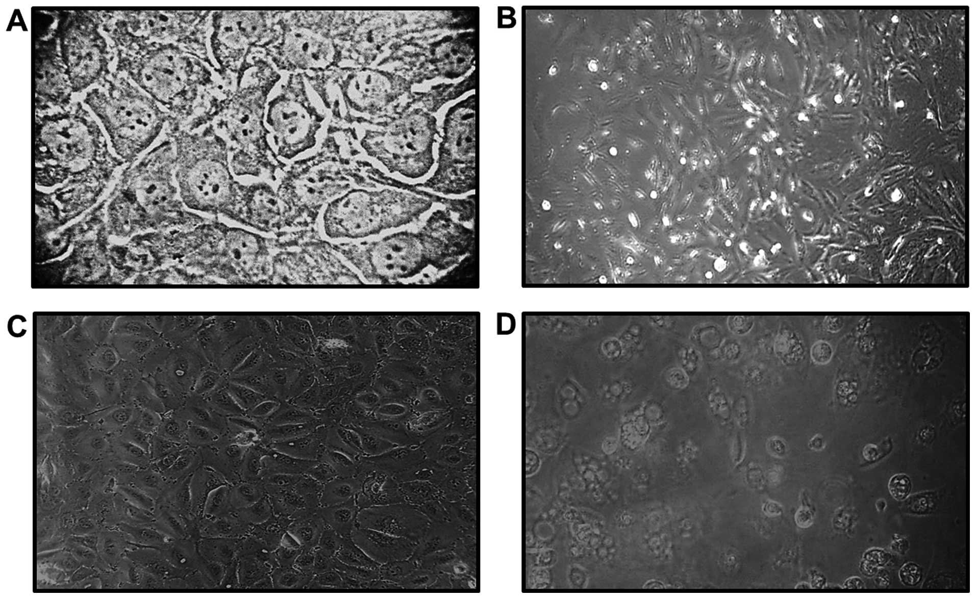
Morphological changes
Direct observation under a light microscope was used
to assess the morphological changes of SCC-4 and SCC-25 cell
cultures following exposure to ECP. The cells treated with ECP were
photographed at 72 h following treatment and morphological changes,
such as vacuolation, bleb formation and loss of cell adhesion were
identified (Fig. 1).
Cell viability
There was a significant inverse association between
ECP concentrations with SCC-4 (β=0.16, P=0.019) and SCC-25 (β=0.24,
P=0.006) cell viability (Table I).
The regression analysis demonstrated that the ECP concentration
explained ∼ 52% of the variations in SCC-4 viability and ∼ 64% of
those in SCC-25 viability.
 | Table I.Regression coefficients (95% CI) for
the association of ECP concentration with SCC-4 and SCC-25 cell
viability. |
Table I.
Regression coefficients (95% CI) for
the association of ECP concentration with SCC-4 and SCC-25 cell
viability.
| Cell lines | β(95%
CI)a | P-value | R2 |
|---|
| SCC-4 | 0.16 (0.03-0.27) |
0.019 |
51.9 |
| SCC-25 | 0.24 (0.09-0.38) |
0.006 |
63.9 |
Discussion
The mechanisms that cause eosinophil recruitment to
malignant neoplasms have not been fully elucidated (18, 20,
25). A complex mixture of the
components of innate and adaptive immune responses are likely to be
involved in this process (21,
26). currently available evidence
suggests that CD4+ T lymphocytes and natural killer
cells are capable of producing the Th2-mediated cytokines IL-4 and
IL-5, thus providing strong eosinophil-specific chemoattractants
and activation signals within the tumor environment (21, 26).
Additionally, the eosinophils are considered to be
recruited to tumors, in part, by the selective eosinophil
chemoattractant eotaxin, which binds to the CCR3 receptor on these
cells (21, 25–27).
The expression of eotaxin in OSCC was investigated by Lorena et
al (25), whose results
demonstrated that eotaxin is mainly produced by tumor-associated
eosinophils, which appears to increase the cell turnover rate in
oral cancer.
It was previously suggested that eosinophils may
affect tumors via direct and/or indirect mechanisms and it appears
that the tumor microenvironment may provide additional signs for
eosinophil degranulation and tumor destruction (19, 21). The antitumor effect of eosinophils
(5, 6, 21,
22) has been associated with the
release of cytotoxic proteins, including ECP, which has been linked
to tumor cell apoptosis.
Furthermore, blood eosinophil counts and the serum
concentration of ECP were found to be significantly higher prior to
and during treatment with IL-2 and IFN- α in patients with renal
cell adenocarcinoma (22). Based
on those results, the authors hypothesized that, although the
precise mechanisms involved in the induction of the release of
eosinophil-derived products are not known, potential candidates are
TNF- α and the direct interaction of the eosinophils with cancer
cells through antibody-dependent mechanisms.
Our results demonstrated that OSCC lines treated
with ECP displayed morphological changes, such as vacuolation, bleb
formation and loss of adhesion (Fig.
1). As we were investigating cell death, a longer period of 72
h was selected. Similar findings were described by Trocmé et
al (28), who exposed primary
human corneal epithelial cell cultures to ECP at concentrations
ranging between 0 and 10 µg/µl for up to 48 h. According to that
study, the morphological changes in the cells observed following
ECP exposure may be partially explained by its effects on the
epithelial actin cytoskeleton. Indeed, the actin filaments have
been implicated in the control of cell shape and cell adhesion to
the substratum.
In our study, the regression analysis demonstrated
that an increase in the ECP concentration was associated with a
significant decrease in SCC-4 and SCC-25 cell viability (Table I). Maeda et al (29) evaluated the effect of ECP on 13
mammalian cell lines and reported that this protein inhibited the
growth of several cell lines, including those derived from skin and
esophageal squamous carcinoma, in a dose-dependent manner.
Glimelius et al (30) investigated the effects of ECP on
Hodgkin's lymphoma cells in vitro. Of note, ECP was
cytotoxic even at low concentrations; however, ECP was unable to
eliminate all tumor cells, particularly not HDLM-2 cells (of T-cell
origin), even at high concentrations and a prolonged exposure time
(72 h). Based on those results, the authors suggested that one
possible mechanism of such selectivity may be the different
sensitivities according to the cell cycle stage of the cell
population. HDLM-2 cells in the G0 phase may be insensitive to ECP
and only cells in active growth phases are eliminated by ECP.
The mechanism of action of ECP is likely due to its
cytotoxic capacity to create pores in the cell membrane, which
allows the passage of water and other small molecules, leading to
osmotic lysis of the target cell (15). The effects of ECP begin with its
binding and aggregation on the cell surface, altering the cell
membrane permeability and modifying the cell ionic equilibrium.
These signals induce cell-specific morphological and biochemical
changes, such as chromatin condensation, reversion of membrane
asymmetry, reactive oxygen species production, activation of
caspase-3-like activity and, eventually, cell death (4).
A cell line is not necessarily a good example of
primary tumor material, as the cells are removed from the normal
tissue environment, which may affect their response to ECP;
however, it is important to emphasize that the present study was
the first to investigate the effects of ECP on OSCC and to
demonstrate a significant inverse association of ECP concentrations
with SCC-4 and SCC-25 cell viability.
Acknowledgements
This study was supported by a research grant (no.
APQ-00558-09) from the Foundation for Research Support of the State
of Minas Gerais (FAPEMIG).
References
|
1
|
Byström J, Garcia RC, Håkansson L, et al:
Eosinophil cationic protein is stored in, but not produced by,
peripheral blood neutrophils. Clin Exp Allergy. 32:1082–1091. 2002.
View Article : Google Scholar : PubMed/NCBI
|
|
2
|
Carreras E, Boix E, Navarro S, Rosenberg
HF, Cuchillo CM and Nogues MV: Surface-exposed amino acids of
eosinophil cationic protein play a critical role in the inhibition
of mammalian cell proliferation. Mol Cell Biochem. 272:1–7. 2005.
View Article : Google Scholar : PubMed/NCBI
|
|
3
|
Molin D: Bystander cells and prognosis in
Hodgkin lymphoma. Review based on a doctoral thesis. Ups J Med Sci.
109:179–228. 2004. View Article : Google Scholar : PubMed/NCBI
|
|
4
|
Navarro S, Aleu J, Jiménez M, Boix E,
Cuchillo CM and Nogués MV: The cytotoxicity of eosinophil cationic
protein/ribonuclease 3 on eukaryotic cell lines takes place through
its aggregation on the cell membrane. Cell Mol Life Sci.
65:324–337. 2008. View Article : Google Scholar : PubMed/NCBI
|
|
5
|
Venge P and Byström J: Eosinophil cationic
protein (ECP). Int J Biochem Cell Biol. 30:433–437. 1998.
View Article : Google Scholar : PubMed/NCBI
|
|
6
|
Venge P, Byström J, Carlson M, et al:
Eosinophil cationic protein (ECP): molecular and biological
properties and the use of ECP as a marker of eosinophil activation
in disease. Clin Exp Allergy. 29:1172–1186. 1999. View Article : Google Scholar : PubMed/NCBI
|
|
7
|
Zhang J and Rosenberg HF: Sequence
variation at two eosinophil-associated ribonuclease loci in humans.
Genetics. 156:1949–1958. 2000.PubMed/NCBI
|
|
8
|
Eriksson J, Woschnagg C, Fernvik E and
Venge P: A SELDI-TOF MS study of the genetic and post-translational
molecular heterogeneity of eosinophil cationic protein. J Leukoc
Biol. 82:1491–1500. 2007. View Article : Google Scholar : PubMed/NCBI
|
|
9
|
Eriksson J, Reimert CM, Kabatereine NB, et
al: The 434 (G>C) polymorphism within the coding sequence of
eosinophil cationic protein (ECP) correlates with the natural
course of Schistosoma mansoni infection. Int J Parasitol.
37:1359–1366. 2007. View Article : Google Scholar : PubMed/NCBI
|
|
10
|
Jönsson UB, Byström J, Stålenheim G and
Venge P: Polymorphism of the eosinophil cationic protein-gene is
related to the expression of allergic symptoms. Clin Exp Allergy.
32:1092–1095. 2002. View Article : Google Scholar : PubMed/NCBI
|
|
11
|
Koh GCH, Shek LPC, Goh DYT, Van Bever H
and Koh DSQ: Eosinophil cationic protein: Is it useful in asthma? A
systematic review. Respira Med. 101:696–705. 2007. View Article : Google Scholar
|
|
12
|
Trulson A, Byström J, Engstrom A, Larsson
R and Venge P: The functional heterogeneity of eosinophil cationic
protein is determined by a gene polymorphism and post-translational
modifications. Clin Exp Allergy. 37:208–218. 2007. View Article : Google Scholar : PubMed/NCBI
|
|
13
|
Byström J, Tenno T, Håkansson L, et al:
Monocytes, but not macrophages, produce the eosinophil cationic
protein. APMIS. 109:507–516. 2001. View Article : Google Scholar : PubMed/NCBI
|
|
14
|
Sugihara R, Kumamoto T, Ito T, Ueyama H,
Toyoshima I and Tsuda T: Human muscle protein degradation in vitro
by eosinophil cationic protein (ECP). Muscle Nerve. 24:1627–1634.
2001. View
Article : Google Scholar : PubMed/NCBI
|
|
15
|
Young JD, Peterson CGB, Venge P and Cohn
ZA: Mechanism of membrane damage mediated by human eosinophil
cationic protein. Nature. 321:613–616. 1986. View Article : Google Scholar : PubMed/NCBI
|
|
16
|
Carreras E, Boix E, Rosenberg HF, Cuchillo
CM and Nogués MV: Both aromatic and cationic residues contribute to
the membrane-lytic and bactericidal activity of eosinophil cationic
protein. Biochemistry. 42:6636–6644. 2003. View Article : Google Scholar : PubMed/NCBI
|
|
17
|
Cormier SA, Taranova AG, Bedient C, et al:
Pivotal advance: Eosinophil infiltration of solid tumors is an
early and persistent inflammatory host response. J Leukoc Biol.
79:1131–1139. 2006. View Article : Google Scholar : PubMed/NCBI
|
|
18
|
Mingomataj EC: Eosinophil-induced
prognosis improvement of solid tumors could be enabled by their
vesicle-mediated barrier permeability induction. Med Hypotheses.
70:582–584. 2008. View Article : Google Scholar : PubMed/NCBI
|
|
19
|
Munitz A and Levi-Schaffer F: Eosinophils:
‘new’ roles for ‘old’ cells. Allergy. 59:268–275. 2004. View Article : Google Scholar : PubMed/NCBI
|
|
20
|
Samoszuk M: Eosinophils and human cancer.
Histol Histopathol. 12:807–812. 1997.PubMed/NCBI
|
|
21
|
Simson L, Ellyard JI, Dent LA, et al:
Regulation of carcinogenesis by IL-5 and CC111: a potential role
for eosinophils in tumor immune surveillance. J Immunol.
178:4222–4229. 2007. View Article : Google Scholar : PubMed/NCBI
|
|
22
|
Trulson A, Nilsson S and Venge P: The
eosinophil granule proteins in serum, but not the oxidative
metabolism of the blood eosinophils, are increased in cancer. Br J
Haematol. 98:312–314. 1997. View Article : Google Scholar : PubMed/NCBI
|
|
23
|
Agostini M, Silva SD, Zecchina KG, et al:
Fatty acid synthase is required for the proliferation of human oral
squamous carcinoma cells. Oral Oncol. 40:728–735. 2004. View Article : Google Scholar : PubMed/NCBI
|
|
24
|
Gomes CC, Moreira LM, Santos VJSV, et al:
Assessment of the genetic risks of a metallic alloy used in medical
implants. Genet Mol Biol. 34:116–121. 2011. View Article : Google Scholar : PubMed/NCBI
|
|
25
|
Lorena SC, Oliveira DT, Dorta RG, Landman
G and Kowalski LP: Eotaxin expression in oral squamous cell
carcinomas with and without tumour associated tissue eosinophilia.
Oral Dis. 9:279–283. 2003. View Article : Google Scholar : PubMed/NCBI
|
|
26
|
Ellyard JI, Simson L and Parish CR:
Th2-mediated anti-tumour immunity: friend or foe? Tissue Antigens.
70:1–11. 2007. View Article : Google Scholar : PubMed/NCBI
|
|
27
|
Murdoch C, Muthana M, Coffelt SB and Lewis
CE: The role of myeloid cells in the promotion of tumour
angiogenesis. Nat Rev Cancer. 8:618–631. 2008. View Article : Google Scholar : PubMed/NCBI
|
|
28
|
Trocmé SD, Hallberg CK, Gill KS, Gleich
GJ, Tyring SK and Brysk MM: Effects of eosinophil granule proteins
on human corneal epithelial cell viability and morphology. Invest
Ophthalmol Vis Sci. 38:593–599. 1997.PubMed/NCBI
|
|
29
|
Maeda T, Kitazoe M, Tada H, et al: Growth
inhibition of mammalian cells by eosinophil cationic protein. Eur J
Biochem. 269:307–316. 2002. View Article : Google Scholar : PubMed/NCBI
|
|
30
|
Glimelius I, Rubin J, Fischer M, et al:
Effect of eosinophil cationic protein (ECP) on Hodgkin lymphoma
cell lines. Exp Hematol. 39:850–858. 2011. View Article : Google Scholar : PubMed/NCBI
|