Introduction
Esophageal squamous cell carcinoma (ESCC) is a
common type of cancer worldwide. Despite the significant
improvements in diagnostic methods, surgical resection and
multidisciplinary therapy, the outcome remains dismal, with a
5-year survival rate of <40% (1,2). Although
the tumor-node-metastasis staging system is well established
(3), patients with the same stage
commonly present with different survival rates. Therefore, it is
crucial to evaluate the molecular and genetic characteristics of
ESCC.
The forkhead box family of transcription factors
comprises evolutionarily conserved DNA-binding proteins that are
present in several organisms (4,5). As a
member of this family, transcription factor forkhead box F2 (FOXF2)
has been well described as an essential signaling molecule for
embryogenesis and metabolism (6–12).
Experiments in vivo have revealed cleft palate and
gastrointestinal defects in FOXF2 knockout mice (6,7). Studies
in humans also demonstrated associations between FOXF2
mutations and congenital diseases (8,9). In
addition, a recent study proved that FOXF2 is involved in the
process of glycose metabolism (10).
Recently, decreased FOXF2 expression was shown to
promote tumor development (13) and
display several functions critical for cancer initiation and
progression. Moreover, reduced FOXF2 mRNA expression was
found to be associated with early-onset metastasis and poor
prognosis in breast cancer patients (14). It was also reported that FOXF2 may be
downregulated by microRNA-182 (15),
which has been proved to accelerate metastasis and promote cell
invasion (16,17). However, the clinicopathological and
prognostic significance of FOXF2 in human ESCC remains unknown.
To elucidate the clinicopathological and prognostic
value of FOXF2 in ESCC, we determined FOXF2 mRNA expression
by quantitative polymerase chain reaction (qPCR) and evaluated its
feasibility as a biomarker for ESCC patients.
Materials and methods
Patient selection
Following approval by the local Institutional Ethics
Committee, a series of 188 consecutive patients with ESCC who
underwent esophagectomy with extended two-field lymphadenectomy at
the Department of Thoracic Oncology of Sun Yat-sen University
Cancer Center between January, 2002 and December, 2008, were
enrolled in this study. Written informed consent was provided by
the participants for their clinical records to be used in this
study. All patient data were anonymized and de-identified in a
confidential manner.
The inclusion criteria were as follows: i)
Pathologically diagnosed ESCC; ii) complete surgical resection;
iii) no distant metastasis; iv) no preexisting/concurrent malignant
disease or a second primary tumor; v) no perioperative mortality;
and vi) availability of fresh samples. Patients receiving
neoadjuvant or adjuvant treatment were excluded. The pretreatment
evaluation included a complete history and physical examination,
complete blood cell count, serum biochemistry, chest radiography,
esophageal barium meal, computed tomography scan of the cervical
region, chest and abdomen, endoscopy and ultrasonography scan of
the abdomen. The pathological staging was reverified based on the
7th American Joint Committee on Cancer (AJCC) staging system
(3).
Following primary treatment, the majority of the
patients were followed up in the outpatient clinic every 3 months
during the first 2 years, every 6 months during years 3–5 and every
12 months thereafter. The survival status was reverified using the
best available method in June, 2014. The median time from surgery
to the last censoring date for the entire cohort was 68.5
months.
qPCR assays
Fresh tumor and non-tumor samples were collected
from regions that were macroscopically assessed as neoplastic and
normal, respectively. The samples were immediately stored on dry
ice after resection and then frozen at −80°C. Total RNA was
extracted from the specimens using the TRIzol reagent (Invitrogen
Life Technologies, Carlsbad, CA, USA) according to the
manufacturer's instructions. Each cDNA was synthesized from 1 µg of
total RNA using RevertAid First Strand cDNA Synthesis kit (Thermo
Fisher Scientific Inc., Waltham, MA, USA) and stored at −80°C.
Glyceraldehyde-3-phosphate dehydrogenase (GAPDH), a
housekeeping gene, was selected as an internal standard to control
for amplification variability. The following primers were used:
FOXF2, forward 5′-CACTACTGGACCATCGACCC-3′; and reverse
5′-CTCACCACGCGGTGGTACAT-3′ (NCBI: NM_001452.1); and GAPDH,
forward 5′-ACT TCAACAGCGACACCCACTC-3′ and reverse
5′-TACCAGGAAATGAGCTTGACAAAG-3′ (NCBI: NM_001256799.1). As the
reverse transcription (RT)-qPCR assays were not performed at the
same time, we utilized FOXF2 mRNA expression of the EC109
ESCC cell line as an internal control (calibrator) to adjust
variation. The PCR mixture of each PCR analysis included 0.12 µl of
cDNA, 5 µl of 2x Power SYBR® Green PCR Master Mix (Applied
Biosystems, Life Technologies, Grand Island, NY, USA), 0.25 µl of
20 mmol/l forward primer, 0.25 µl of reverse primer and 4.38 µl of
distilled water. RT-qPCR was performed using LightCycler 480 (Roche
Applied Science, Penzberg, Germany) with the following thermal
cycling profile: 95°C for 10 min, followed by 40 cycles of
amplification (95°C for 10 sec and 60°C for 20 sec) and then 72°C
for 30 sec. Each assay was performed at least three times. Any
samples with a coefficient of variance >10% were retested. The
relative expression level of FOXF2 mRNA for each sample was
calculated as 2−ΔΔCtsample as follows:
ΔΔCtsample = ΔCtcalibrator -
ΔCtsample, where ΔCtcalibrator of
FOXF2 mRNA = ΔCtcalibrator of FOXF2 -
Ctcalibrator of GAPDH; ΔCtsample =
Ctsample of FOXF2 - Ctsample of
GAPDH. The value of the Ct difference is equal to a
2n-fold difference.
Statistical analysis
The statistical analysis was performed using SPSS
19.0 software (SPSS, Inc., Chicago, IL, USA). A paired two-tailed
t-test was employed to analyze the difference in FOXF2 mRNA
expression between the paired tumor and non-tumor tissues. A
receiver operating characteristic (ROC) curve was generated to
select the optimal cut-off value and, therefore, divided the
FOXF2 mRNA expression into two groups, namely low- and
high-expression groups. The χ2 test was used to
determine the associations between FOXF2 mRNA expression and
categorized clinicopathological parameters. To determine the
factors associated with an increased risk of lymph node metastasis
(LNM), crude and adjusted analyses were performed using univariate
and multivariate logistic regression.
The cancer-specific survival (CSS) was calculated
from the date of surgery to either the date of death from ESCC or
the last follow-up. The survival analysis was performed using the
Kaplan-Meier method and the differences between curves were
assessed by the log-rank test. Univariate and multivariate Cox
regression analyses were used to identify the factors associated
with prognosis. For selecting variables for the multivariate
logistic/Cox regression model, a cut-off value of P=0.10 was used.
P<0.05 was considered to indicate a statistically significant
difference.
Results
Human FOXF2 mRNA expression in tumor
and non-tumor tissues
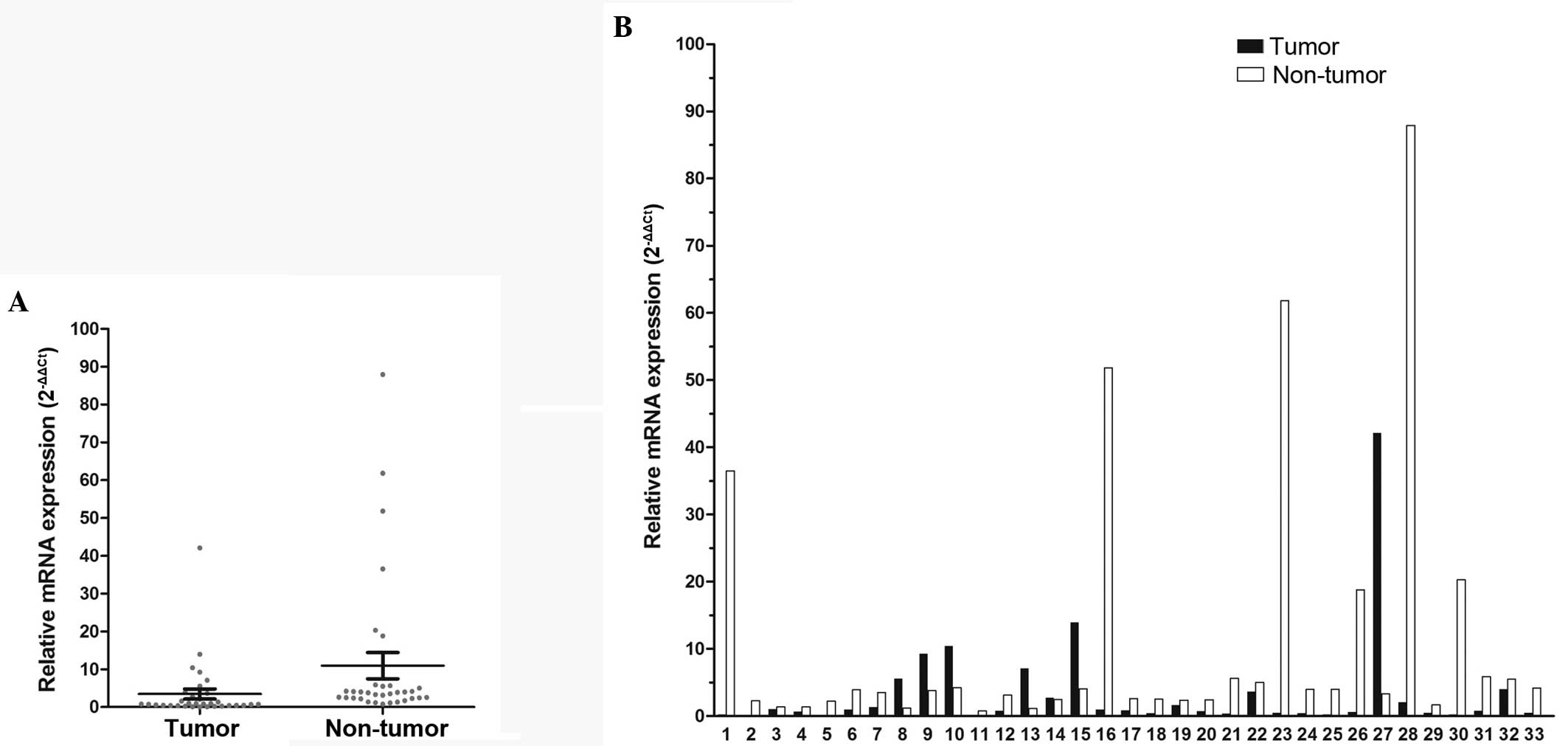
In total, 33 patients with paired tumor and
non-tumor tissues were enrolled in this study. FOXF2 was
found to be significantly downregulated in tumor compared to
non-tumor tissues from the same patient (P=0.048, Fig. 1A). The median value of the
normal/tumor (N/T) ratio of FOXF2 mRNA expression was 4.1;
the N/T ratio was >two-fold in 66.7% of the patients (22/33) and
the highest ratio was ≤165.91-fold (Fig.
1B).
Association between FOXF2 mRNA
expression level and clinicopathological parameters
A total of 188 patients with available tumor tissue
samples were enrolled in this study, with a median age of 59 years
(range, 34–88 years). The patient characteristics are summarized in
Table I. The median value of
FOXF2 mRNA expression was 0.70 in the tumor tissue samples
(range, 0.02–42.10). According to the ROC curve (Fig. 2), the optimal cut-off value of
FOXF2 mRNA with the best discriminatory power was 1.2. Using
this cut-off value, the entire cohort was classified into two
groups, namely high-level (>1.2; n=125) and low-level (≤1.2;
n=63) FOXF2 mRNA expression. There was no significant
association between FOXF2 mRNA expression and age, gender,
tumor location, tumor length, tumor cell differentiation,
pathological T stage, AJCC stage and the number of resected lymph
nodes. However, we observed that the rate of LNM was significantly
associated with a low level of FOXF2 mRNA expression
(P=0.044) (Table I).
 | Table I.Correlations between FOXF2
expression and clinicopathologicalcharacteristics. |
Table I.
Correlations between FOXF2
expression and clinicopathologicalcharacteristics.
|
Characteristics | Total (n=188) | FOXF2 mRNA
expression |
P-valuea |
|---|
|
|---|
| High (n=63) | Low (n=125) |
|---|
| Age, years |
| 0.198 |
|
≤59b | 98 | 37 (58.7) | 61 (48.8) |
|
>59 | 90 | 26 (41.3) | 64 (51.2) |
| Gender |
| 0.468 |
|
Female | 51 | 15 (23.8) | 36 (28.8) |
|
Male | 137 | 48 (76.2) | 89 (71.2) |
| Tumor location |
| 0.374 |
|
Upper | 36 | 15 (23.8) | 21 (16.8) |
|
Middle | 104 | 35 (55.6) | 69 (55.2) |
|
Lower | 48 | 13 (20.6) | 35 (28.0) |
| Tumor length,
cm | | 0.380 |
|
≤4.2b | 90 | 33 (52.4) | 57 (45.6) |
|
>4.2 | 98 | 30 (47.6) | 68 (54.4) |
| Tumor cell
differentiation |
| 0.888 |
|
High | 45 | 14 (22.2) | 31 (24.8) |
|
Moderate | 97 | 34 (54.0) | 63 (50.4) |
|
Poor | 46 | 15 (23.8) | 31 (24.8) |
| pT stage |
| 0.353 |
|
T1b-T2 | 46 | 18 (28.6) | 28 (22.4) |
|
T3-T4a | 142 | 45 (71.4) | 97 (77.6) |
| pN stage |
| 0.044 |
| N0 | 100 | 40 (63.5) | 60 (48.0) |
|
N1/N2/N3 | 88 | 23 (36.5) | 65 (52.0) |
| AJCC stage |
| 0.088 |
|
I–II | 106 | 41 (65.1) | 65 (52.0) |
|
III | 82 | 22 (34.9) | 60 (48.0) |
| Resected lymph | | 0.656 |
| nodes, no. ±
SD | 24.0±11.8 | 23.8±11.9 | 24.6±11.9 |
Evaluation of FOXF2 mRNA expression as
a risk factor for LNM
In the univariate logistic regression analysis,
patients with a low level of FOXF2 mRNA expression exhibited
a significantly higher risk of LNM compared to those with a high
level of expression, with a hazard ratio (HR) of 1.884 [95%
confidence interval (CI): 1.012–3.507]. This effect was further
observed in the multivariate logistic analysis, with a marginal
significance (P=0.060) and an adjusted HR of 1.828 (95% CI:
0.975–3.430). The independent risk factor for LNM was advanced
pathological T stage (P=0.028) (Table
II).
| Table II.Univariate and multivariate logistic
regression analyses of factors associated with lymph node
metastasis. |
Table II.
Univariate and multivariate logistic
regression analyses of factors associated with lymph node
metastasis.
| Factors | Univariate
analysis | Multivariate
analysis |
|---|
|
|
|---|
| HR (95% CI) | P-value | HR (95% CI) | P-value |
|---|
| Age, years
(≤59/>59) | 1.078
(0.607–1.912) | 0.799 | – | NAa |
| Gender (F/M) | 1.915
(0.985–3.724) | 0.055 | 1.834
(0.921–3.655) | 0.085 |
| Tumor location
(U/Md/L) | 1.178
(0.765–1.814) | 0.458 | – | NAa |
| Tumor
differentiation (high/moderate/poor) | 1.337
(0.882–2.027) | 0.171 | – | NAa |
| Tumor length, cm
(≤4.2/>4.2) | 1.425
(0.801–2.546) | 0.228 | – | NAa |
| pT stage
(T1b-T2/T3-T4a) | 2.186
(1.087–4.398) | 0.028 | 2.186
(1.087–4.398) | 0.028 |
| resected lymph
nodes, no. (≤21/>21) | 0.984
(0.554–1.747) | 0.955 | – | NAa |
| FOXF2 mRNA
expression (high/low) | 1.884
(1.012–3.507) | 0.046 | 1.828
(0.975–3.430) | 0.060 |
Association between FOXF2 mRNA
expression level and CSS
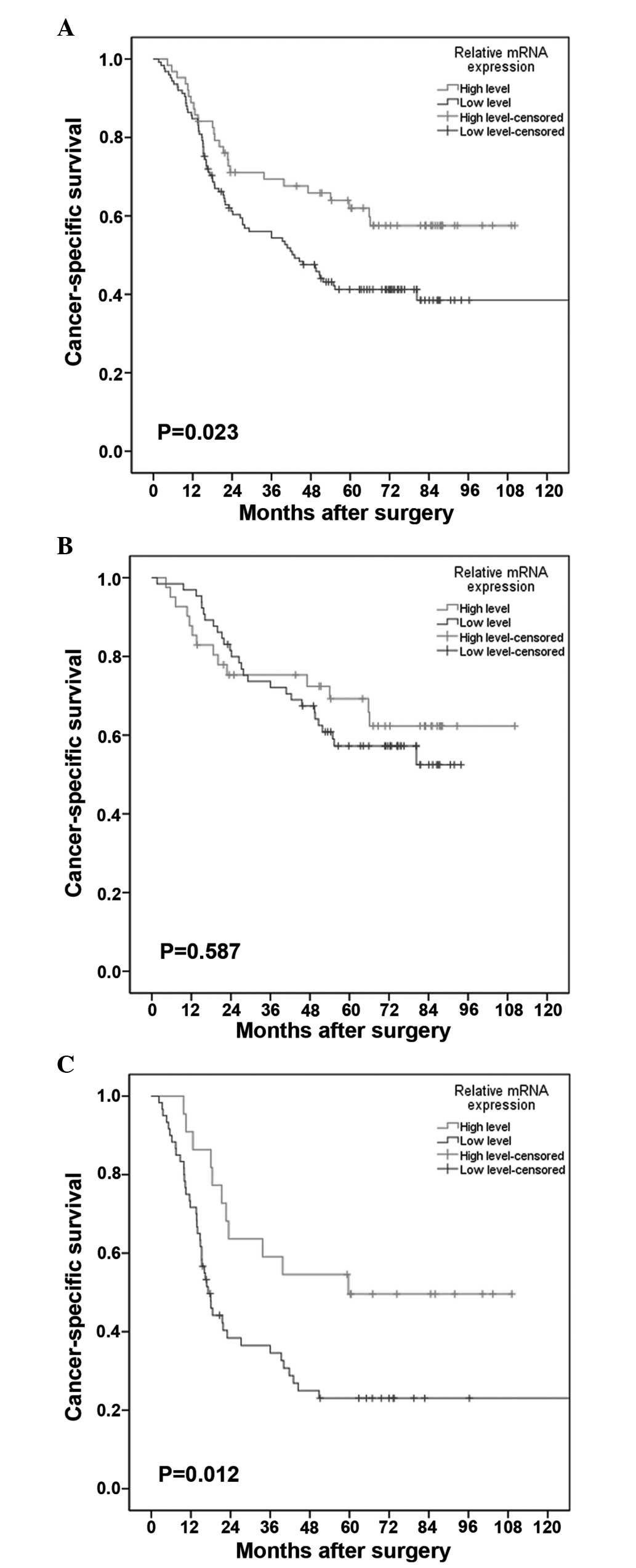
In this study, the median survival time was 54.0
months, with an estimated 5-year CSS of 48.0%. In the univariate
Cox analysis, patients with low-level FOXF2 mRNA expression
exhibited a significantly enhanced mortality risk compared to those
with high-level expression (HR=1.700, 95% CI: 1.077–2.681), with a
5-year CSS of 41.1 and 61.9%, respectively (Fig. 3A). This effect was further verified in
the multivariate Cox analysis, with an adjusted HR of 1.714 (95%
CI: 1.085–2.708). Other negative prognostic factors with
independent significance included advanced AJCC stage (P<0.001)
and a resected lymph node number of ≤21 (P=0.016). The details of
the univariate and multivariate analyses are shown in Table III.
| Table III.Univariate and multivariate Cox
analyses of factors associated with cancer-specific survival. |
Table III.
Univariate and multivariate Cox
analyses of factors associated with cancer-specific survival.
| Factors | Univariate Cox
analysis | Multivariate Cox
analysis |
|---|
|
|
|---|
| HR (95% CI) | P-value | HR (95% CI) | P-value |
|---|
| Age, years
(≤59/>59) | 1.300
(0.872–1.936) | 0.198 | – | NAa |
| Gender (F/M) | 1.119
(0.717–1.745) | 0.621 | – | NAa |
| Tumor location
(U/Md/L) | 1.131
(0.837–1.527) | 0.422 | – | NAa |
| Tumor
differentiation (high/moderate/poor) | 1.352
(1.009–1.812) | 0.043 | 1.276
(0.939–1.735) | 0.119 |
| Tumor length, cm
(≤4.2/>4.2) | 0.913
(0.613–1.360) | 0.654 | – | NAa |
| AJCC stage
(I/II/III) | 2.384
(1.660–3.424) | <0.001 | 2.482
(1.728–3.566) | <0.001 |
| resected lymph
nodes, no. (≤21/>21) | 0.646
(0.431–0.971) | 0.035 | 0.606
(0.403–0.910) | 0.016 |
| FOXF2 mRNA
expression (high/low) | 1.700
(1.077–2.681) | 0.023 | 1.714
(1.085–2.708) | 0.021 |
Prognostic significance of FOXF2 mRNA
expression level in the subgroup analysis
The association between FOXF2 mRNA expression
and CSS across strata of other potential predictors of patient
outcome were assessed. As shown in Table
IV, following adjustment for known prognostic factors, the
increased risk of cancer-related mortality conferred by low-level
FOXF2 mRNA expression was unchanged in male patients
(adjusted HR=1.790, 95% CI: 1.044–3.072) and patients with
advanced-stage disease (adjusted HR=2.924, 95% CI: 1.466–5.833).
However, this association was insignificant in other subgroup
analyses. The CSS curves stratified by FOXF2 mRNA expression
level in patients with stage I/II and patients with stage III
disease are shown in Fig. 3B and C,
respectively.
| Table IV.Subgroup analysis for FOXF2
mRNA expression. |
Table IV.
Subgroup analysis for FOXF2
mRNA expression.
| Factors | 5-year OS (%) | FOXF2 mRNA
expression (no. of events/no. at risk) |
|
|---|
| Multivariate Cox
analysis |
|---|
|
|
|
|---|
| High | Low | HR | 95% CI | P-value |
|---|
| Age, years |
|
≤59a | 53.1 | 13/37 | 34/61 | 1.862 | 0.980–3.539 | 0.058 |
|
>59 | 42.4 | 12/26 | 38/64 | – | – | NAb |
| Gender |
|
Female | 46.6 |
7/15 | 20/36 | – | – | NAb |
|
Male | 49.9 | 18/48 | 52/89 | 1.790 | 1.044–3.072 | 0.034 |
| Tumor location |
|
Upper | 56.4 |
5/15 | 11/21 | – | – | NAb |
|
Middle | 46.0 | 15/35 | 41/69 | 1.667 | 0.921–3.017 | 0.091 |
|
Lower | 47.1 |
5/13 | 20/35 | – | – | NAb |
| Tumor length,
cm |
|
≤4.2a | 47.4 | 14/33 | 35/57 | – | – | NAb |
|
>4.2 | 49.8 | 11/30 | 37/68 | 1.777 | 0.945–3.340 | 0.074 |
| AJCC stage |
|
I–II | 61.3 | 14/41 | 28/65 | – | – | NAb |
|
III | 20.7 | 11/22 | 44/60 | 2.924 | 1.466–5.833 | 0.002 |
| Resected lymph
nodes, no. |
|
≤21a | 41.7 | 15/32 | 43/68 | – | – | NAb |
|
>21 | 55.6 | 10/31 | 29/57 | 2.038 | 0.987–4.205 | 0.054 |
Discussion
It was previously demonstrated that FOXF2 acts as a
tumor suppressor in breast and prostate cancer (14,15). In
this study, we observed that FOXF2 mRNA expression was
significantly higher in normal esophageal tissue compared to that
in tumor specimens. Thus, FOXF2 may act as a tumor suppressor in
ESCC. We hypothesized that FOXF2 maintains a normal expression
level in normal esophageal epithelial cells and plays an important
role in balancing cell behavior, such as proliferation,
differentiation, mitotic cycle and apoptosis, which was also
supported by previous studies (7,11–13). However, FOXF2 expression may be
compromised in ESCC during the course of tumorigenesis, similar to
prostate cancer (18,19). Although the precise mechanism is not
known, one possible explanation involves FOXF2 downregulation by
microRNA-182 (15), the
overexpression of which has been observed in various human cancers,
such as colorectal (20), cervical
(21) and non-small-cell lung cancer
(22). In addition, we observed that
a low level of FOXF2 mRNA expression was associated with a
high rate of LNM; the same result was obtained in patients with
breast cancer (14).
When our analyses focused exclusively on patient
survival, the level of FOXF2 mRNA expression was found to be
an independent and significant predictive variable. The 5-year CSS
was significantly higher in patients with high-level FOXF2
mRNA expression compared to that in patients with low-level
expression. The effect of FOXF2 on prognosis may be attributed to
its intrinsic nature as an anti-oncogene.
Several studies have been conducted to elucidate how
FOXF2 exerts its tumor suppressor effect. It was reported that
FOXF2 may downregulate matrix metalloproteinases (MMPs) and
upregulate tissue inhibitor of metalloproteinase-3, a known
inhibitor of MMPs (15,19). Due to their ability to degrade the
extracellular matrix, activated MMPs accelerate metastasis and
decrease survival in patients with ESCC (23), colorectal (24), breast (25) and gastric cancer (26). In addition, FOXF2 was reported to
inhibit the Wnt pathway (7,13), the activation of which has been
associated with a high risk of metastasis and a poor outcome in
patients with pancreatic (27),
breast (28) and prostate cancer
(29). However, the potential
mechanisms of FOXF2 as a tumor suppressor in ESCC remain unknown
and require further investigation.
In our subgroup analysis, we observed that the
increased risk conferred by low-level FOXF2 mRNA expression
was limited to patients with stage III disease. A possible
explanation may be associated with the fact that the survival rate
is generally relatively low in patients with advanced ESCC;
therefore, there may be a tendency to observe significant survival
differences according to a certain factor. Second, the superior
outcomes in the high FOXF2 mRNA expression group may be
partially associated with the role of FOXF2 in suppressing
metastasis (7,13,15,19); this
effect is expected to be more prominent in patients with a high
predisposition for metastasis, which was also indicated by Kong
et al (14); in that study,
the poor effect of decreased FOXF2 mRNA expression on
survival in breast cancer was limited to patients with a
triple-negative profile, a well-known subtype characterized by
early-onset metastasis and a dismal outcome (30). Additionally, we found FOXF2
mRNA expression to be associated with survival in male patients.
Since the interaction between gender and FOXF2 remains unknown, we
hypothesized that this phenomenon may be partially attributed to
the high risk of LNM in male patients (adjusted HR=1.834, 95% CI:
0.921–3.655) in this study.
To date, advances in molecular biology have led to
the rapid development of individualized management in various human
cancers. Based on a cohort of patients treated by surgery alone, we
observed that patients with low-level FOXF2 mRNA expression
had a significantly lower CSS compared to those with high-level
expression. Furthermore, this effect was independent of the
aggressiveness of lymphadenectomy and patient characteristics. In
this sense, surgery alone may be insufficient for patients with a
low level of FOXF2 mRNA expression and multidisciplinary
therapy should be recommended. Future studies should focus on the
interaction between chemotherapy/chemoradiotherapy and FOXF2
expression to verify our hypotheses.
In conclusion, FOXF2 may be an anti-oncogene
in ESCC. Decreased FOXF2 mRNA expression was found to be
associated with poor prognosis in patients with completely resected
ESCC. However, the clinical value of the changes in FOXF2
mRNA levels in ESCC require further validation by large multicenter
studies.
Acknowledgements
This study was supported by grants from the
Fundamental Research Funds for the Central Universities (no.
13ykpy49) and the National Natural Science Foundation of China (no.
81402003).
References
|
1
|
Rice TW, Rusch VW, Apperson-Hansen C, et
al: Worldwide esophageal cancer collaboration. Dis Esophagus.
22:1–8. 2009. View Article : Google Scholar : PubMed/NCBI
|
|
2
|
Allum WH, Stenning SP, Bancewicz J, Clark
PI and Langley RE: Long-term results of a randomized trial of
surgery with or without preoperative chemotherapy in esophageal
cancer. J Clin Oncol. 27:5062–5067. 2009. View Article : Google Scholar : PubMed/NCBI
|
|
3
|
Edge S, Byrd DR, Compton CC, Fritz AG,
Greene FL and Trotti A: AJCC Cancer Staging Manual. 7th. Springer;
Chicago: pp. 67–72. 2010
|
|
4
|
Carlsson P and Mahlapuu M: Forkhead
transcription factors: Key players in development and metabolism.
Dev Biol. 250:1–23. 2002. View Article : Google Scholar : PubMed/NCBI
|
|
5
|
Katoh M and Katoh M: Human FOX gene family
(Review). Int J Oncol. 25:1495–1500. 2004.PubMed/NCBI
|
|
6
|
Wang T, Tamakoshi T, Uezato T, Shu F,
Kanzaki-Kato N, Fu Y, Koseki H, Yoshida N, Sugiyama T and Miura N:
Forkhead transcription factor FoxF2 (LUN)-deficient mice exhibit
abnormal development of secondary palate. Dev Biol. 259:83–94.
2003. View Article : Google Scholar : PubMed/NCBI
|
|
7
|
Ormestad M, Astorga J, Landgren H, Wang T,
Johansson BR, Miura N and Carlsson P: Foxf1 and Foxf2 control
murine gut development by limiting mesenchymal Wnt signaling and
promoting extracellular matrix production. Development.
133:833–843. 2006. View Article : Google Scholar : PubMed/NCBI
|
|
8
|
Jochumsen U, Werner R, Miura N,
Richter-Unruh A, Hiort O and Holterhus PM: Mutation analysis of
FOXF2 in patients with disorders of sex development (DSD) in
combination with cleft palate. Sex Dev. 2:302–308. 2008. View Article : Google Scholar : PubMed/NCBI
|
|
9
|
Yu L, Wynn J, Ma L, et al: De novo copy
number variants are associated with congenital diaphragmatic
hernia. J Med Genet. 49:650–659. 2012. View Article : Google Scholar : PubMed/NCBI
|
|
10
|
Westergren R, Nilsson D, Heglind M, Arani
Z, Grände M, Cederberg A, Ahrén B and Enerbäck S: Overexpression of
Foxf2 in adipose tissue is associated with lower levels of IRS1 and
decreased glucose uptake in vivo. Am J Physiol Endocrinol Metab.
298:E548–E554. 2010. View Article : Google Scholar : PubMed/NCBI
|
|
11
|
Aitola M, Carlsson P, Mahlapuu M, Enerbäck
S and Pelto-Huikko M: Forkhead transcription factor FoxF2 is
expressed in mesodermal tissues involved in epithelio-mesenchymal
interactions. Dev Dyn. 218:136–149. 2000. View Article : Google Scholar : PubMed/NCBI
|
|
12
|
Nik AM, Reyahi A, Pontén F and Carlsson P:
Foxf2 in intestinal fibroblasts reduces numbers of Lgr5+
stem cells and adenoma formation by inhibiting Wnt signaling.
Gastroenterology. 144:1001–1011. 2013. View Article : Google Scholar : PubMed/NCBI
|
|
13
|
van den Brink GR and Rubin DC: Foxf2: A
mesenchymal regulator of intestinal adenoma development.
Gastroenterology. 144:873–876. 2013. View Article : Google Scholar : PubMed/NCBI
|
|
14
|
Kong PZ, Yang F, Li L, Li XQ and Feng YM:
Decreased FOXF2 mRNA expression indicates early-onset metastasis
and poor prognosis for breast cancer patients with histological
grade II tumor. PLoS ONE. 8:e615912013. View Article : Google Scholar : PubMed/NCBI
|
|
15
|
Hirata H, Ueno K, Shahryari V, Deng G,
Tanaka Y, Tabatabai ZL, Hinoda Y and Dahiya R: MicroRNA-182-5p
promotes cell invasion and proliferation by down regulating FOXF2,
RECK and MTSS1 genes in human prostate cancer. PLoS ONE.
8:e555022013. View Article : Google Scholar : PubMed/NCBI
|
|
16
|
Sachdeva M, Mito JK, Lee CL, et al:
MicroRNA-182 drives metastasis of primary sarcomas by targeting
multiple genes. J Clin Invest. 124:4305–4319. 2014. View Article : Google Scholar : PubMed/NCBI
|
|
17
|
Lei R, Tang J, Zhuang X, et al:
Suppression of MIM by microRNA-182 activates RhoA and promotes
breast cancer metastasis. Oncogene. 33:1287–1296. 2014. View Article : Google Scholar : PubMed/NCBI
|
|
18
|
van der Heul-Nieuwenhuijsen L, Dits NF and
Jenster G: Gene expression of forkhead transcription factors in the
normal and diseased human prostate. BJU Int. 103:1574–1580. 2009.
View Article : Google Scholar : PubMed/NCBI
|
|
19
|
van der Heul-Nieuwenhuijsen L, Dits N, Van
Ijcken W, de Lange D and Jenster G: The FOXF2 pathway in the human
prostate stroma. Prostate. 69:1538–1547. 2009. View Article : Google Scholar : PubMed/NCBI
|
|
20
|
Yang MH, Yu J, Jiang DM, Li WL, Wang S and
Ding YQ: microRNA-182 targets special AT-rich sequence-binding
protein 2 to promote colorectal cancer proliferation and
metastasis. J Transl Med. 12:1092014. View Article : Google Scholar : PubMed/NCBI
|
|
21
|
Tang T, Wong HK, Gu W, Yu MY, To KF, Wang
CC, Wong YF, Cheung TH, Chung TK and Choy KW: MicroRNA-182 plays an
onco-miRNA role in cervical cancer. Gynecol Oncol. 129:199–208.
2013. View Article : Google Scholar : PubMed/NCBI
|
|
22
|
Ning FL, Wang F, Li ML, Yu ZS, Hao YZ and
Chen SS: MicroRNA-182 modulates chemosensitivity of human non-small
cell lung cancer to cisplatin by targeting PDCD4. Diagn Pathol.
9:1432014. View Article : Google Scholar : PubMed/NCBI
|
|
23
|
Gu ZD, Li JY, Li M, Gu J, Shi XT, Ke Y and
Chen KN: Matrix metalloproteinases expression correlates with
survival in patients with esophageal squamous cell carcinoma. Am J
Gastroenterol. 100:1835–1843. 2005. View Article : Google Scholar : PubMed/NCBI
|
|
24
|
Murray GI, Duncan ME, O'Neil P, Melvin WT
and Fothergill JE: Matrix metalloproteinase-1 is associated with
poor prognosis in colorectal cancer. Nat Med. 2:461–462. 1996.
View Article : Google Scholar : PubMed/NCBI
|
|
25
|
Pellikainen JM, Ropponen KM, Kataja VV,
Kellokoski JK, Eskelinen MJ and Kosma VM: Expression of matrix
metalloproteinase (MMP)-2 and MMP-9 in breast cancer with a special
reference to activator protein-2, HER2 and prognosis. Clin Cancer
Res. 10:7621–7628. 2004. View Article : Google Scholar : PubMed/NCBI
|
|
26
|
Koskensalo S, Mrena J, Wiksten JP,
Nordling S, Kokkola A, Hagström J and Haglund C: MMP-7
overexpression is an independent prognostic marker in gastric
cancer. Tumour Biol. 31:149–155. 2010. View Article : Google Scholar : PubMed/NCBI
|
|
27
|
Jiang H, Li Q, He C, et al: Activation of
the Wnt pathway through Wnt2 promotes metastasis in pancreatic
cancer. Am J Cancer Res. 4:537–544. 2014.PubMed/NCBI
|
|
28
|
Dey N, Barwick BG, Moreno CS, et al: Wnt
signaling in triple negative breast cancer is associated with
metastasis. BMC Cancer. 13:5372013. View Article : Google Scholar : PubMed/NCBI
|
|
29
|
Yamamoto H, Oue N, Sato A, Hasegawa Y,
Yamamoto H, Matsubara A, Yasui W and Kikuchi A: Wnt5a signaling is
involved in the aggressiveness of prostate cancer and expression of
metalloproteinase. Oncogene. 29:2036–2046. 2010. View Article : Google Scholar : PubMed/NCBI
|
|
30
|
Uhm JE, Park YH, Yi SY, et al: Treatment
outcomes and clinicopathologic characteristics of triple-negative
breast cancer patients who received platinum-containing
chemotherapy. Int J Cancer. 124:1457–1462. 2009. View Article : Google Scholar : PubMed/NCBI
|