


S1PR3 suppresses the inflammatory response and extracellular matrix degradation in human nucleus pulposus cells
- Authors:
- Published online on: April 25, 2024 https://doi.org/10.3892/etm.2024.12553
- Article Number: 265
-
Copyright: © Tian et al. This is an open access article distributed under the terms of Creative Commons Attribution License.
Abstract
Introduction
Chronic low back pain (CLBP) is considered the leading cause of disability and repeated visits for patients with lumbar degenerative disease, contributing to the global socioeconomic burden (1-3). Intervertebral disc tissue is composed of three predominant structures: Nucleus pulposus (NP), annulus fibrosus (AF) and cartilage endplate (EP) (4). NP is a flexible sphere located in the center of the disc, surrounded by AF in the front, back, left and right directions (5). EP is attached to the superior and lower NP (6). Disc degeneration is an age-related biological process characterized by decreased hydration and extracellular matrix (ECM) deposition, increased inward growth of neurovascular structures and release of inflammation-related cytokines within NP tissues, which causes spinal instability along with CLBP (4,7,8). Proinflammatory factors can inhibit ECM production in human nucleus pulposus cells (HNPC) (9). On the other hand, it can promote the production of degrading enzymes such as matrix metalloproteinase (MMP) (10). Therefore, inhibiting inflammation is an effective way to alleviate disc degeneration.
Sphingosine 1-phosphate (S1P) is a bioactive phospholipid that regulates numerous cellular physiological processes such as proliferation, survival and cytoskeletal rearrangement by binding to a family of five G protein-coupled receptors (GPCRs) (11). S1P receptor 3, (S1PR3) belonging to the GPCR family, is necessary for the promotion of bone formation in response to S1P (12). The S1P-S1PR3 axis has been reported to have anti-inflammatory functions (13). S1P lyase inhibition improves sepsis by increasing the S1P-S1PR3 signaling axis and reducing production of cytokines, including tumor necrosis factor (TNF)-α and interleukin (IL)-6(14). In addition, S1P3-/- mice showed the lower survival rate of and the higher levels of TNF-α and IL-6(15).
However, the biological roles of S1PR3 in intervertebral disc degeneration and the underlying mechanism are not well understood. The current study focused on the roles of S1PR3 in disc degeneration and in vitro functional studies were performed in LPS-induced HNPCs to further elucidate the mechanism underlying the regulation of S1PR3 in inflammation-related disc degeneration. The current study provided novel insights into the functional roles of S1PR3 in the pathogenesis of disc degeneration.
Materials and methods
Cell culture
Immortalized human NP cell line HNPC (cat. no. iCell-0028a) provided by Cellverse Bioscience Technology Co., Ltd. were cultured in DMEM (Gibco; Thermo Fisher Scientific, Inc.) supplied with 10% FBS and 1% penicillin/streptomycin (both Gibco; Thermo Fisher Scientific, Inc.) at 5% CO2 and at 37˚C. Lipopolysaccharide (LPS; Sigma-Aldrich; Merck KGaA) with concentration range of 0.01-100 µg/ml was used to treat HNPCs for 24-48 h at 37˚C, aiming to trigger cell inflammation in vitro.
Cell transfection
To overexpress S1PR3 and Toll-like receptor (TLR) 2, the pc-DNA3.1 vector containing the whole length of S1PR3 (over-S1PR3) or TLR2 (over-TLR2), and the empty vector [over-negative control (NC)] were synthesized by Genepharm, Inc. With the application of Lipofectamine® 2000 (Invitrogen; Thermo Fisher Scientific, Inc.), 100 nM recombinants were introduced to HNPCs following the manufacturer's instructions. After 48 h of transfection, cells were used for follow-up experiments.
Cell counting kit-8 (CCK-8) assay
The CCK-8 assay was used for the estimation of HNPC viability. Cells were cultured in DMEM with 10% FBS for 24 h at 37˚C, followed by incubation with 10 µl WST-8 (Beyotime Institute of Biotechnology) for 2 h. A microplate reader (Bio-Rad Laboratories, Inc.) was used, and optical density was determined at 450 nm.
ELISA
The levels of IL-1β, IL-6 and TNF-α in cell culture supernatants were investigated using ELISA kits (cat. nos. #DLB50, #D6050B, #DTA00D, respectively; R&D Systems, Inc.) following the manufacturer's instructions. A microplate reader (Bio-Rad Laboratories, Inc.) was used, and optical density was determined at 450 nm.
Apoptosis assay
Apoptosis was investigated using the FITC Annexin V/PI Apoptosis Detection Kit I (cat. no. 556547; BD Biosciences) following the manufacturer's instructions. Briefly, the collection of cells was carried out following the indicated treatment. Afterward, PBS-rinsed cells were resuspended in binding buffer. Subsequently, cells were incubated with Annexin V-FITC for 15 min and propidium iodide (PI; 10 mg/ml) for 5 min away from the light using BD FACSVerse™ System (BD Biosciences), all steps at room temperature. FlowJo_V10 (FlowJo LLC ) was used for analysis.
Reverse transcription-quantitative PCR (RT-qPCR)
Total RNAs were extracted from HNPC using TRIzol (Invitrogen; Thermo Fisher Scientific, Inc.) following the manufacturer's protocol. Then the RNA was reverse transcribed into cDNA using PrimeScript RT Master Mix (Perfect Real Time; Takara Bio, Inc.) following the manufacturer's instructions. The amplification of cDNA was carried out by qPCR using the SYBR Premix Ex Taq™ II kit (Takara Bio, Inc.). The PCR program was 95˚C for 3 min, followed by 35 cycles of denaturation at 95˚C for 30 sec, annealing at 60˚C for 30 sec and extension at 72˚C for 1 min. A final extension step at 72˚C for 7 min was performed. The primer sequences for PCR are presented as below: A disintegrin and metalloproteinase with thrombospondin motifs 4 (Adamts-4) forward, 5'-GGAAATTCAGATGTGGTACTGCC-3', and reverse 5'-GCCACTAGGACTTGCAGTGT-3'; Adamts-5 forward, 5'-CCATGGCAACTGGGGATCTT-3', and reverse, 5'-TCTCCTCCACATACTCCGCA-3'; Aggrecan forward, 5'-AGGGCGAGTGGAATGATGTT-3', and reverse, 5'-GCGTTTGTAGGTGGTGGCTG-3'; Collagen II forward, 5'-CTTCCCCCTCCTGCTCCAAG-3', and reverse, 5'-TCTCCGAAGGGGATCTCAGG-3'; MMP3 forward, 5'-TGAGGACACCAGCATGAACC-3', and reverse, 5'-ACTTCGGGATGCCAGGAAAG-3'; MMP13 forward, 5'-GCACTTCCCACAGTGCCTAT-3', and reverse 5'-AGTTCTTCCCTTGATGGCCG-3'; TLR2 forward, 5'-CCAAGTGAAGGCAGGAAGACA-3', and reverse 5'-GGAAACTCGAGGCAGACCAA-3'; GAPDH forward, 5'-GGGAAACTGTGGCGTGAT-3', and reverse, 5'-GAGTGGGTGTCGCTGTTGA-3'. The primer sequences were synthesized from Sangon Biotech Co., Ltd., China. Relative gene expression was quantified using the 2-ΔΔCq method normalized to GAPDH (16,17).
Western blotting
Total protein from sample cells was isolated using RIPA buffer (Beyotime Institute of Biotechnology) and quantification was completed using the BCA Protein Assay kit (Beijing Dingguo Changsheng Biotechnology Co., Ltd.) following the manufacturer's instructions. A total of 30 µg protein per well were separated by 10% SDS-polyacrylamide gels and transferred to PVDF membranes. Subsequently, membranes were blocked using 5% non-fat milk for 2 h at room temperature and incubated with the following primary antibodies: S1PR3 (cat. no. ab126622); Bcl-2 (cat. no. ab182858); Bax (cat. no. ab32503); Adamts-5 (cat. no. ab41037); Adamts-4 (cat. no. ab314856); Aggrecan (cat. no. ab315486); Collagen II (cat. no. ab307674); MMP3 (cat. no. ab52915); MMP13 (cat. no. ab39012); TLR2 (cat. no. ab68159); phosphorylated (p)-STAT3 (cat. no. ab267373); STAT3 (cat. no. ab68153); p-JNK (cat. no. ab124956); JNK (cat. no. ab179461); p-ERK (cat. no. ab201015); ERK (cat. no. ab18469); p-p38 (cat. no. ab308038); p38 (cat. no. ab182453); and GAPDH (cat. no. ab8245) (all dilutions 1:1,000; Abcam) overnight at 4˚C and the HRP-conjugated goat anti-rabbit or mouse secondary antibodies (cat. nos. sc-2004 or sc-2005; 1:5,000; Santa Cruz Biotechnology, Inc.) for 1 h at room temperature. The visualization of protein bands was carried out using an enhanced chemiluminescence (ECL) reagent kit (Amersham Biosciences), while protein density was analyzed using ImageJ (version 1.49; National Institutes of Health).
Statistical analysis
SPSS (version 22.0; IBM Corp.) and GraphPad Prism (version 6; Dotmatics) were used for data analysis. Data are shown as mean ± SD of results derived from three independent experiments performed in triplicate. One-way ANOVA with Bonferroni post hoc test were used for comparisons among multiple groups. P<0.05 was considered to indicate a statistically significant difference.
Results
Expression of S1PR3 in LPS-induced HNPCs is decreased
To investigate the biological roles of S1PR3 in LPS-induced HNPCs, the effects of LPS on the viability of HNPCs were detected. Treatment with 10-100 µg/ml LPS reduced HNPC viability compared with the control group (Fig. 1A and B). In addition, the western blotting results demonstrated that LPS concentration-dependently declined S1PR3 protein content in HNPCs (Fig. 1C).

Overexpression of S1PR3 inhibits LPS-induced cell damage, inflammatory release and apoptosis of HNPCs
With the aim of investigating the role that S1PR3 plays in intervertebral disc degeneration, S1PR3 was overexpressed in LPS-induced HNPCs, and transfection efficiency is shown in Fig. 2A. The production of TNFα, IL-1β and IL-6 were significantly decreased after transfection with over-S1PR3 compared with the LPS + over-NC group (Fig. 2B). The CCK-8 assay results revealed that the viability in HNPCs with S1PR3 overexpression increased 30% compared with the LPS + over-NC group (Fig. 2C). In addition, flow cytometry showed a significant reduction of 21% in the rate of apoptosis after transfection with over-S1PR3 compared with the LPS + over-NC group (Fig. 2D). Moreover, S1PR3 overexpression increased the level of Bcl-2, while it decreased the level of Bax in LPS-treated cells (Fig. 2E).

Overexpression of S1PR3 promotes LPS-induced deposition of ECM proteins in HNPCs
RT-qPCR and western blotting results indicated that the levels of Adamts-5 and -4 were notably decreased after transfection with over-S1PR3 compared with those in the LPS + over-NC group (Fig. 3A and B). Moreover, the contents of the polyproteoglycan aggrecan and collagen II were found to be elevated after overexpressing S1PR3 in LPS-induced HNPCs (Fig. 3C and D). Additionally, the levels of the matrix degrading enzymes MMP3 and MMP13 were reduced in LPS-induced HNPCs transfected with over-S1PR3 compared with those in the LPS + over-NC group (Fig. 3E and F).

Upregulation of S1PR3 represses the expression of TLR2-regulated STAT3 and MAPK signaling in LPS-induced HNPCs
Next, the mechanism involved in the function of S1PR3 in LPS-induced HNPCs was further explored. LPS elevated the protein level of TLR2 by 5-fold compared with the control, while S1PR3 overexpression reduced the production of TLR2 by 1.8-fold compared with the LPS-over-NC group in HNPCs (Fig. 4A). Upregulation of S1PR3 significantly reduced the phosphorylation of STAT3 induced by LPS in HNPCs (Fig. 4B). Additionally, the expression of p-JNK, p-ERK and p-p38 were increased by 2.3-, 2.7- and 3.9-fold in the LPS group, but S1PR3 overexpression reduced the expression levels of p-JNK, p-ERK and p-p38 by 1.2-, 1.5- and 1.8-fold (Fig. 4C). With the aim of exploring the biological role that TLR2 plays in HNPCs, TLR2 was overexpressed and transfection efficiency is shown in Fig. 4D. The significantly decreased p-STAT3 and p-JNK expression levels in S1PR3-overexpressed HNPCs were found to be elevated after overexpressing TLR2 (Fig. 4E).
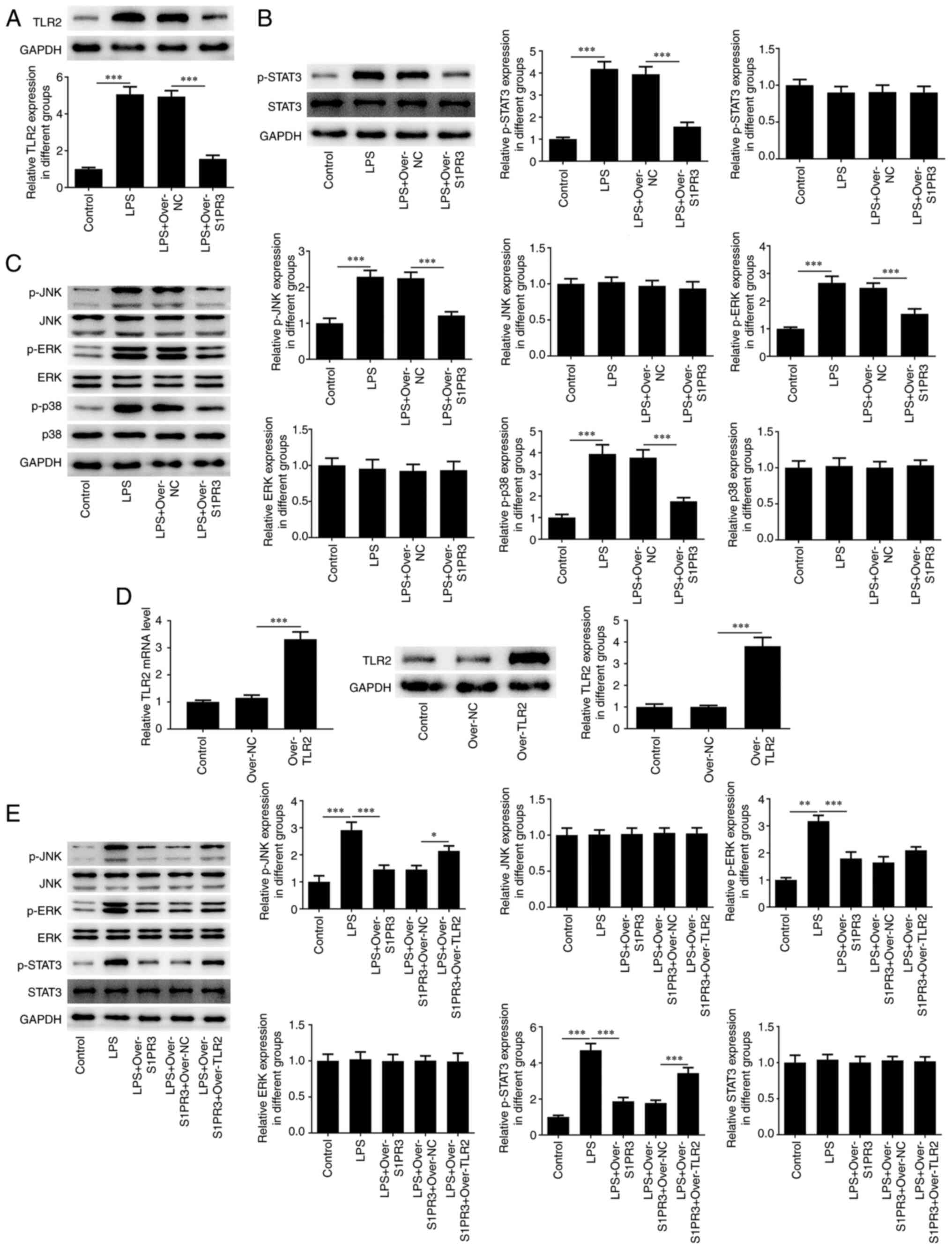
S1PR3 alleviates LPS-induced inflammatory release, apoptosis and ECM degradation of HNPCs through the TLR2/STAT3 and TLR2/MAPK pathways
TLR2 overexpression elevated the production of TNFα, IL-1β and IL-6 in S1PR3-overexpressed HNPCs (Fig. 5A). Moreover, the CCK-8 assay data showed that TLR2 overexpression decreased the viability of S1PR3-overexpressed HNPCs (Fig. 5B). In addition, the apoptosis rate in cells co-transfected with over-S1PR3 + over-TLR2 was increased by 26% compared with that in cells transfected with over-S1PR3 alone (Fig. 5C), which is in line with western blotting data, as evidenced by the reduced Bcl-2 and elevated Bax contents (Fig. 5D). Furthermore, RT-qPCR and western blotting revealed that TLR2 overexpression significantly increased the levels of Adamts-5, Adamts-4, MMP3 and MMP13, while it reduced the levels of aggrecan and collagen II in LPS-induced HNPCs transfected with over-S1PR3 (Fig. 6).


Discussion
Intervertebral disc degeneration, characterized by progressive failure of structure and ageing of the intervertebral disc, leads to pain in the lower back and even global disability (18). Existing studies have revealed that aberrant functions in HNPCs, including senescence, cytokine secretion, apoptosis and ECM degradation, are associated with IDD pathogenesis (19-21). HNPCs can generate ECM components, such as aggrecan, type II and X collagen, playing a crucial function in maintaining the integrity of intervertebral discs (22). Additional evidence indicates that the aberrant functions of HNPCs may be the key to the degeneration pathogenesis of intervertebral discs (23). In the current study, the aim was to identify the potential role of S1PR3 in HNPC viability, inflammation, apoptosis and ECM degradation, as well as disclose the molecular mechanisms underlying its function.
S1P, a bioactive signaling agent, is derived from mammalian membrane sphingolipids, and has roles in immune and inflammatory responses and cardiovascular systems (24). Most of the biological functions of S1P are regulated by S1PR1-5. Among these receptors, S1PR3 mediates multiple biological activities such as inflammation and vascular barrier function (25-27). It has been reported that pFTY720 activates S1PR3 and inhibits the TLR2/4-PI3K-NFκB signaling pathway to protect astrocytes from neuroinflammation induced by oxygen-glucose deprivation (28). S1PR3 expression has also been shown to be reduced in tissue with intervertebral disc degeneration (29). In the current study, it was shown that S1PR3 expression was low in LPS-induced HNPCs. S1PR3 overexpression restrained the cell inflammation response by reducing the levels of TNFα, IL-1β and IL-6, and repressed the level of apoptosis in LPS-induced HNPCs. It was also observed that S1PR3 overexpression repressed the levels of Adamts-5, Adamts-4, MMP3 and MMP13, while it increased the levels of aggrecan and collagen II, indicating that S1PR3 overexpression suppressed ECM degradation of LPS-stimulated HNPCs.
A previous study found that S1PR3 can inhibit the expression of the TLR family protein TLR2(28). Activation of the TLR2/JNK/mitochondrial-mediated pathway can promote apoptosis of nucleus pulpocytes and induce intervertebral disc degeneration (30). Nevertheless, Yang et al (31) reported that TSG-6 from bone marrow mesenchymal stem cells can protect against intervertebral disc degeneration via the TLR2/NF-κB signaling pathway. Moreover, TLR2 takes part in the inflammation response by activating the STAT3 and MAPK pathways (32,33). Additionally, Wu et al (34) showed that resveratrol blocks the IL-6/JAK/STAT3 pathway to protect HNPCs from degeneration. Another study has showed that the MAPK signaling proteins JNK, ERK and p38 are involved in the apoptosis and activation of HNPCs (35). In the current study, S1PR3 overexpression led to decreased TLR2 level and reduced levels of STAT3 and MAPK pathways. When TLR2 was overexpressed, it was shown that TLR2 overexpression reversed the effects of S1PR3 overexpression on LPS-induced NP cell inflammation and apoptosis, along with ECM degradation.
There are several limitations in the present study. Since the environment for cellular experiments is different from that in living organisms, thus the result of this study might not be suitable for in vivo experiments and clinical trials. The animal experiments and clinical trials will be performed in the further study. In addition, the current experiments only demonstrated that S1PR3 regulated TLR2, thus mediating TLR2/STAT3 and TLR2/MAPK signaling, but the specific mechanism by which S1PR3 regulates TLR2 remains to be explored.
To conclude, the results of the present study indicated that S1PR3 overexpression alleviated the inflammatory response and ECM degradation induced by LPS in HNPCs via suppression of the STAT3 and MAPK signaling pathways, which might provide novel sights into the exploration of S1PR3 as a promising candidate for intervertebral disc degeneration.
Acknowledgements
Not applicable.
Funding
Funding: No funding was received.
Availability of data and materials
The datasets used and/or analyzed during the current study are available from the corresponding author on reasonable request.
Authors' contributions
ZT and ZL designed the study, drafted and revised the manuscript. HG and WX analyzed the data and searched the literature. ZT, ZL, HG and WX performed the experiments. All authors read and approved the final manuscript. ZT and ZL confirm the authenticity of all the raw data.
Ethics approval and consent to participate
Not applicable.
Patient consent for publication
Not applicable.
Competing interests
The authors declare that they have no competing interests.
References
|
Urits I, Burshtein A, Sharma M, Testa L, Gold PA, Orhurhu V, Viswanath O, Jones MR, Sidransky MA, Spektor B and Kaye AD: Low back pain, a comprehensive review: Pathophysiology, diagnosis, and treatment. Curr Pain Headache Rep. 23(23)2019.PubMed/NCBI View Article : Google Scholar | |
|
Will JS, Bury DC and Miller JA: Mechanical low back pain. Am Fam Physician. 98:421–428. 2018.PubMed/NCBI | |
|
Vlaeyen JWS, Maher CG, Wiech K, Van Zundert J, Meloto CB, Diatchenko L, Battié MC, Goossens M, Koes B and Linton SJ: Low back pain. Nat Rev Dis Primers. 4(52)2018.PubMed/NCBI View Article : Google Scholar | |
|
Xin J, Wang Y, Zheng Z, Wang S, Na S and Zhang S: Treatment of intervertebral disc degeneration. Orthop Surg. 14:1271–1280. 2022.PubMed/NCBI View Article : Google Scholar | |
|
Chen S, Fu P, Wu H and Pei M: Meniscus, articular cartilage and nucleus pulposus: A comparative review of cartilage-like tissues in anatomy, development and function. Cell Tissue Res. 370:53–70. 2017.PubMed/NCBI View Article : Google Scholar | |
|
Bhujel B, Shin HE, Choi DJ and Han I: Mesenchymal stem cell-derived exosomes and intervertebral disc regeneration: Review. Int J Mol Sci. 23(7306)2022.PubMed/NCBI View Article : Google Scholar | |
|
Kirnaz S, Capadona C, Wong T, Goldberg JL, Medary B, Sommer F, McGrath LB Jr and Härtl R: Fundamentals of intervertebral disc degeneration. World Neurosurg. 157:264–273. 2022.PubMed/NCBI View Article : Google Scholar | |
|
Risbud MV and Shapiro IM: Role of cytokines in intervertebral disc degeneration: Pain and disc content. Nat Rev Rheumatol. 10:44–56. 2014.PubMed/NCBI View Article : Google Scholar | |
|
Vergroesen PP, Kingma I, Emanuel KS, Hoogendoorn RJ, Welting TJ, van Royen BJ, van Dieën JH and Smit TH: Mechanics and biology in intervertebral disc degeneration: A vicious circle. Osteoarthritis Cartilage. 23:1057–1070. 2015.PubMed/NCBI View Article : Google Scholar | |
|
Krut Z, Pelled G, Gazit D and Gazit Z: Stem cells and exosomes: New therapies for intervertebral disc degeneration. Cells. 10(2241)2021.PubMed/NCBI View Article : Google Scholar | |
|
Cartier A and Hla T: Sphingosine 1-phosphate: Lipid signaling in pathology and therapy. Science. 366(eaar5551)2019.PubMed/NCBI View Article : Google Scholar | |
|
Meshcheryakova A, Mechtcheriakova D and Pietschmann P: Sphingosine 1-phosphate signaling in bone remodeling: Multifaceted roles and therapeutic potential. Expert Opin Ther Targets. 21:725–737. 2017.PubMed/NCBI View Article : Google Scholar | |
|
Hu Y, Yang C, Shen G, Yang S, Cheng X, Cheng F, Rao J and Wang X: Hyperglycemia-triggered sphingosine-1-phosphate and sphingosine-1-phosphate receptor 3 signaling worsens liver ischemia/reperfusion injury by regulating M1/M2 polarization. Liver Transpl. 25:1074–1090. 2019.PubMed/NCBI View Article : Google Scholar | |
|
Weigel C, Hüttner SS, Ludwig K, Krieg N, Hofmann S, Schröder NH, Robbe L, Kluge S, Nierhaus A, Winkler MS, et al: S1P lyase inhibition protects against sepsis by promoting disease tolerance via the S1P/S1PR3 axis. EBioMedicine. 58(102898)2020.PubMed/NCBI View Article : Google Scholar | |
|
Hou J, Chen Q, Wu X, Zhao D, Reuveni H, Licht T, Xu M, Hu H, Hoeft A, Ben-Sasson SA, et al: S1PR3 signaling drives bacterial killing and is required for survival in bacterial sepsis. Am J Respir Crit Care Med. 196:1559–1570. 2017.PubMed/NCBI View Article : Google Scholar | |
|
Livak KJ and Schmittgen TD: Analysis of relative gene expression data using real-time quantitative PCR and the 2(-Delta Delta C(T)) method. Methods. 25:402–408. 2001.PubMed/NCBI View Article : Google Scholar | |
|
Yurube T, Takada T, Hirata H, Kakutani K, Maeno K, Zhang Z, Yamamoto J, Doita M, Kurosaka M and Nishida K: Modified house-keeping gene expression in a rat tail compression loading-induced disc degeneration model. J Orthop Res. 29:1284–1290. 2011.PubMed/NCBI View Article : Google Scholar | |
|
Francisco V, Pino J, González-Gay M, Lago F, Karppinen J, Tervonen O, Mobasheri A and Gualillo O: A new immunometabolic perspective of intervertebral disc degeneration. Nat Rev Rheumatol. 18:47–60. 2022.PubMed/NCBI View Article : Google Scholar | |
|
Li Z, Chen X, Xu D, Li S, Chan MTV and Wu WKK: Circular RNAs in nucleus pulposus cell function and intervertebral disc degeneration. Cell Prolif. 52(e12704)2019.PubMed/NCBI View Article : Google Scholar | |
|
He R, Wang Z, Cui M, Liu S, Wu W, Chen M, Wu Y, Qu Y, Lin H, Chen S, et al: HIF1A Alleviates compression-induced apoptosis of nucleus pulposus derived stem cells via upregulating autophagy. Autophagy. 17:3338–3360. 2021.PubMed/NCBI View Article : Google Scholar | |
|
Shao Z, Wang B, Shi Y, Xie C, Huang C, Chen B, Zhang H, Zeng G, Liang H, Wu Y, et al: Senolytic agent Quercetin ameliorates intervertebral disc degeneration via the Nrf2/NF-κB axis. Osteoarthritis Cartilage. 29:413–422. 2021.PubMed/NCBI View Article : Google Scholar | |
|
Li Z, Yu X, Shen J, Chan MT and Wu WK: MicroRNA in intervertebral disc degeneration. Cell Prolif. 48:278–283. 2015.PubMed/NCBI View Article : Google Scholar | |
|
Lin J, Du J, Wu X, Xu C, Liu J, Jiang L, Cheng X, Ge G, Chen L, Pang Q, et al: SIRT3 mitigates intervertebral disc degeneration by delaying oxidative stress-induced senescence of nucleus pulposus cells. J Cell Physiol. 236:6441–6456. 2021.PubMed/NCBI View Article : Google Scholar | |
|
Kunkel GT, Maceyka M, Milstien S and Spiegel S: Targeting the sphingosine-1-phosphate axis in cancer, inflammation and beyond. Nat Rev Drug Discov. 12:688–702. 2013.PubMed/NCBI View Article : Google Scholar | |
|
Muppidi JR, Lu E and Cyster JG: The G protein-coupled receptor P2RY8 and follicular dendritic cells promote germinal center confinement of B cells, whereas S1PR3 can contribute to their dissemination. J Exp Med. 212:2213–2222. 2015.PubMed/NCBI View Article : Google Scholar | |
|
Bajwa A, Huang L, Kurmaeva E, Gigliotti JC, Ye H, Miller J, Rosin DL, Lobo PI and Okusa MD: Sphingosine 1-phosphate receptor 3-deficient dendritic cells modulate splenic responses to ischemia-reperfusion injury. J Am Soc Nephrol. 27:1076–1090. 2016.PubMed/NCBI View Article : Google Scholar | |
|
Nussbaum C, Bannenberg S, Keul P, Gräler MH, Gonçalves-de-Albuquerque CF, Korhonen H, von Wnuck Lipinski K, Heusch G, de Castro Faria Neto HC, Rohwedder I, et al: Sphingosine-1-phosphate receptor 3 promotes leukocyte rolling by mobilizing endothelial P-selectin. Nat Commun. 6(6416)2015.PubMed/NCBI View Article : Google Scholar | |
|
Dong YF, Guo RB, Ji J, Cao LL, Zhang L, Chen ZZ, Huang JY, Wu J, Lu J and Sun XL: S1PR3 is essential for phosphorylated fingolimod to protect astrocytes against oxygen-glucose deprivation-induced neuroinflammation via inhibiting TLR2/4-NFκB signalling. J Cell Mol Med. 22:3159–3166. 2018.PubMed/NCBI View Article : Google Scholar | |
|
Yang K, Li H and Li C: Expression and role of Sphingosine 1-phosphate receptors in intervertebral disc degeneration. J Back Musculoskelet Rehabil. 33:255–262. 2020.PubMed/NCBI View Article : Google Scholar | |
|
Lin Y, Jiao Y, Yuan Y, Zhou Z, Zheng Y, Xiao J, Li C, Chen Z and Cao P: Propionibacterium acnes induces intervertebral disc degeneration by promoting nucleus pulposus cell apoptosis via the TLR2/JNK/mitochondrial-mediated pathway. Emerg Microbes Infect. 7(1)2018.PubMed/NCBI View Article : Google Scholar | |
|
Yang H, Tian W, Wang S, Liu X, Wang Z, Hou L, Ge J, Zhang X, He Z and Wang X: TSG-6 secreted by bone marrow mesenchymal stem cells attenuates intervertebral disc degeneration by inhibiting the TLR2/NF-κB signaling pathway. Lab Invest. 98:755–772. 2018.PubMed/NCBI View Article : Google Scholar | |
|
Jin Y, Nguyen TLL, Myung CS and Heo KS: Ginsenoside Rh1 protects human endothelial cells against lipopolysaccharide-induced inflammatory injury through inhibiting TLR2/4-mediated STAT3, NF-κB, and ER stress signaling pathways. Life Sci. 309(120973)2022.PubMed/NCBI View Article : Google Scholar | |
|
Xu Z, Hao X, Li M and Luo H: Rhodococcus equi-derived extracellular vesicles promoting inflammatory response in macrophage through TLR2-NF-κB/MAPK pathways. Int J Mol Sci. 23(9742)2022.PubMed/NCBI View Article : Google Scholar | |
|
Wu C, Ge J, Yang M, Yan Q, Wang Y, Yu H, Yang H and Zou J: Resveratrol protects human nucleus pulposus cells from degeneration by blocking IL-6/JAK/STAT3 pathway. Eur J Med Res. 26(81)2021.PubMed/NCBI View Article : Google Scholar | |
|
Sun K, Zhu J, Yan C, Li F, Kong F, Sun J, Sun X, Shi J and Wang Y: CGRP regulates nucleus pulposus cell apoptosis and inflammation via the MAPK/NF-κB signaling pathways during intervertebral disc degeneration. Oxid Med Cell Longev. 2021(2958584)2021.PubMed/NCBI View Article : Google Scholar |


