Introduction
Since the volume of the orbit is a significant
determinant of the facial appearance (1), and is influenced by many diseases and
situations, understanding the volumetric values and changes with
respect to body parameters and gender is likely to be of guidance
in the resolution of serious clinical issues, such as when studying
the mechanisms of exophthalmos in Graves' orbitopathy, planning
orbital decompression, orbital reconstruction following fractures,
determining the size of orbital implants following enucleation, or
conducting esthetic procedures such as blepharoplasty. Previous
studies have examined factors affecting the orbital or eyeball
volume by computed tomography (CT) or magnetic resonance imaging
(MRI) (2–4). Chau et al (4) reported that MRI provided more accurate
results than CT in orbital and eyeball volume measurements. Ranly
(5) observed that the orbital volume
is proportional to eyeball volume since the orbit is associated
with the growth of the bone and eyeball. Kahn et al
(6) reported the change of orbital
size with respect to age, but did not study orbital volumes.
Previous studies have examined volumetric measurements of the orbit
and their variability (7–9) with gender or body parameters; however,
volumetric parameters were examined separately in these studies,
and in a limited number of cases. The aim of the present study was
to examine the total orbital volume (TOV) and total orbital fat
volume (TOFV) together, and to investigate their correlation with
clinical parameters, such as age, gender, height and weight, in a
large group of patients in order to the determine the effect of
these parameters on orbital appearance, as well as to identify
racial differences in orbital volume.
Materials and methods
Subjects
Ethical approval for the present study was obtained
from the Bozok Medical Faculty Ethics Committee (Yozgat, Turkey).
Informed consent was obtained from each patient prior to their
details being used in the current study. A retrospective review was
undertaken in the radiology department. Subjects were selected from
the patients who had been referred to the radiology departments of
the Bozok and Cumhuriyet Universities (Yozgat and Sivas, Turkey,
respectively) in the previous 3 years for orbital or maxillofacial
evaluation. Thin-slice cranial magnetic resonance (MR) images of
patients obtained for various reasons, such as frontal mass or
vascular lesions, which did not directly affect the orbital
structures, were also selected for orbital measurements. Patients
with a history of previous surgery, trauma or infection of ocular,
orbital or periorbital structures, and who had any refractive
errors, heterotropia, mass lesions involving the maxilla or
adjacent structures, and osteomyelitis of the maxilla, and patients
with known thyroid-associated orbitopathy or hyperthyroidism were
excluded to rule out the potential effects of these factors on
measurements. A total of 1,453 Anatolian subjects with ages ranging
from 20 to 69 years (705 males and 748 females) were selected and
grouped into five age groups with 10-year intervals. Table I shows the range of ages and the mean
± standard deviation (SD) age for each group of patients.
 | Table I.Mean ± standard deviation (SD) ages of
the participants and the number of subjects in each group. |
Table I.
Mean ± standard deviation (SD) ages of
the participants and the number of subjects in each group.
|
|
| Male | Female | Male + female |
|---|
|
|
|
|
|
|
|---|
| Groups | Age range, years | n | Age, years (mean ±
SD) | n | Age, years (mean ±
SD) | n | Age, years (mean ±
SD) |
|---|
| Group I | 20–29 | 162 | 27.8±1.1 | 175 | 24.8±3.3 | 337 | 26.3±2.9 |
| Group II | 30–39 | 151 | 36.5±3.3 | 168 | 32.1±2.1 | 319 | 34.3±3.0 |
| Group III | 40–49 | 149 | 45.2±3.9 | 166 | 43.8±5.1 | 315 | 44,5±4.6 |
| Group IV | 50–59 | 163 | 56.1±4.1 | 148 | 56.9±3.9 | 311 | 56.5±4.1 |
| Group V | 60–69 | 80 | 66.9±4.1 | 91 | 64.9±4.0 | 171 | 65.9±4.0 |
| Total | 20–69 | 705 | 46.5±13.8 | 748 | 44.5±13.4 | 1,453 | 45.5±13.5 |
Image acquisition
All MRI examinations were performed using a whole
body 1.5-T unit (Magnetom Symphony; Siemens, Germany) with a
16-channel sensitivity encoding (SENSE) head coil. T1-weighted
(T1W) axial, coronal and sagittal images were analyzed. T1W-turbo
spin echo (TSE) images were obtained with the following parameters:
repetition time (TR)/echo time (TE)/number of signal averages
(NSA), 114 msec/3,810 msec/1; flip angle, 150°; matrix, 256×192;
field of view (FOV), 17.8×17.4 cm; bandwidth, 130 kHz; echo train
length, 13; and slice thickness, 3 mm.
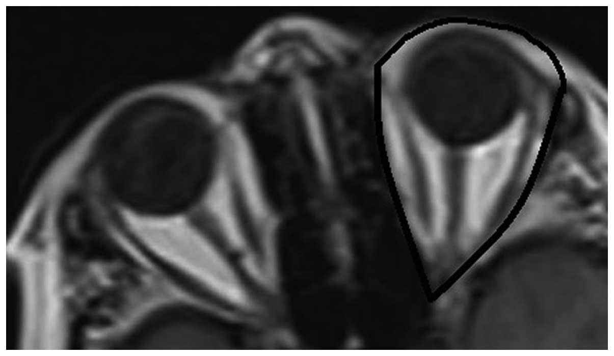
Volumetry
For the determination of TOV in axial slices, the
posterior boundary of the orbit was defined as the crossing line of
the medial and lateral walls of the optic foramen. The anterior
boundary of the orbit was defined as the line that connects the two
end points of the front medial and lateral walls and which lies
down the cornea (Fig. 1). The
eyelids were included, since they are challenging to separate from
the anterior boundary of the cornea and sclera. Following
assignment of the region of interest (ROI) by two radiologists in
consensus, and the area of the ROI multiplied by slice thickness,
TOV was calculated by summing these volumes in each slice in T1W
axial images.
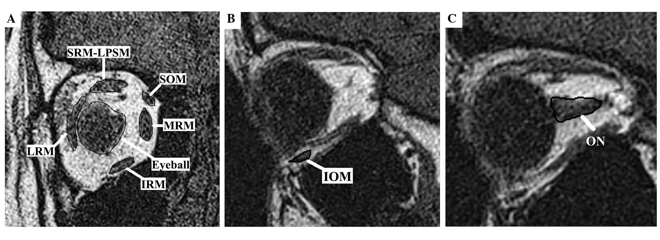
Since the orbital cavity volume inside the bony
orbit includes the eyeball, orbital muscles, optic nerve and
orbital fatty tissues, when determining the TOFV, the eyeball,
optic nerve, lacrimal gland and orbital muscle volumes were
subtracted from the TOV, with the exception of the vasculature
which is almost impossible to outline volumetrically; the
difference was recorded as the TOFV. In order to do this, the
volumes of the medial rectus muscle (MRM), inferior rectus muscle
(IRM), lateral rectus muscle (LRM), superior rectus-levator
palpebrae superioris muscle complex (SRM-LPSM) and superior oblique
muscle (SOM) were measured in T1W coronal images by summing the
cross-sectional areas in every section and multiplying by the image
plane thickness. Inferior oblique muscle (IOM) and optic nerve (ON)
volumes were calculated similarly on T1W sagittal images, as
described by Detorakis et al (10,11)
(Fig. 2).
Statistical analysis
Clinical information (age, gender, height and
weight) was obtained for each subject. Continuous variables were
expressed as the mean ± SD. To investigate the effect of age and
gender on the TOV and TOFV, two-way analysis of variance (ANOVA)
was employed using SPSS software (version 15.0; SPSS, Inc.,
Chicago, IL, USA), in which age and gender (with two levels) were
used as two independent variables. A multiple regression analysis
was performed to determine the effect of the body parameters
(height and weight) on the change in TOV and TOFV with respect to
age and gender. P-values less than 0.05 were considered to be
statistically significant.
Results
TOV
The mean TOV of all men was 32.21±1.55
cm3, and that of all women was 31.11±1.87 cm3
and the overall average for the study population was 31.66±1.91
cm3. The mean TOV of men was significantly greater than
that of women (P<0.001), and the mean TOV of all subjects in
group V was greater than that of all other groups (P<0.001),
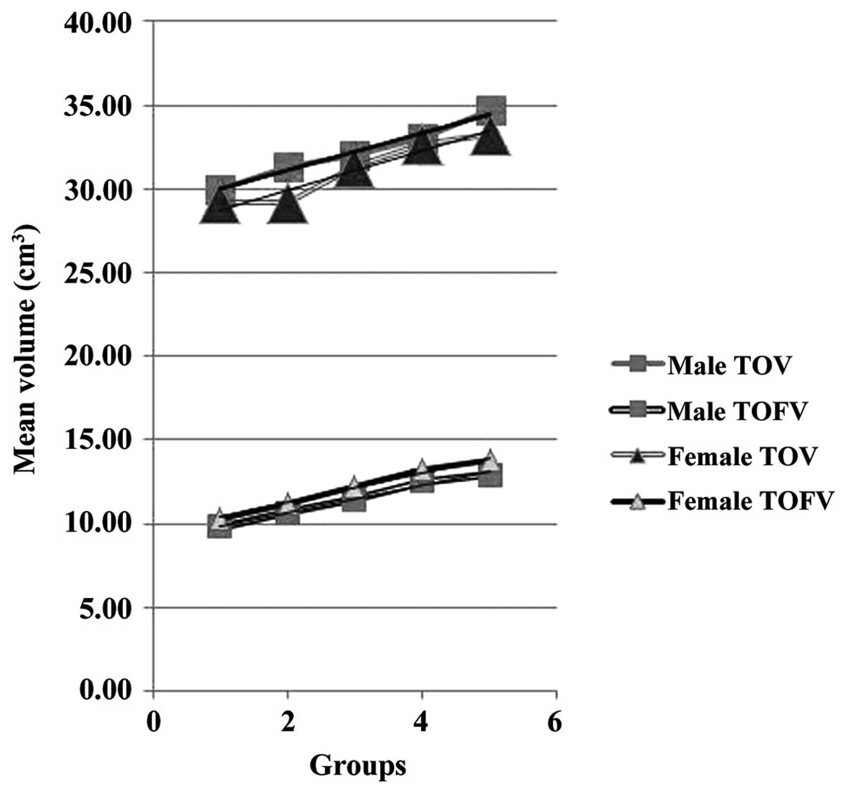
demonstrating that TOV increased with age (Table II). There was no significant
difference between the right and left orbital volumes for either
men or women. As shown in Table
III and Fig. 3, there was an
interactive effect between age and gender (P<0.01); as age
increased, the TOV of men increased more rapidly than that of
women.
| Table II.Average TOV and TOFV (in
cm3) with respect to gender and age. |
Table II.
Average TOV and TOFV (in
cm3) with respect to gender and age.
|
| Male | Female | Male + female |
|---|
|
|
|
|
|
|---|
| Group | TOV | TOFV | TOV | TOFV | TOV | TOFV |
|---|
| Group I | 29.99±1.47 | 9.83±1.32 | 29.13±1.74 | 10.34±1.73 | 29.56±1.47 | 10.08±1.64 |
| Group II | 31.33±1.88 | 10.65±1.45 | 29.17±1.65 | 11.24±1.35 | 30.25±1.28 | 10.94±1.26 |
| Group III | 31.99±1.28 | 11.46±1.74 | 31.33±1.88 | 12.22±1.40 | 31.66±1.35 | 11.84±1.59 |
| Group IV | 33.02±1.33 | 12.54±1.82 | 32.66±1.36 | 13.18±1.82 | 32.84±1.25 | 12.86±1.41 |
| Group V | 34.75±1.38 | 12.93±1.34 | 33.29±1.82 | 13.77±1.33 | 34.02±1.88 | 13.35±1.49 |
| Total | 32.21±1.55 | 11.48±1.81 | 31.11±1.87 | 12.15±1.66 | 31.66±1.91 | 11.81±1.88 |
| Table III.Results of TOV and TOFV analysis with
ANOVA, using age and gender as independent variables. |
Table III.
Results of TOV and TOFV analysis with
ANOVA, using age and gender as independent variables.
|
| Type III sum of
squares |
| Mean square | F | P-value |
|---|
|
|
|
|
|
|
|
|---|
| Source | TOV | TOFV | d.f | TOV | TOFV | TOV | TOFV | TOV | TOFV |
|---|
| Corrected
model | 382.397 | 256.198 | 3 | 132.655 | 111.583 | 49.587 | 42.314 |
0.000 |
0.000 |
| Age | 165.765 | 132.161 | 1 | 135.478 | 132.161 | 58.563 | 50.858 | <0.001 | <0.001 |
| Gender | 184.008 | 129.810 | 1 | 152.145 | 129.810 | 69.532 | 61.545 | <0.001 | <0.001 |
| Age × gender | 23.667 | 21.667 | 1 | 23.667 | 21.667 | 13.666 | 11.213 | <0.01 | <0.01 |
TOFV
The mean TOFV was 11.48±1.81 cm3 for men,
and 12.15±1.66 cm3 for women, the overall mean for the
study population being 11.81±1.88 cm3. The mean TOFV of
women was significantly greater than that of men (P<0.001). The
mean TOFV of all subjects in group V was greater than that of the
mean TOFV values of all other groups (P<0.001), which showed
that TOFV also increased with age (Table II). There was no significant
difference between the right and left orbital volumes for either
gender. There was an interactive effect between age and gender
(P<0.01); as age increased, the TOFV of women increased more
rapidly than that of men (Table
III and Fig. 3).
Correlations
While both weight and height showed a positive
correlation with TOV in male and female subjects in groups I-III,
only weight showed a positive correlation with TOV in the female
subjects in groups IV and V. In all groups, TOFV showed a positive
correlation with weight, but no significant correlation with height
was observed. The effects of the body parameters (height and
weight) on the changes in TOV and TOFV with respect to age and
gender are shown in Tables IV and
V.
| Table IV.Results of multiple regression
analysis for TOV, using body parameters (height and weight) as
independent variables. |
Table IV.
Results of multiple regression
analysis for TOV, using body parameters (height and weight) as
independent variables.
|
|
| Unstandardized
coefficients |
|
|
|
|---|
|
|
|
|
|
|
|
|---|
| Age | Predictors | B | SE | Standardized
coefficient β | t | P-value |
|---|
| Group I |
|
|
|
|
|
|
|
Male | Constant | 5.706 | 6.816 |
| 0.637 | 0.529 |
| Height | 0.057 | 0.055 | 2.411 | 1.027 | 0.003a |
| Weight | 0.091 | 0.031 | 2.498 | 2.981 | 0.006a |
|
Female | Constant | 0.068 | 5.413 |
| 0.009 | 0.993 |
| Height | 0.061 | 0.049 | 2.385 | 2.173 | 0.005a |
| Weight | 0.064 | 0.045 | 2.419 | 1.422 | 0.004a |
| Group II |
|
|
|
|
|
|
|
Male | Constant | 11.254 | 5.833 |
| 0.699 | 0.502 |
| Height | 0.081 | 0.049 | 2.481 | 1.311 | 0.048a |
| Weight | 0.074 | 0.029 | 2.433 | 2.989 | 0.006a |
|
Female | Constant | 8.866 | 6.312 |
| 0.003 | 0.859 |
| Height | 8.303 | 0.039 | 2.311 | 2.215 | 0.004a |
| Weight | 0.061 | 0.035 | 2.851 | 1.328 | 0.008a |
| Group III |
|
|
|
|
|
|
|
Male | Constant | 21.185 | 19.664 |
| 2.809 | 0.009 |
| Height | 0.098 | 0.089 | 2.307 | 1.738 | 0.008a |
| Weight | 0.086 | 0.031 | 2.487 | 2.753 | 0.007a |
|
Female | Constant | 9.505 | 6.434 |
| 2.099 | 0.420 |
| Height | 0.006 | 0.041 | 2.021 | 0.141 | 0.004a |
| Weight | 0.065 | 0.024 | 2.405 | 2.758 | 0.009a |
| Group IV |
|
|
|
|
|
|
|
Male | Constant | 26.214 | 22.545 |
| 2.211 | 0.569 |
| Height | -0.434 | -0.387 | -0.255 | 1.782 | 0.299 |
| Weight | 0.124 | 0.198 | 0.359 | 1.958 | 0.485 |
|
Female | Constant | 6.426 | 8.102 |
| 2.013 | 0.682 |
| Height | 0.312 | 0.299 | 0.019 | 0.155 | 0.799 |
| Weight | 0.063 | 0.021 | 2.898 | 2.598 | 0.009a |
| Group V |
|
|
|
|
|
|
|
Male | Constant | 28.216 | 23.663 |
| 2.809 | 0.419 |
| Height | -0.155 | 0.098 | -0.207 | 1.738 | 0.313 |
| Weight | 0.148 | 0.131 | 0.487 | 1.753 | 0.411 |
|
Female | Constant | 9.505 | 6.434 |
| 2.099 | 0.630 |
| Height | 0.214 | 0.141 | 0.021 | 0.141 | 0.711 |
| Weight | 0.065 | 0.024 | 2.316 | 2.758 | 0.009a |
| Table V.Results of multiple regression
analysis for TOFV, using body parameters (height and weight) as
independent variables. |
Table V.
Results of multiple regression
analysis for TOFV, using body parameters (height and weight) as
independent variables.
|
|
| Unstandardized
coefficients |
|
|
|
|---|
|
|
|
|
|
|
|
|---|
| Age | Predictors | B | SE | Standardized
coefficient β | t | P-value |
|---|
| Group I |
|
|
|
|
|
|
|
Male | Constant | 11.214 | 9.213 |
| 0.582 | 0.503 |
| Height | 0.051 | 0.056 | 2.864 | 1.386 | 0.697 |
| Weight | 0.082 | 0.038 | 2.194 | 2.107 | 0.006a |
|
Female | Constant | 0.044 | 4.832 |
| 0.089 | 0.993 |
| Height | 0.037 | 0.041 | 2.657 | 2.547 | 0.857 |
| Weight | 0.048 | 0.039 | 2.189 | 1.875 | 0.004a |
| Group II |
|
|
|
|
|
|
|
Male | Constant | 18.315 | 16.212 |
| 1.341 | 0.614 |
| Height | 0.068 | 0.086 | 1.954 | 1.378 | 0.313 |
| Weight | 0.041 | 0.034 | 2.965 | 2.656 | 0.006a |
|
Female | Constant | 10.265 | 8.205 |
| 3.012 | 0.391 |
| Height | 0.006 | 0.061 | 2.861 | 0.398 | 0.424 |
| Weight | 0.090 | 0.021 | 2.613 | 2.265 | 0.008a |
| Group III |
|
|
|
|
|
|
|
Male | Constant | 21.315 | 18.542 |
| 2.341 | 0.216 |
| Height | 0.072 | 0.078 | 1.832 | 1.378 | 0.548 |
| Weight | 0.039 | 0.038 | 2.818 | 2.656 | 0.007a |
|
Female | Constant | 12.365 | 9.268 |
| 3.012 | 0.256 |
| Height | 0.058 | 0.069 | 2.317 | 0.398 | 0.854 |
| Weight | 0.099 | 0.019 | 2.326 | 2.265 | 0.009a |
| Group IV |
|
|
|
|
|
|
|
Male | Constant | 22.498 | 20.128 |
| 2.785 | 0.212 |
| Height | 0.098 | 0.066 | 1.919 | 1.585 | 0.366 |
| Weight | 0.049 | 0.031 | 2.625 | 1.965 | 0.006a |
|
Female | Constant | 13.294 | 10.025 |
| 3.444 | 0.125 |
| Height | 0.098 | 0.081 | 0.825 | 0.325 | 0.568 |
| Weight | 0.065 | 0.023 | 2.448 | 2.265 | 0.004a |
| Group V |
|
|
|
|
|
|
|
Male | Constant | 23.214 | 22.838 |
| 2.425 | 0.554 |
| Height | 0.001 | 0.098 | 0.189 | 1.284 | 0.197 |
| Weight | 0.148 | 0.0184 | 0.397 | 1.697 | 0.005a |
|
Female | Constant | 17.212 | 11.232 |
| 2.536 | 0.265 |
| Height | 0.291 | 0.182 | 0.029 | 0.261 | 0.265 |
| Weight | 0.086 | 0.018 | 1.093 | 2.256 | 0.009a |
Statistical analysis of the results confirms that as
age increases TOV and TOFV also increase; however, the correlation
of these volumetric parameters was not found to be statistically
significant (Pearson's bivariate correlation coefficient, 0.58,
P=0.02).
Discussion
This study evaluated orbital volumetric parameters
and the correlation of these measurements with age, gender and body
parameters (height and weight) in a group of 1,453 subjects with
exclusion of any previous orbital disease history, using MRI, and
is one of the largest series in the literature. The high number of
patients may be attributable to the high number of patients with
multiple sclerosis in this region of the country, who frequently
present with visual symptoms and undergo MRI for them. The orbit is
a limited anatomical structure filled with eyeball,
non-compressible fluid and orbital cavity soft tissues. Atchison
et al and Chau et al (3,4) reported
that MRI provided more accurate results than CT in orbital and
eyeball volume measurements and that the average orbital volume was
20.9 cm3 in an Asian population, and Forbes et al
(12) identified an upper limit of
30.1 cm3 for normal bony orbit in Western individuals.
These values are smaller than those in the present study since they
were obtained only from the bony orbit, and did not include
anterior soft tissues of the orbit. A study carried out in the
United States using CT, which also included all of the soft tissues
around the eye showed similar volumetric values as the present
study, which suggests that the size of the orbits in the Anatolian
population resemble those of Western individuals, in contrast to
those of the Far Eastern world (13).
In the literature, the average orbital volumetric
changes with respect to gender are controversial. Certain studies
found larger orbital volumes in men than women (3,14), while
another (12) found no statistically
significant difference. In the present study, it was observed that
the average TOV was larger in men than in women. This result may be
attributed to the fact that the axial skeleton is greater in size
in men than in women, which would affect the bony orbit as well. It
has been reported that orbital size correlates well with the bone
size of the whole body skeleton as a result of the growth pattern,
and also with skull length (3,5), which
may explain the difference between genders. There was also an
interactive effect between age and gender: As age increased, the
TOV of men increased more rapidly than that of women. Since the
bony elements decrease with age in the whole body (15), the finding that decrements in orbital
angles and the volume of the bony orbit also occur with age, in a
similar manner to those in the axial skeleton (16,17) is
not surprising. However, considering the results of the present
study, the total volume loss of the orbital structures appears to
be less than that of the bony orbital volume, which may be due to
replacement of the lost volume with adipose tissue.
Regarding the changes in TOV with respect to the
body parameters, positive correlations were observed with both
weight and height in male and female subjects in group I-II. As
orbital volume has an association with bony elements, Shaw et
al (17) pointed out that
orbital bony changes are similar to those of the axial skeleton.
Kirchengast et al (18)
suggested that a lower weight status and a low amount of lean body
mass may be associated with increased bone loss, and Gracia-Marco
et al (19) showed that
adolescents with a low level of fitness had a lower bone mineral
content. Therefore, it can be concluded that the orbital volume has
a positive correlation with weight as well as height. However, in
the present study, only weight showed a positive correlation with
TOV in female subjects older than 50 years of age. Kirchengast
et al (18) suggested that
the absolute fat mass had a significant impact on bone mineral
density in females, due to the diminished conversion rates from
androgens to estrogens in a low amount of adipose tissue. Since
high amounts of fat tissue are particularly present in elderly
women, this can explain our results. However, further studies based
on physiological and biological analysis are required to clarify
this issue.
TOFV is an important indicator of eye movements,
eyelid configuration and function. It is known that loss of TOFV
due to any cause causes eyelid retraction or proptosis that
requires reconstruction to restore functional anatomy and aesthetic
facial appearance, and in several studies (20,21)
orbital fat volume augmentation is discussed. In the present study,
the average TOFV of women was significantly greater than that of
men. Additionally, the average TOFV increased as age increased.
This is in contrast with the speculation of Ahmadi et al
(22) that the orbital adipocytes
undergo atrophy with advancing age. The significant correlation of
age with TOFV may be attributable to a relative reduction in
muscular bulk and volume of the globe itself with increasing age
(23,24). In addition, Darcy et al
(25) observed an increase in total
orbital fat and periocular soft tissue with aging, which they
claimed was due to fat expansion, which is consistent with the
results of the present study, and which may be the rationale for
fat excision from the eyelids in plastic and aesthetic surgery.
Although the present study revealed an increase in TOFV and TOV
with respect to age, there was no statistically significant
correlation between the changes in TOFV and TOV, which should be
further investigated. There was a positive interactive effect with
age and gender regarding TOFV, showing that as age increased the
TOFV of women increased more rapidly than that of men. In contrast
to some studies (26,27), TOFV showed a positive correlation
with weight in the present study. It is suggested that with
increment of weight, as the distribution of fatty tissues varies in
the body, this also affects the TOFV values. When age is
considered, following hormonal changes in the postmenopausal
period, the abnormal increase of fatty tissues in women may explain
the results of the present study. Further studies based on body
mass index are required to support this hypothesis. Another
limitation of this study is that it was carried out in a certain
limited population, despite its large size, and, therefore, may not
be valid in various parts of the world, such as eastern Asia.
To conclude, the study findings provide basic
information about orbital volume changes due to gender and body
parameters and may aid in clinical practice, such as when studying
the mechanisms of exophthalmos in Graves' orbitopathy, planning
orbital decompression or orbital reconstruction following
fractures, or the size of orbital implants following enucleation,
blepharoplasty and other esthetic procedures involving eyelids and
retroorbital fatty tissue. In addition, knowing the normal size and
proportions of the contents of the orbit may help diagnosing
unilateral ophthalmopathy.
Acknowledgements
Main topics of this study have been accepted for
oral presentation, and were presented at ECR 2014, Vienna, Austria
with presentation No. B-0171.
References
|
1
|
Quatrehomme G and Gérard S: Classical
noncomputer-assisted craniofacial reconstructionComputer-Graphic
Facial Reconstruction. Clement JG and Marks MK: Elsevier;
Burlington, MA: pp. 15–32. 2005
|
|
2
|
Bentley RP, Sgouros S, Natarajan K, et al:
Normal changes in orbital volume during childhood. J Neurosurg.
96:742–746. 2002. View Article : Google Scholar : PubMed/NCBI
|
|
3
|
Atchison DA, Jones CE, Schmid KL, et al:
Eye shape in emmetropia and myopia. Invest Ophthalmol Vis Sci.
45:3380–3386. 2004. View Article : Google Scholar : PubMed/NCBI
|
|
4
|
Chau A, Fung K and Yap M: Evaluation of
the accuracy of volume determination on the orbit and eyeball using
MRI. Radiography. 11:35–39. 2005. View Article : Google Scholar
|
|
5
|
Ranly DM: Craniofacial growth. Dent Clin
North Am. 44:457–470. 2000.PubMed/NCBI
|
|
6
|
Kahn DM and Shaw RB Jr: Aging of the bony
orbit: A three-dimensional computed tomographic study. Aesthet Surg
J. 28:258–264. 2008. View Article : Google Scholar : PubMed/NCBI
|
|
7
|
Kim SP, Lee BY, Lee SJ, et al: A study on
orbital volume of Korean people in their 20s or 40s. Ophthalmic
Res. 47:98–102. 2012. View Article : Google Scholar : PubMed/NCBI
|
|
8
|
Detorakis ET, Drakonaki E, Papadaki E,
Pallikaris IG and Tsilimbaris MK: Effective orbital volume and
eyeball position: An MRI study. Orbit. 29:244–249. 2010. View Article : Google Scholar : PubMed/NCBI
|
|
9
|
Darcy SJ, Hakimi M, Miller TA, Goldberg
RA, Evans GR, Demer JL and Rudkin GH: Magnetic resonance imaging
characterization of orbital soft-tissue volume dynamics with age.
Plast Reconstr Surg. 124:1362–1364. 2009. View Article : Google Scholar : PubMed/NCBI
|
|
10
|
Detorakis ET, Engstrom RE, Straatsma BR
and Demer JL: Functional anatomy of the anophthalmic socket:
Insights from magnetic resonance imaging. Invest Ophthalmol Vis
Sci. 44:4307–4313. 2003. View Article : Google Scholar : PubMed/NCBI
|
|
11
|
Detorakis ET, Drakonaki EE, Papadaki E,
Tsilimbaris MK and Pallikaris IG: Evaluation of globe position
within the orbit: Clinical and imaging correlations. Br J
Ophthalmol. 94:135–136. 2010. View Article : Google Scholar : PubMed/NCBI
|
|
12
|
Forbes G, Gehring DG, Gorman CA, Brennan
MD and Jackson IT: Volume measurements of normal orbital structures
by computed tomographic analysis. AJR Am J Roentgenol. 145:149–154.
1985. View Article : Google Scholar : PubMed/NCBI
|
|
13
|
Özkan AY, Karmonik C, Chaudhry IA, Arat A,
Totan S and Yüksel E: Measurement of eyelid and orbital fat volume
in different age groups by computed tomography. Turkiye Klinikleri
J Med Sci. 30:995–1001. 2010. View Article : Google Scholar
|
|
14
|
Furuta M: Measurement of orbital volume by
computed tomography: Especially on the growth of the orbit. Jpn J
Ophthalmol. 45:600–606. 2001. View Article : Google Scholar : PubMed/NCBI
|
|
15
|
Shaw RB Jr and Kahn DM: Aging of the
midface bony elements: A three-dimensional computed tomographic
study. Plast Reconstr Surg. 119:675–683. 2007. View Article : Google Scholar : PubMed/NCBI
|
|
16
|
Richard MJ, Morris C, Deen BF, Gray L and
Woodward JA: Analysis of the anatomic changes of the aging facial
skeleton using computer-assisted tomography. Ophthal Plast Reconstr
Surg. 25:382–386. 2009. View Article : Google Scholar : PubMed/NCBI
|
|
17
|
Shaw RB Jr, Katzel EB, Koltz PF, Kahn DM,
Puzas EJ and Langstein HN: Facial bone density: Effects of aging
and impact on facial rejuvenation. Aesthet Surg J. 32:937–942.
2012. View Article : Google Scholar : PubMed/NCBI
|
|
18
|
Kirchengast S, Peterson B, Hauser G and
Knogler W: Body composition characteristics are associated with the
bone density of the proximal femur end in middle- and old-aged
women and men. Maturitas. 39:133–145. 2001. View Article : Google Scholar : PubMed/NCBI
|
|
19
|
Gracia-Marco L, Vicente-Rodríguez G,
Casajús JA, Molnar D, Castillo MJ and Moreno LA: Effect of fitness
and physical activity on bone mass in adolescents: The HELENA
Study. Eur J Appl Physiol. 111:2671–2680. 2011. View Article : Google Scholar : PubMed/NCBI
|
|
20
|
Lee JY, Lee KH, Shin HM, Chung KH, Kim GI
and Lew H: Orbital volume augmentation after injection of human
orbital adipose-derived stem cells in rabbits. Invest Ophthalmol
Vis Sci. 54:2410–2416. 2013. View Article : Google Scholar : PubMed/NCBI
|
|
21
|
Park J, Cho HK and Moon JI: Changes to
upper eyelid orbital fat from use of topical bimatoprost,
travoprost, and latanoprost. Jpn J Ophthalmol. 55:22–27. 2011.
View Article : Google Scholar : PubMed/NCBI
|
|
22
|
Ahmadi H, Shams PN, Davies NP, Joshi N and
Kelly MH: Age-related changes in the normal sagittal relationship
between globe and orbit. J Plast Reconstr Aesthet Surg. 60:246–250.
2007. View Article : Google Scholar : PubMed/NCBI
|
|
23
|
Hahn FJ and Chu WK: Ocular volume measured
by CT scans. Neuroradiology. 26:419–420. 1984. View Article : Google Scholar : PubMed/NCBI
|
|
24
|
Smith P: On the size of the cornea in
relation to age, sex, refraction and primary glaucoma. Trans
Ophthalmol Soc UK. 10:68–78, 18p0.
|
|
25
|
Darcy SJ, Miller TA, Goldberg RA,
Villablanca JP, Demer JL and Rudkin GH: Magnetic resonance imaging
characterization of orbital changes with age and associated
contributions to lower eyelid prominence. Plast Reconstr Surg.
122:921–931. 2008. View Article : Google Scholar : PubMed/NCBI
|
|
26
|
Castanares S: Blepharoplasty for herniated
intraorbital fat; anatomical basis for a new approach. Plast
Reconstr Surg (1946). 8:46–58. 1951. View Article : Google Scholar : PubMed/NCBI
|
|
27
|
Sires BS, Lemke BN, Dortzbach RK and
Gonnering RS: Characterization of human orbital fat and connective
tissue. Ophthal Plast Reconstr Surg. 14:403–414. 1998. View Article : Google Scholar : PubMed/NCBI
|