Introduction
Human epidermal growth factor receptor 2 (HER2) is
amplified in 20–25% of breast cancer cases and is associated with a
shorter time to relapse and a reduced overall survival time
(1,2). In combination with chemotherapy, the
humanized HER2-targeting monoclonal antibody, trastuzumab,
demonstrates a marked therapeutic efficacy for treating patients
with HER2-expressing metastatic breast cancer. Despite this, a
considerable proportion of metastatic HER2-amplified breast cancers
fail to respond to, or demonstrate a limited beneficial response
to, trastuzumab (3,4). Therefore, an obligation exists to
identify novel biomarkers that may predict the response of
metastatic breast cancers to trastuzumab.
Previous studies have demonstrated that
phosphoinositol-3 kinase (PI3K) activation may be associated with
trastuzumab resistance in breast cancer (5). The activation of the PI3K pathway is
usually determined by phosphatase and tensin homolog (PTEN) loss
and/or activating mutations of phosphoinositol-3 kinase α catalytic
subunit (PIK3CA). A number of preclinical studies have demonstrated
that PTEN loss and PIK3CA mutation are significantly associated
with a poorer trastuzumab-based treatment efficacy in HER2-positive
metastatic breast cancers (6).
Therefore, the activation status of the PI3K pathway may be used in
clinical practice to predict HER2-targeted therapy resistance.
Andersen et al (7) revealed that the proline-rich Akt
substrate of 40 kDa (PRAS40) phosphorylated at threonine 246
(phospho-PRAS40Thr246) was positively correlated with
PI3K pathway activation. This identified
phospho-PRAS40Thr246 as a potential marker of PI3K
pathway activation. In addition, the
phospho-PRAS40Thr246 epitope is highly stable in tissue
samples and is therefore suitable for immunohistochemistry (IHC)
techniques (7). The present study
aimed to investigate whether phospho-PRAS40Thr246, as a
novel immunostaining marker for PI3K pathway activation, has the
potential to predict trastuzumab efficacy in patients with
HER2-positive metastatic breast cancer.
Materials and methods
Patient materials
Subsequent to obtaining written informed consent,
formalin-fixed, paraffin-embedded (FFPE) HER2-overexpressing
primary breast carcinoma specimens were retrospectively collected
from 55 patients who had developed metastatic breast cancer and had
received trastuzumab treatment alone (6 mg/kg alone once every
three weeks for one year; n=34) or in combiantion with paclitaxel
(175 mg/m2, once every three weeks for three months;
n=21) between January 2007 and January 2011 at the Shandong
Provincial Qianfoshan Hospital (Jinan, China). This study was
approved by the ethics committee of Shandong Provincial Qianfoshan
Hospital, Shandong University (Jinan, China). Complete data on
tumor characteristics, treatment details and follow-up results of
disease progression were obtained for all cases. Prior to the
study, no patient had received trastuzumab-based neoadjuvant
therapy. The tumors were identified as HER2-positive by the
overexpression of HER2, as detected by IHC, and/or HER2 gene
amplification using fluorescence in situ hybridization.
According to standard clinical instructions, the expression of the
estrogen and progesterone receptors was evaluated by IHC, and
expression data were retrospectively reviewed and extracted from
the medical records. An increase in the diameters of the existing
lesions by ≥20%, or the appearance of any new lesion, was defined
as progressive disease (8). The
time-to-progression (TTP) was calculated starting from the
initiation of trastuzumab-based treatment until the time of disease
progression. The clinical and pathological characteristics are
shown in Table I.
 | Table IAssociation between
phospho-PRAS40Thr246 expression, PI3K activation markers
and clinicopatholgical characteristics. |
Table I
Association between
phospho-PRAS40Thr246 expression, PI3K activation markers
and clinicopatholgical characteristics.
|
Phospho-PRAS40Thr246 | |
|---|
|
| |
|---|
| Characteristics | Negative, n | Positive, n | P-value |
|---|
| Age, years | | | 0.319 |
| Premenopausal | 13 | 5 | |
| Postmenopausal | 20 | 17 | |
| Grade | | | 0.869 |
| 1–2 | 17 | 10 | |
| 3 | 16 | 12 | |
| ER status | | | 0.407 |
| Negative | 20 | 10 | |
| Positive | 13 | 12 | |
| PR status | | | 0.860 |
| Negative | 23 | 14 | |
| Positive | 10 | 8 | |
| PIK3CA mutation | | | 0.284 |
| Negative | 29 | 16 | |
| Positive | 4 | 6 | |
| PTEN loss | | | 0.024 |
| Negative | 26 | 10 | |
| Positive | 7 | 12 | |
| PIK3CA mutants or low
PTEN | | | 0.001 |
| No | 24 | 4 | |
| Yes | 9 | 18 | |
IHC
IHC was performed using 4-μm thick FFPE breast
cancer sections. Subsequent to a 10-min incubation in
microwave-based antigen-retrieval solutions (0.1 M sodium citrate),
immunostaining was performed using a primary monoclonal rabbit
anti-human PTEN antibody (dilution 1:50; Cell Signaling Technology,
Beverly, MA, USA) and a polyclonal rabbit anti-human
phospho-PRAS40Thr246 antibody (dilution 1:100; Cell
Signaling Technology). The negative control group received rabbit
immunoglobulin G (1:50 for PTEN; 1:100 for
phospho-PRAS40Thr246; Cell Signaling Technology) at the
same concentration. The immunohistochemical results of PTEN
expression were evaluated semi-quantitatively using the
immunoreactive score (IRS). The IRS was achieved by multiplying the
staining intensity (0, negative; <1, weak; 2, moderate; and 3,
strong) by the percentage of positive cells (0, 1%; 1, 1–10%; 2,
11–50%; 3, 51–80%; and 4, >80%) within each whole tissue
section. An IRS score of ≤3 was defined as PTEN loss (9). The phospho-PRAS40Thr246
immunostaining intensity of each tumor sample was interpreted by an
H score system, which was derived by multiplying the fraction of
positively-stained tissue (%) by the intensity of the staining (1,
weak; 2, moderate; and 3, strong). An H score of ≥100 defined cases
as phospho-PRAS40Thr246-positive (7).
Detection of PIK3CA mutations
The genomic DNA of FFPE sections consisting of ≥50%
tumor cells was extracted using an E.Z.N.A FFPE DNA Isolation kit
(Omega Bio-Tek, Norcross, GA, USA), according to the manufacturer’s
instructions. In total, five common PIK3CA mutations, namely E542K,
E545K in exon 9, E545D, H1047R and H1047L in exon 20, were detected
by the amplification-refractory mutation system, allele-specific
polymerase chain reaction, using an AmoyDx™ PIK3CA Five Mutations
Detection kit (Amoy Diagnostics Co., Ltd., Haicang, China), as
previously described (10).
Statistics
The association between
phospho-PRAS40Thr246 expression and other clinical or
pathological characteristics was determined using the χ2
test. The Kaplan-Meier method was used to plot the TTP data. The
differences between the groups were analyzed using the log-rank
test. The univariate and multivariate analyses of predictive
factors were performed using Cox’s proportional hazard regression.
All tests were performed using MedCalc 11.4 software (MedCalc,
Ostend, Belgium). P<0.05 was considered to indicate a
statistically significant difference.
Results
In the present study, IHC was used to detect
phospho-PRAS40Thr246 expression in HER2-positive
metastatic breast cancer tissues. The specificity and suitability
of the phospho-PRAS40Thr246 antibody used for the IHC
has been validated by previous studies (11,12).
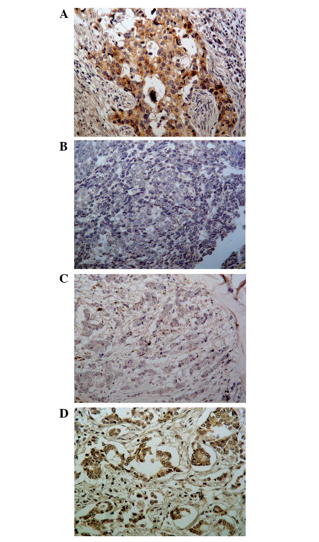
As revealed in Fig. 1,
phospho-PRAS40Thr246 was expressed diffusely throughout
the cytoplasm of the breast cancer cells at varying intensities,
and demonstrated an occasional nuclear location. Based on previous
studies, an H score of ≥100 was used as the criterion for the
positive expression of phospho-PRAS40Thr246. Using this
range, a total of 22 cases (40.0%) of HER2-positive breast cancer
tumors were defined as phospho-PRAS40Thr246-positive.
The clinicopathological variables of the patients were not
significantly associated with the phospho-PRAS40Thr246
expression levels (Table I).
In addition, the PI3K pathway activation status of
the same set of HER2-positive breast cancer tumors was
investigated. In total, PTEN loss was demonstrated in 34.5% (19/55)
of the HER2-positive breast cancer tissues. Furthermore, seven
mutations in exon 20 (H1047R) and three in exon 9 (E542K and E545K)
were identified, which corresponded to a PIK3CA mutation frequency
of 18.2%. In agreement with previous findings (5,13),
with the exception of two cases, PTEN loss and PIK3CA mutation
status were not present within the same tumor samples in the
present study. According to the previously established criteria
(5), patients were classified as
having an activated (with either PIK3CA mutants or low PTEN
expression; n=27) or an unactivated PI3K pathway status (with
PIK3CA mutant type and high PTEN expression; n=28). A significant
association was identified between phospho-PRAS40Thr246
expression and PI3K pathway status, and PTEN loss alone, but not
with PIK3CA mutation alone (Table
II).
| Table IIUnivariate and multivariate analysis
for overall survival. |
Table II
Univariate and multivariate analysis
for overall survival.
| Univariate
analysis | Multivariate
analysis |
|---|
|
|
|
|---|
| Characteristics | P-value | HR (95% CI) | P-value |
|---|
| Age, years | 0.805 | | |
| Grade | 0.016 | 2.253
(1.187–4.274) | 0.013 |
| ER status | 0.411 | | |
| PR status | 0.487 | | |
| PIK3CA mutation | 0.163 | | |
| PTEN loss | 0.070 | | |
| PI3K activation | 0.013 | | |
|
Phospho-PRAS40Thr246 | 0.031 | 2.081
(1.113–3.890) | 0.022 |
Univariate analysis was used to determine the
association between disease progression and PI3K pathway activation
status following trastuzumab-based treatment in HER2-positive
metastatic breast cancers, as determined by different marker sets.
As revealed in Table II, patients
with PIK3CA mutations and/or PTEN loss were at an increased risk
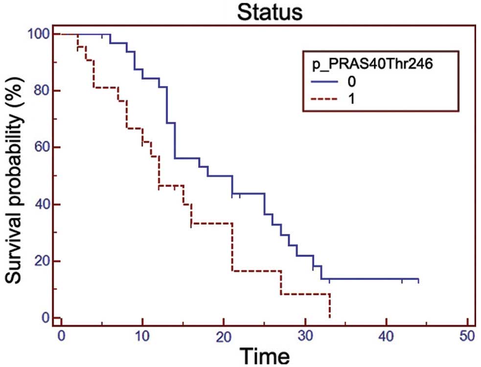
for disease progression compared with other subgroups. Furthermore,
patients with positive phospho-PRAS40Thr246 expression
exhibited a significantly shorter TTP following trastuzumab-based
treatment than those with negative expression (Fig. 2). The hazard ratios (HR), which were
based on multivariate Cox regression analysis, and were adjusted
for other significant predictors (grade), revealed that positive
phospho-PRAS40Thr246 expression was an independent and
significant risk factor for disease progression (HR, 2.081; 95%
confidence interval, 1.113–3.890; P=0.022; Table II)
Discussion
Hyperactivation of the PI3K pathway, as determined
by PIK3CA mutations or low PTEN expression, was observed in ~50% of
the HER2-positive metastatic breast cancers in the present study.
In agreement with previous studies (14,15),
PI3K hyperactivation contributed to an increased risk of disease
progression following trastuzumab treatment.
In addition to PIK3CA mutations and low PTEN
expression, a panel of biomarkers [including topoisomerase IIα
(16), MET oncogene, hepatocyte
growth factor (17),
phospho-AKTSer473 and phospho-p70S6KThr389
(8)] have been revealed to be
associated with PI3K pathway activation, and were proposed as
candidate biomarkers to predict trastuzumab resistance in
HER2-positive metastatic breast cancers. Despite this, the markers
demonstrate limited associations with trastuzumab efficacy in
clinical sets.
Phospho-PRAS40Thr246 is a novel
downstream target of the PI3K/Akt signaling pathway (18). In a recent study,
phospho-PRAS40Thr246 expression was positively
correlated with PI3K pathway activation and predicted PI3K pathway
inhibitor sensitivity in triple-negative breast tumor tissues and a
PTEN-deficient mouse prostate tumor model (7). Due to a high epitope stability,
phospho-PRAS40Thr246 has an improved clinical
translation potential compared with other PI3K pathway downstream
targets, such as AKTThr308 (7). A previous study identified that
phospho-PRAS40Thr246 was overexpressed in ~40% of
primary breast cancer samples, and as an independent prognostic
factor, was associated with slower disease progression (12). The present study indicated that
phospho-PRAS40Thr246 expression was significantly
associated with PIK3CA mutations and low PTEN expression. In
addition, more than twice as many patients were identified at an
increased risk for disease progression. Therefore, the analysis of
phospho-PRAS40Thr246 expression levels could reflect
PI3K pathway activation status and present a novel biomarker to
identify HER2-amplified breast cancers that are unlikely to respond
to trastuzumab-based therapy.
A previous study demonstrated that the PI3K
inhibitor, GDC-0941, decreased the phosphorylation of PRAS40 and
was highly efficacious in the treatment of trastuzumab-resistant
cells and tumors (19). These
results, together with the association between
phospho-PRAS40Thr246 expression and trastuzumab
resistance identified in the present study, suggest that
phospho-PRAS40Thr246 may be a candidate for predicting
trastuzumab responses. However, the potential mechanism has not
previously been investigated.
The present study revealed the significance of
phospho-PRAS40Thr246 in identifying HER2-positive breast
cancer patients with a high risk of disease progression following
trastuzumab-based therapy. In addition,
phospho-PRAS40Thr246 may possess an important role in
predicting the development of trastuzumab resistance. The exact
molecular mechanism underlying the effect of
phospho-PRAS40Thr246 expression levels on trastuzumab
resistance requires further investigation in order to be clinically
beneficial.
References
|
1
|
Slamon DJ, Clark GM, Wong SG, et al: Human
breast cancer: correlation of relapse and survival with
amplification of the HER-2/neu oncogene. Science. 235:177–182.
1987. View Article : Google Scholar : PubMed/NCBI
|
|
2
|
Slamon DJ, Godolphin W, Jones LA, et al:
Studies of the HER-2/neu proto-oncogene in human breast and ovarian
cancer. Science. 244:707–712. 1989. View Article : Google Scholar : PubMed/NCBI
|
|
3
|
Huang Y, Fu P and Fan W: Novel targeted
therapies to overcome trastuzumab resistance in HER2-overexpressing
metastatic breast cancer. Current Drug Targets. 14:889–898. 2013.
View Article : Google Scholar : PubMed/NCBI
|
|
4
|
Vogel CL, Cobleigh MA, Tripathy D, et al:
Efficacy and safety of trastuzumab as a single agent in first-line
treatment of HER2-overexpressing metastatic breast cancer. J Clin
Oncol. 20:719–726. 2002. View Article : Google Scholar : PubMed/NCBI
|
|
5
|
Berns K, Horlings HM, Hennessy BT, et al:
A functional genetic approach identifies the PI3K pathway as a
major determinant of trastuzumab resistance in breast cancer.
Cancer Cell. 12:395–402. 2007. View Article : Google Scholar : PubMed/NCBI
|
|
6
|
Razis E, Bobos M, Kotoula V, et al:
Evaluation of the association of PIK3CA mutations and PTEN loss
with efficacy of trastuzumab therapy in metastatic breast cancer.
Breast Cancer Res Treat. 128:447–456. 2011. View Article : Google Scholar : PubMed/NCBI
|
|
7
|
Andersen JN, Sathyanarayanan S, Di Bacco
A, et al: Pathway-based identification of biomarkers for targeted
therapeutics: personalized oncology with PI3K pathway inhibitors.
Science Transl Med. 2:43ra552010. View Article : Google Scholar
|
|
8
|
Esteva FJ, Guo H, Zhang S, et al: PTEN,
PIK3CA, p-AKT, and p-p70S6K status: association with trastuzumab
response and survival in patients with HER2-positive metastatic
breast cancer. Am J Pathol. 177:1647–1656. 2010. View Article : Google Scholar : PubMed/NCBI
|
|
9
|
Nagata Y, Lan KH, Zhou X, et al: PTEN
activation contributes to tumor inhibition by trastuzumab, and loss
of PTEN predicts trastuzumab resistance in patients. Cancer Cell.
6:117–127. 2004. View Article : Google Scholar : PubMed/NCBI
|
|
10
|
Zhang L, Shi L, Zhao X, Wang Y and Yue W:
PIK3CA gene mutation associated with poor prognosis of lung
adenocarcinoma. Onco Targets Ther. 6:497–502. 2013.PubMed/NCBI
|
|
11
|
Lu YZ, Deng AM, Li LH, et al: Prognostic
role of phospho-PRAS40 (Thr246) expression in gastric cancer. Arch
Med Sci. 10:149–153. 2014. View Article : Google Scholar : PubMed/NCBI
|
|
12
|
Fu R: Clinicopathological and prognostic
significance of phosphorylated PRAS40 (Thr246) in breast cancer.
Asian Biomedicine (Research Reviews and News). 6:573–577. 2012.
|
|
13
|
Saal LH, Holm K, Maurer M, et al: PIK3CA
mutations correlate with hormone receptors, node metastasis, and
ERBB2, and are mutually exclusive with PTEN loss in human breast
carcinoma. Cancer Res. 65:2554–2559. 2005. View Article : Google Scholar : PubMed/NCBI
|
|
14
|
Eichhorn PJ, Gili M, Scaltriti M, et al:
Phosphatidylinositol 3-kinase hyperactivation results in lapatinib
resistance that is reversed by the mTOR/phosphatidylinositol
3-kinase inhibitor NVP-BEZ235. Cancer Res. 68:9221–9230. 2008.
View Article : Google Scholar : PubMed/NCBI
|
|
15
|
Wang Q, Ding H, Liu B, et al: Addition of
the Akt inhibitor triciribine overcomes antibody resistance in
cells from ErbB2/Neu-positive/PTEN-deficient mammary tumors. Int J
Oncol. 44:1277–1283. 2014.PubMed/NCBI
|
|
16
|
Fountzilas G, Christodoulou C, Bobos M, et
al: Topoisomerase II alpha gene amplification is a favorable
prognostic factor in patients with HER2-positive metastatic breast
cancer treated with trastuzumab. J Transl Med. 10:2122012.
View Article : Google Scholar : PubMed/NCBI
|
|
17
|
Minuti G, Cappuzzo F, Duchnowska R, et al:
Increased MET and HGF gene copy numbers are associated with
trastuzumab failure in HER2-positive metastatic breast cancer. Br J
Cancer. 107:793–799. 2012. View Article : Google Scholar : PubMed/NCBI
|
|
18
|
Wang H, Zhang Q, Wen Q, et al:
Proline-rich Akt substrate of 40kDa (PRAS40): a novel downstream
target of PI3k/Akt signaling pathway. Cell Signal. 24:17–24. 2012.
View Article : Google Scholar
|
|
19
|
Junttila TT, Akita RW, Parsons K, et al:
Ligand-independent HER2/HER3/PI3K complex is disrupted by
trastuzumab and is effectively inhibited by the PI3K inhibitor
GDC-0941. Cancer Cell. 15:429–440. 2009. View Article : Google Scholar : PubMed/NCBI
|