Introduction
Canine breast cancer (BC) is the most significant
tumor in female dogs, whereas human BC is the most prevalent type
of cancer in females worldwide (1–2). There
is much evidence indicating that canine BC mimics the
characteristics of human BC with regards to incidence,
histomorphological characteristics, gene profiles, molecular
signaling pathways and clinical course (3). Numerous experts believe that canine BC
can be a powerful translation research model for human BC (4). Carcinomas account for the majority of
histological classifications of canine BC and sarcomas are less
frequent (5). However, to the best
of our knowledge no case of canine primary lymphoma of the breast
has been reported. However, human primary breast lymphoma (PBL) is
a rare malignancy and statistics show that <0.5% of human BCs
and 2% of all extranodal non-Hodgkin lymphomas (NHLs) are PBL
(6).
The current study presents a case of canine BC of
the primary NHL type for the first time and subsequently the theory
of considering it as a model for human PBL is raised.
Materials and methods
A 6-year-old female terrier with a history of
ovariohysterectomy at the age of 1 was referred to a veterinary
clinic with a lump in the left caudal abdominal mammary gland. The
primary diagnosis of the veterinary surgeon was canine BC. Thoracic
imaging and abdominal ultrasonography (according to the diagnostic
guidelines of metastatic canine BC) were performed to check for
metastasis, but no symptom was observed (7). Finally, simple mastectomy was
performed and a sample of the 2-cm tumor was sent to the pathology
laboratory of cancer institute of Iran.
The tumor section was fixed by 4% formaldehyde in
0.1 M phosphate-buffered saline (PBS) solution. The fixed tissue
was dehydrated by graded concentrations of ethanol and embedded in
paraffin wax, and subsequently stained with hematoxylin and eosin
(H&E). The slides were reviewed by a pathologist. For
immunohistochemical examination, paraffin-embedded blocks were
cooled in an ice-water mixture for 30 min before sectioning. A
total of 4 µm sections were cut and placed on slides. After a brief
drying period of ~15 min, the sections were heat-fixed to the slide
at 37°C. The sections were deparaffinized and rehydrated in graded
ethanol concentrations. The prepared slides were stained
immunohistochemically by cytokeratin 7 (CK7), CK5/6, cluster of
differentiation 3 (CD3), CD10, CD15, CD19, CD20, and Bcl-6
according to the manufacturer's instructions for the kits (Dako
Denmark A/S, Glostrup, Denmark).
Results
No cytology or histology assessments were performed
prior to the surgery. No evidence of carcinoma was observed during
the microscopic study of H&E slides, and the arrangement of
malignant cells indicated lymphoproliferative neoplasms.
Immunohistochemistry (IHC) staining with CK7 and CK5/6 markers
confirmed that the tumor did not have an epithelial nature.
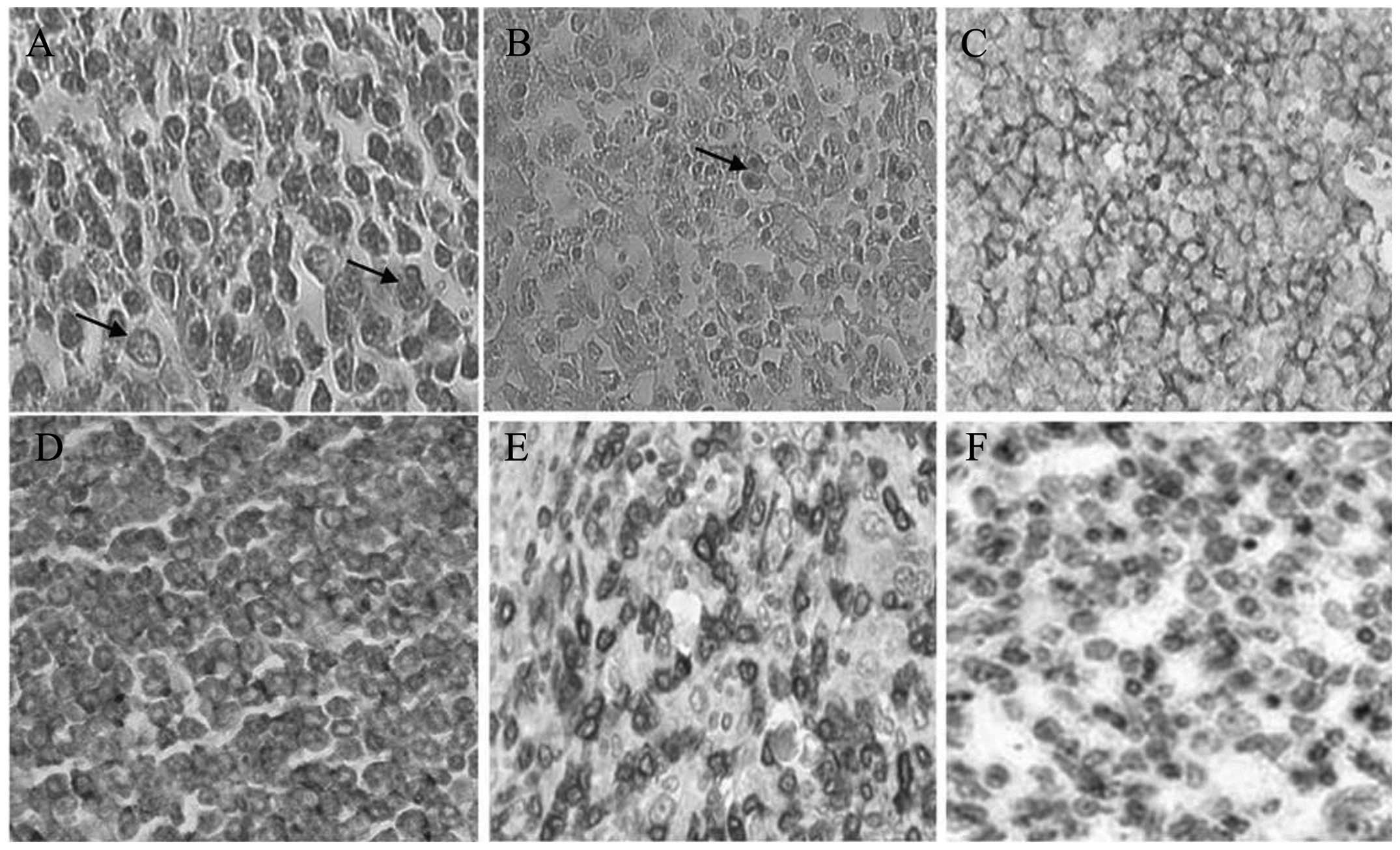
Prominent histomorphological characteristics of the malignant cells
are described as follows: Cell sizes were larger than normal
lymphocytes and they had round and elliptical vesicular nuclei.
Nucleoli were often isolated and they were observed near the
nuclear membrane (Fig. 1). The
cytoplasm of the cells was often basophilic and certain anaplastic
cells appeared to be multinucleate and showed the pathological view
of Reed-Sternberg-like cells (Fig.
1). According to histomorphological characteristics, the
pattern of malignant cells showed diffuse large B-cell lymphoma
(DLBCL), however, the Reed-Sternberg-like cells view was misleading
and could show a Hodgkin's lymphoma (HL) pattern. In order to
determine the immunophenotype of tumor cells, slides were prepared
from paraffin blocks and subsequently stained with the IHC method
using CD3, CD10, CD15, CD19, CD20 and B-cell lymphoma 6 markers
(Fig. 1). IHC results are observed
in Table I. In addition, Ki-67
marker staining showed that cell cycle activity was ~50% and could
reject the possibility of Burkitt's lymphoma (starry sky pattern
was not observed with regards to histomorphology).
 | Table IImmunohistochemistry (IHC) test
results assessed by CD markers. |
Table I
Immunohistochemistry (IHC) test
results assessed by CD markers.
| IHC staining | CD marker result |
|---|
| CD3 | Negative |
| CD10 | Positive |
| CD15 | Negative |
| CD19 | Positive |
| CD20 | Positive |
| Bcl-6 | Scatter |
With regard to the histomorphological findings and
IHC, the final diagnosis was extranodal-NHL of the DLBCL type and
considering the clinical findings that showed no evidence of a
primary lymphoma having invaded the breast area, the final
diagnosis for this case was canine PBL.
Discussion
As it was indicated, the animal had canine BC of the
primary NHL type. Canine NHL is a common malignancy (8). However, this disease comprises a
heterogeneous group of canine malignancies and has an extremely
varied prognosis (9). The major
cause is attributed to various NHL subtypes. Canine NHL subtypes
were examined with immunophenotyping assessments, and considering
their immunophenotyping similarities and adaptations with human
NHL, canine NHL was classified according to World Health
Organization guidelines (10).
DLBCL comprises the most significant group of canine NHL, as
statistics show that 48% of all canine lymphomas belong to this
category (11). Extensive evidence
suggests canine NHL to be a model for human NHL. Following the
separation of canine lymphoma cells, the study by Ito et al
(8) cultured and passaged them and
developed a xenograft model. Subsequently, their molecular profile
was examined and it was concluded that not only is canine NHL
morphologically and behaviorally similar to human NHL, but its
molecular changes also mimic human lymphoma. Pawlak et al
(10) had previously addressed this
theory.
In human DLBCL it is known that tumor growth is
rapid and prognosis is poor (12,13).
Bienzle and Vernau (9) stated that
the survival time of canine DLBCL is short, and that similar to
human DLBCL, its prognosis is bad. Using molecular techniques,
Richards et al (11) divided
canine DLBCL into two subcategories. After assessing their survival
time, the study concluded that the course of the disease is similar
to human DLBCL and its prognosis is poor.
Human PBL is a relatively rare form of human BC and
the majority of its types are DLBCL (6). No previous study of canine breast
lymphoma can be found in the databases of PubMed and Google
scholar. However, the histomorphological characteristics, molecular
pathology and clinical data of the present case confirmed breast
lymphoma. Observation of Reed-Sternberg-like cells may be
suggestive of canine HL, but the surface marker of CD15 was
negative. Such a microscopic view appears to be due to the presence
of large inclusion-like nucleoli in highly anaplastic cells,
whereas this surface marker is positive in >90% of typical
Reed-Sternberg cells (14). The
results of the present case report indicate that canine NHL in the
mammary gland area can mimic the properties of HBL.
In general, modeling in the area of oncology
research requires reliable evidence showing that the model is
powerful regarding translational research, as the results of the
pre-clinical phase require extension to the clinical phase
(15). Although reliable scientific
communities have suggested xenograft for the pre-clinical phase of
the tumor, the canine model has received increasing attention in
oncology research during recent years (3,10).
Three main causes have been cited for this: Firstly, these tumors
are spontaneous, as they were not experimentally induced; secondly,
the life of dogs are so that the clinical course of the disease is
extremely well shown and the disease shifts from early to advance
stage. As a result, invasion and metastasis can be followed in this
model. Thirdly, with regard to the fact that dogs live alongside
humans, they are exposed to the same risk factors and so the course
of molecular changes and genetic mutations can also be studied
(4,8,15).
Although no studies of canine breast lymphoma have
been reported thus far, and to the best of our knowledge, this is
the first report of canine NHL in the mammary area of dog, when
considering previous evidence emphasizing the similarities in
histomorphology, immunophenotyping and clinical course of canine
and human NHL, the theory can be raised that canine PBL can also be
a model for research on human PBL. Performing case series studies
in this area in the future is required. Not only can canine cancer
models be considered a study phase in pre-clinical research, but
they can also be useful for afflicted dogs, as they may be helpful
in developing novel (ethical) treatments and also reduce the
suffering caused by canine cancer.
Acknowledgements
The present study had no financial sponsor and all
the costs of pathology tests were paid by the authors. The authors
would like to express their gratitude to Dr Taghizadeh-jahed,
veterinary surgeon, for surgically removing the tumor. They would
also like to thank Ms. Morsali and the Pathobiology Laboratory of
Dr E'temad Moghaddam for helping prepare the pathological
slides.
References
|
1
|
Sleeckx N, de Rooster H, Veldhuis Kroeze
EJ, Van Ginneken C and Van Brantegem L: Canine mammary tumours, an
overview. Reprod Domest Anim. 46:1112–1131. 2011. View Article : Google Scholar : PubMed/NCBI
|
|
2
|
Redig AJ and McAllister SS: Breast cancer
as a systemic disease: a view of metastasis. J Intern Med.
274:113–126. 2013. View Article : Google Scholar : PubMed/NCBI
|
|
3
|
Pinho SS, Carvalho S, Cabral J, Reis CA
and Gartner F: Canine tumors: a spontaneous animal model of human
carcinogenesis. Transl Res. 159:165–172. 2012. View Article : Google Scholar : PubMed/NCBI
|
|
4
|
Rivera P and von Euler H: Molecular
biological aspects on canine and human mammary tumors. Vet Pathol.
48:132–146. 2011. View Article : Google Scholar : PubMed/NCBI
|
|
5
|
Goldschmidt M, Peña L, Rasotto R and
Zappulli V: Classification and grading of canine mammary tumors.
Vet Pathol. 48:117–131. 2011. View Article : Google Scholar : PubMed/NCBI
|
|
6
|
Cheah CY, Campbell BA and Seymour JF:
Primary breast lymphoma. Cancer Treat Rev. 40:900–908. 2014.
View Article : Google Scholar
|
|
7
|
Cassali GD, Lavalle GE, De Nardi AB, et
al: Consensus for the diagnosis, prognosis and treatment of canine
mammary tumors. Braz J Vet Pathol. 4:153–180. 2011.
|
|
8
|
Ito D, Frantz AM and Modiano JF: Canine
lymphoma as a comparative model for human non-Hodgkin lymphoma:
recent progress and applications. Vet Immunol Immunopathol.
159:192–201. 2014. View Article : Google Scholar : PubMed/NCBI
|
|
9
|
Bienzle D and Vernau W: The diagnostic
assessment of canine lymphoma: implications for treatment. Clin Lab
Med. 31:21–39. 2011. View Article : Google Scholar : PubMed/NCBI
|
|
10
|
Pawlak A, Obminska-Mrukowicz B and Rapak
A: The dog as a model for comparative studies of lymphoma and
leukemia in humans. Postepy Hig Med Dosw (Online). 67:471–480.
2013.(In Polish).
|
|
11
|
Richards KL, Motsinger-Reif AA, Chen HW,
et al: Gene profiling of canine B-cell lymphoma reveals germinal
center and postgerminal center subtypes with different survival
times, modeling human DLBCL. Cancer Res. 73:5029–5039. 2013.
View Article : Google Scholar
|
|
12
|
Aviv A, Tadmor T and Polliack A: Primary
diffuse large B-cell lymphoma of the breast: looking at
pathogenesis, clinical issues and therapeutic options. Ann Oncol.
24:2236–2244. 2013. View Article : Google Scholar : PubMed/NCBI
|
|
13
|
Roschewski M, Dunleavy K and Wilson WH:
Diffuse large B cell lymphoma: molecular targeted therapy. Int J
Hematol. 96:552–561. 2012. View Article : Google Scholar : PubMed/NCBI
|
|
14
|
Hansmann ML and Willenbrock K: WHO
classification of Hodgkin's lymphoma and its molecular pathological
relevance. Pathologe. 23:207–218. 2002.(In German).
|
|
15
|
Vail DM and MacEwen EG: Spontaneously
occurring tumors of companion animals as models for human cancer.
Cancer Invest. 18:781–792. 2000. View Article : Google Scholar : PubMed/NCBI
|