Introduction
The first laparoscopic cholecystectomy was performed
by Mouret (1) in 1987. Since then,
the laparoscopic technique has been used widely in every type of
common surgery. While the majority of surgeons have focused on
reducing the number and size of surgical incisions, other surgeons
are taking a more challenging surgical approach, characterized by
making the incisions more covert to cause fewer scars. This
advanced technology, which was put forward by Navarra et al
in 1992 (2), is termed
laparoendoscopic single-site surgery (LESS). A different surgical
approach using a natural orifice in the body such as the
gastrointestinal tract or vagina, known as natural orifice
transluminal endoscopic surgery (NOTES), was reported by Marescaux
et al (3) in 2007. NOTES is
closer to the concept of minimally invasive surgery (4–9). In
China, NOTES has been performed by numerous medical centers.
However, NOTES requires higher surgical skill and expensive devices
and may also result in intra-abdominal infection and puncture mouth
leakage (10–12), which have limited its use in China.
In actual clinical work, LESS is easier to popularize due to the
lower costs and surgical skill required. A number of medical
centers have implemented modified LESS. A number of medical centers
have implemented modified LESS techniques, including transumbilical
endoscopic surgery and single-port LESS (13,14).
These techniques are locally implemented improvements that have
been made to the traditional laparoscopic technology.
Single-port laparoscopic technology is widely used
in minimally invasive surgery. Due to the expensive instrumentation
and (15,16) higher surgical skills that are
required, as well as the intra-abdominal infection and puncture
mouth leakage produced, the promotion of its use is challenging.
For this reason, a minimally invasive technique, periumbilical
laparoscopic surgery through triple channels using common
instrumentation, is introduced in the present study. Our
periumbilical laparoscopic surgery through triple channels has been
demonstrated to be a safe and effective laparoscopic procedure,
with successful experiences in 106 cases of laparoscopic
cholecystectomy. These results motivated us to perform the present
study. The results achieved by the technique are comparable with
those of single-port laparoscopic cholecystectomy.
Materials and methods
Ethics
The study was conducted in accordance with the
declaration of Helsinki and with approval from the Ethics Committee
of the Second Affiliated Hospital of Kunming Medical University.
Written informed consent was obtained from all participants.
General information
Based on the reports from B-ultrasound, 108 cases of
completed periumbilical laparoscopic cholecystectomy (78 cases of
simple cholecystolithiasis and 30 cases of gallbladder polyposis)
treated using common instrumentation through triple channels were
selected for the present study. Comparative analysis was performed
with a control group consisting of 356 patients who had undergone
traditional laparoscopic cholecystectomy between June 2009 and May
2011. In the experimental group, the youngest patient was 32 years
old and the oldest was 66 years old. The mean age was 52.96±6.72
years old. In the control group, the youngest patient was 28 years
old and the oldest was 76 years old. The mean age was 54.87±7.81
years old. The tests and preparations for all patients prior to
surgery were the same. No statistically significant differences
were observed between the two groups with regard to age, gender and
type of disease (Table I).
 | Table IPatient characteristics. |
Table I
Patient characteristics.
| Factor | Experimental
group | Control group | t-value
(P-value) | χ2-value
(P-value) |
|---|
| Age | 52.96±6.72 | 54.87±7.81 | −1.689 (0.092) | - |
| Gender
(male/female) | 44/64 | 137/219 | - | 1.545 (0.214) |
| Type of disease
(stone/polyp) | 78/30 | 287/69 | - | 0.069 (0.793) |
Surgical methods
Conventional laparoscopic cholecystectomy was used
for the control group and periumbilical laparoscopic
cholecystectomy through triple channels was performed for the
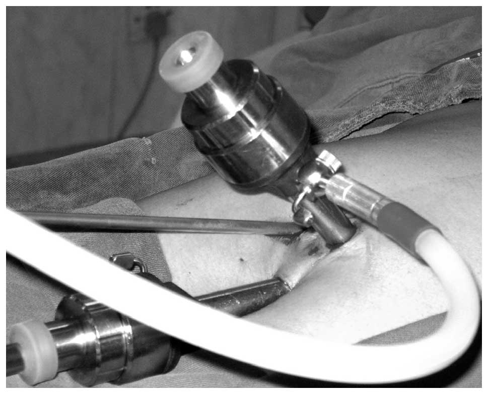
experimental group. In the experimental group, we used common
instrumentation and a method that reduced the interference between
troca and air leakage. In addition, this method does not require
the operator to wipe the lens repeatedly (Fig. 1).
Surgical position
The surgical positions of the 2 groups were the
same, wherein the patients were turned left 30° and the head was
turned 25° upwards. However, for the experimental group, the main
surgeon stood behind the assistant, as it was more convenient.
Surgical instruments
The two techniques used the same surgical equipment,
including a laparoscope (model, Xenon Nova 300; Code, 20134020;
KARL STORZ, Tuttlingen, Germany), gallbladder grasper, separating
pliers, dissecting scissors, electrical separating hook, puncture
needle, suction tube, gasless machine and television pickup system.
No special laparoscopic instruments were employed.
Experimental method
Experimental data, including the amount of surgical
bleeding, surgery time, intestinal function recovery time,
hospitalization time and hospitalization cost were analyzed using
statistical tests (Table II).
 | Table IIExperimental data. |
Table II
Experimental data.
| Factor | Experimental
group | Control group | t-value | P-value |
|---|
| Amount of bleeding
(ml) | 67.81±9.03 | 65.58±9.15 | 1.648 | 0.100 |
| Surgery time
(min) | 110.31±14.57 | 43.98±7.64 | 46.673 | 0.000 |
| Recovery time of
intestinal function (h) | 28.88±5.69 | 30.37±6.78 | −1.529 | 0.128 |
| Hospitalization time
(days) | 4.00±0.94 | 3.79±0.66 | −1.911 | 0.057 |
| Hospitalization cost
(thousand yuan) | 7.4±0.6 | 7.6±0.7 | −1.867 | 0.063 |
Statistical analysis
A Student’s t-test was used to compare differences
between the groups using SPSS 10.0 software (SPSS Inc., Chicago,
IL, USA). P<0.000312 was considered to indicate a statistically
significant result.
Results
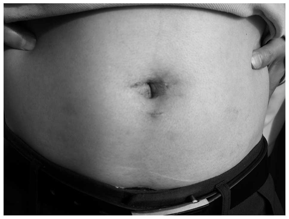
Surgery was successfully completed in the 106 cases
from the experimental group using common instrumentation without
complications. However, in 2 cases a change to the traditional
laparoscopic cholecystectomy approach was required due to
insufficient gallbladder artery clipping. No statistically
significant differences in the amount of surgical bleeding,
intestinal function recovery time, hospitalization time and
hospitalization cost were observed between the groups. However, a
statistically significant difference was observed in the surgery
time (Fig. 2).
Out of the 106 cases in the experimental group, 84
were followed up for 1 to 28 months, with a mean of 17.4 months. Of
the cases followed up, 79 showed no specific discomfort, while 3
experienced abdominal pain without jaundice at 3 to 6 months after
surgery. The pain was diagnosed as gallbladder stump inflammation
through magnetic resonance cholangiopancreatography (MRCP) tests.
The symptoms were relieved following anti-inflammatory allopathic
therapy and the patients subsequently reported no discomfort. A
further 2 cases experienced unbearable abdominal pain without
jaundice at 9 to 13 months after surgery. The pain was diagnosed as
residual stones. The residual stones of the 2 patients were removed
using endoscopic sphincterotomy (EST) and followed up.
Discussion
With the rapid development of minimally invasive
techniques, the ambitions of numerous surgeons have become
achievable. LESS and NOTES are typical examples of such techniques.
A considerable number of studies have been performed (17–19)
and advancements have been achieved. In our case, however, problems
arose during the process of popularization mainly due to the
disadvantages, including the use of specialized instrumentation and
higher surgical costs. A number of medical centers (20,21)
have used special instruments for periumbilical laparoscopic
cholecystectomy and attained positive results. However,
popularization of the method is problematic due to regional
economic differences and non-conformity of health care policy.
Consideration of the reported experiences of these medical centers
and our own experience suggests the following advantages for our
particular laparoscopic cholecystectomy technique.
Firstly, only common laparoscopic instruments are
required to perform laparoscopic cholecystectomy. Despite the
cost-saving measures, no abdominal scarring is produced. Surgeons
with relevant experience of laparoscopic cholecystectomy may
perform this surgery, even if they belong to a basic-level medical
unit.
Secondly, the problem of interference between the
trocar and air leakage was considerably reduced due to the use of 3
channels. The ‘big triangle’ from the traditional laparoscopic
cholecystectomy was replaced by a small triangle around the
umbilical opening. In cases of uncontrolled bleeding during
surgery, one or two punctures may be established below the xiphoid
process or rib bow to stop the bleeding. This was carried out in 2
cases from the experimental group.
Thirdly, this technique is more suitable for
basic-level medical units. The technique may be performed as long
as a laparoscopic device and a surgeon with experience of
laparoscopic cholecystectomy are available in the hospital. This
approach involves an improvement in the laparoscopic technique
only, so the use of large medical equipment, particularly MRI or
special instrumentation, such as flexional separating pliers, is
unnecessary and the approach is suitable for use in basic-level
medical units. Ultimately, this technique is likely to be be
beneficial in less economically developed regions.
However, certain concerns require consideration.
These include the full exposure of the Calot’s triangle to prevent
bile duct injury and bleeding. Damage due to the electrical
conductivity of instruments in a narrow space is another concern.
Moreover, suturing the periumbilical incision to prevent umbilical
herniation is necessary. Therefore, anesthetic drugs, such as 2%
lidocaine, are highly recommended prior to suturing to reduce
postoperative analgesic dosage. Absorbable suture materials and
careful suturing are also important for cosmesis. As surgical
experience increases, the surgery time of this technique may be
reduced. At present, it takes 45 to 80 min to perform each
operation after experiencing 30 cases. According to the literature,
this technique has already been utilized in appendectomy,
sigmoidectomy and liver cyst decortication. Furthermore, 12 cases
of acute appendicitis have been treated using this technique and
satisfactory results were obtained. Lastly, the management of
complications is similar to that of the traditional laparoscopic
technique.
The current results indicate that the technique is
safe and feasible for use in laparoscopic cholecystectomy. It does
not produce abdominal scars and preserves an unblemished
appearance. Furthermore, the technique is particularly suitable for
use in China and only requires common laparoscopic instrumentation.
Therefore, popularization of the technique should be
implemented.
References
|
1
|
Litynski GS: Profiles in laparoscopy:
Mouret, Dubois, and Perissat: the laparoscopic breakthrough in
Europe (1987–1988). JSLS. 3:163–167. 1999.PubMed/NCBI
|
|
2
|
Navarra G, Pozza E, Occhionorelli S,
Carcoforo P and Donini I: One-wound laparoscopic cholecystectomy.
Br J Surg. 84:6951997. View Article : Google Scholar : PubMed/NCBI
|
|
3
|
Marescaux J, Dallemagne B, Perretta S,
Wattiez A, Mutter D and Coumaros D: Surgery without scars: report
of transluminal cholecystectomy in a human being. Arch Surg.
142:823–826. 2007. View Article : Google Scholar : PubMed/NCBI
|
|
4
|
Kalloo AN, Singh VK, Jagannath SB, et al:
Flexible transgastric peritoneoscopy: a novel approach to
diagnostic and therapeutic interventions in the peritoneal cavity.
Gastrointest Endosc. 60:114–117. 2004. View Article : Google Scholar
|
|
5
|
Perretta S, Dallemagne B, Coumaros D and
Marescaux J: Natural orifice transluminal endoscopic surgery:
transgastric cholecystectomy in a survival porcine model. Surg
Endosc. 22:1126–1130. 2008. View Article : Google Scholar
|
|
6
|
Jacob DA and Raakow R: Single-port
transumbilical endoscopic cholecystectomy: a new standard? Dtsch
Med Wochenschr. 135:1363–1367. 2010.(In German).
|
|
7
|
Carvalho GL, Silva FW, Silva JS, et al:
Needlescopic clipless cholecystectomy as an efficient, safe, and
cost-effective alternative with diminutive scars: the first 1000
cases. Surg Laparosc Endosc Percutan Tech. 19:368–372. 2009.
View Article : Google Scholar : PubMed/NCBI
|
|
8
|
Kuon Lee S, You YK, Park JH, Kim HJ, Lee
KK and Kim DG: Single-port transumbilical laparoscopic
cholecystectomy: a preliminary study in 37 patients with
gallbladder disease. J Laparoendosc Adv Surg Tech A. 19:495–499.
2009.PubMed/NCBI
|
|
9
|
Tsimoyiannis EC, Tsimogiannis KE,
Pappas-Gogos G, et al: Different pain scores in single
transumbilical incision laparoscopic cholecystectomy versus classic
laparoscopic cholecystectomy: a randomized controlled trial. Surg
Endosc. 24:1842–1848. 2010. View Article : Google Scholar
|
|
10
|
Rawlings A, Hodgett SE, Matthews BD,
Strasberg SM, Quasebarth M and Brunt LM: Single-incision
laparoscopic cholecystectomy: initial experience with critical view
of safety dissection and routine intraoperative cholangiography. J
Am Coll Surg. 211:1–7. 2010. View Article : Google Scholar
|
|
11
|
Ohdaira T, Ikeda K, Tajiri H, Yasuda Y and
Hashizume M: Usefulness of a flexible port for natural orifice
transluminal endoscopic surgery by the transrectal and transvaginal
routes. Diagn Ther Endosc. 2010:4730802010. View Article : Google Scholar : PubMed/NCBI
|
|
12
|
Hensel M, Schernikau U, Schmidt A and Arlt
G: Comparison between transvaginal and laparoscopic cholecystectomy
- a retrospective case-control study. Zentralbl Chir. 137:48–54.
2012.(In German).
|
|
13
|
Zhu JF, Hu H, Ma YZ, et al: Preliminary
clinical report on transumbilical endoscopic surgery. Chin J Min
Inv Surg. 1:75–77. 2008.(In Chinese).
|
|
14
|
Hernandez M, Morton CA, Ross S, et al:
Laparoendoscopic single site cholecystectomy: the first 100
patients. Am Surg. 75:681–685. 2009.PubMed/NCBI
|
|
15
|
Chamberlain RS and Sakpal SV: A
comprehensive review of single-incision laparoscopic surgery (SILS)
and natural orifice transluminal endoscopic surgery (NOTES)
techniques for cholecystectomy. J Gastrointest Surg. 13:1733–1740.
2009. View Article : Google Scholar
|
|
16
|
Al-Tayar H, Nielsen PE and Jørgensen LN:
Transumbilical cholecystectomy. Ugeskr Laeger. 172:1508–1511.
2010.(In Danish).
|
|
17
|
Bucher P, Ostermann S, Pugin F and Morel
P: Female population perception of conventional laparoscopy,
transumbilical LESS, and transvaginal NOTES for cholecystectomy.
Surg Endosc. 25:2308–2315. 2011. View Article : Google Scholar : PubMed/NCBI
|
|
18
|
Pagano D, Echeverri GJ, Gridelli B, Spada
M, Botrugno I and Bartoccelli C: Natural orifice transumbilical
cholecystectomy using a tri-port trocar and conventional
instruments. J Am Coll Surg. 210:1013–1014. 2010. View Article : Google Scholar : PubMed/NCBI
|
|
19
|
Roy P and De A: Transumbilical
multiple-port laparoscopic cholecystectomy (TUMP-LC): a prospective
analysis of 50 initial patients. J Laparoendosc Adv Surg Tech A.
20:211–217. 2010. View Article : Google Scholar : PubMed/NCBI
|
|
20
|
Yu WB, Zhang GY, Li F, Yang QY and Hu SY:
Transumbilical single port laparoscopic cholecystectomy with a
simple technique: initial experience of 33 cases. Minim Invasive
Ther Allied Technol. 19:340–344. 2010. View Article : Google Scholar : PubMed/NCBI
|
|
21
|
Cao LP, Que RS, Zhou F, Ding GP and Jing
DX: Transumbilical single-port laparoscopic cholecystectomy using
traditional laparoscopic instruments: a report of thirty-six cases.
J Zhejiang Univ Sci B. 12:862–866. 2011. View Article : Google Scholar
|


