Introduction
Nocardia spp. are gram-positive aerobic
bacteria, which are commonly found in soil, water and air (1). Contaminated dust, soil and food can
enter the body through the respiratory tract, gastrointestinal
tract and skin and proceed to cause corresponding symptoms, such as
coughing, expectoration, hemoptysis and skin abscesses, which are
considered as nocardiosis (2).
Nocardiosis is a vital, but often ignored, infectious disease in
immunocompromised hosts, which is particularly serious in the
absence of timely diagnosis and therapy. Nocardiosis can be caused
by various Nocardia spp., including Nocardia
asteroides, Nocardia brasiliensis, Nocardia
cyriacigeorgica, Nocardia farcinica and Nocardia
otitidiscaviarum (3). As
compared with the other Nocardia spp., N.
otitidiscaviarum appears to be rare (3%) which was first
recognized in samples taken from a Sumatran cavy or guinea pig with
ear disease (3). The present case
report describes a case of disseminated nocardiosis induced by
infection with N. otitidiscaviarum in an immunocompetent
host. Furthermore, in order to summarize the clinical features of
this disease and improve understanding, a review of the existing
literature of the disease induced by this species is also
presented.
Case report
A 37 year-old Chinese man presented at Beijing
Shijitan Hospital (Beijing, China) with an intermittent cough with
little sputum, which had lasted 50 days, and a mass on the right
side of his neck, which had first presented 1 month previously. The
patient initially attributed the neck pain to a neck sprain,
however the mass gradually increased in size over a period of 30
days. The patient complained of a 2-month history of unintentional
weight loss (≥8 kg) and mild pallor of the conjunctiva, skin and
palms was detected. No fever, fatigue or night sweats had been
experienced since the onset of disease. The patient had no past
history of tuberculosis, diabetes mellitus or steroid therapy. The
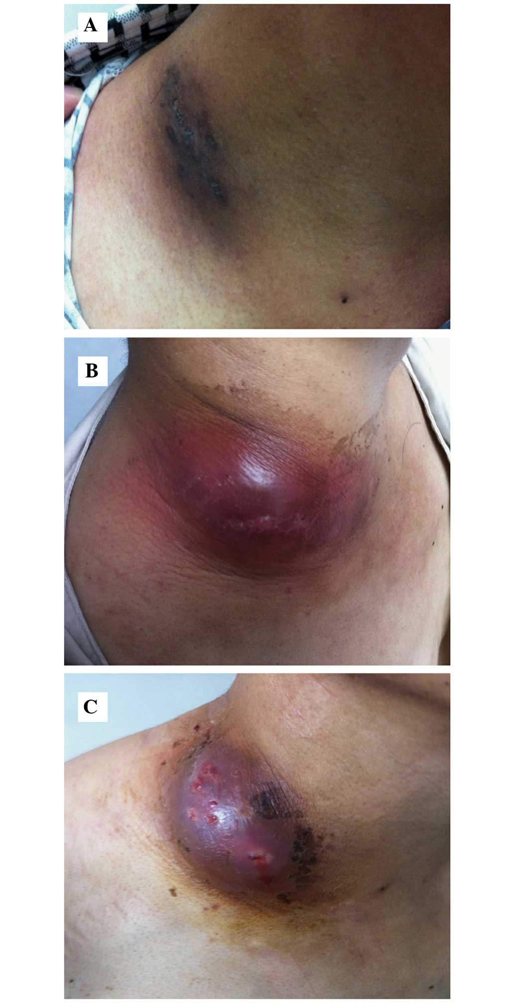
right-hand side of the patient's neck exhibited a marked swelling
(12×8 cm; Fig. 1), the temperature
of which was slightly elevated; however, no cervical or
supraclavicular lymphadenopathy was detected. Following
examination, percussion dullness was detected in the patient's
chest from the first to sixth intercostal spaces on the right
mid-clavicular line with a faint expiratory wheeze on the right.
The heart rhythm of the patient was regular with no gallops or
murmurs and abdominal examination raised no concerns.
Laboratory examination detected mild anemia with a
hemoglobin concentration of 6.9 g/dl (reference, 130–175 g/dl), in
addition to the following: Red blood count, 3.89×10¹²
cells/l (reference, 4.3–5.8×10¹² cells/l); mean
corpuscular volume, 65.8 fl (reference, 82–100 fl); mean hemoglobin
content, 18 pg (reference, 27–34 pg); mean hemoglobin
concentration, 270 g/l (reference, 316–354 g/l); and white blood
cell (WBC) count, 8.91×10⁹ cells/l (reference,
3.5–9.5×10⁹ cells/l. Serum tumor marker levels were:
Cancer antigen-125, 68.3 U/ml (reference, 0–35 U/ml), Cyfra21-1,
4.19 ng/ml (reference, 0–3.3 ng/ml); and neuron-specific enolase,
49.78 ng/ml (reference, 0–17 ng/ml). Abnormal thyroid function was
detected: Serum total thyroxine (T4), 6.65 µg/dl (reference,
5.1–14.1 µg/dl); total triiodothyronine (T3), 85.3 ng/dl
(reference, 80–200 ng/dl); thyrotropin, 9.68 IU/ml (reference,
0.27–4.2 g/dl); free T3, 1.67 pg/ml (reference, 2–4.4 pg/ml); and
free T4, 0.94 ng/dl (reference, 0.93–1.7 ng/dl).
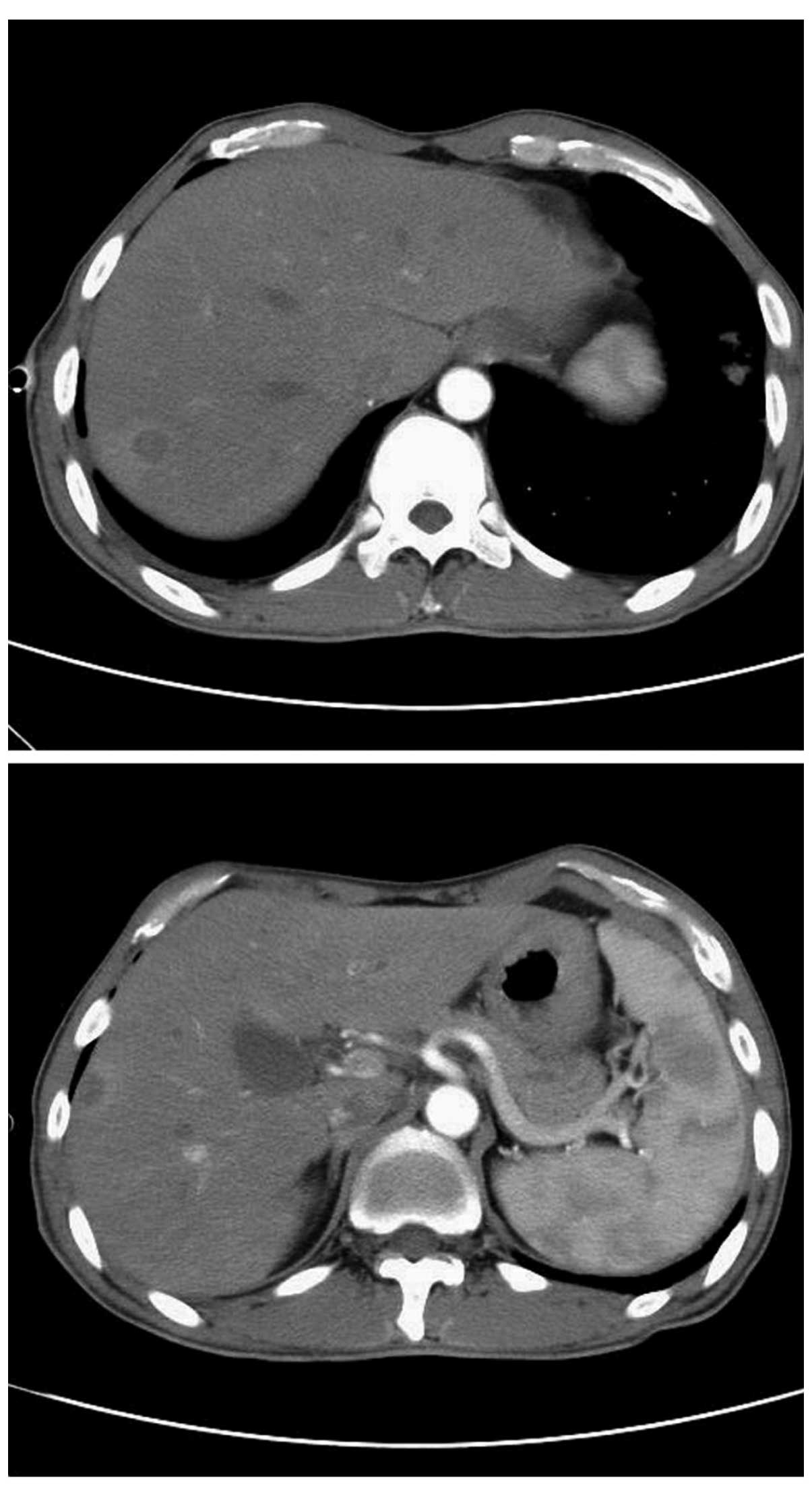
Ultrasound investigation (ACUSON Sequoia 512;
Siemens Healthineers, Erlangen, Germany) indicated that the
right-hand side of the patient's neck was hyperechoic and computed
tomography (CT) of the chest (Somatom Definition AS; Siemens
Healthineers) demonstrated a mass in the upper lobe of the right
lung, multiple nodules in both lungs and pleural effusion in the
right lung (Fig. 2).
The initial diagnosis was right upper pulmonary
neoplasm with right cervical lymph node and pleural metastases;
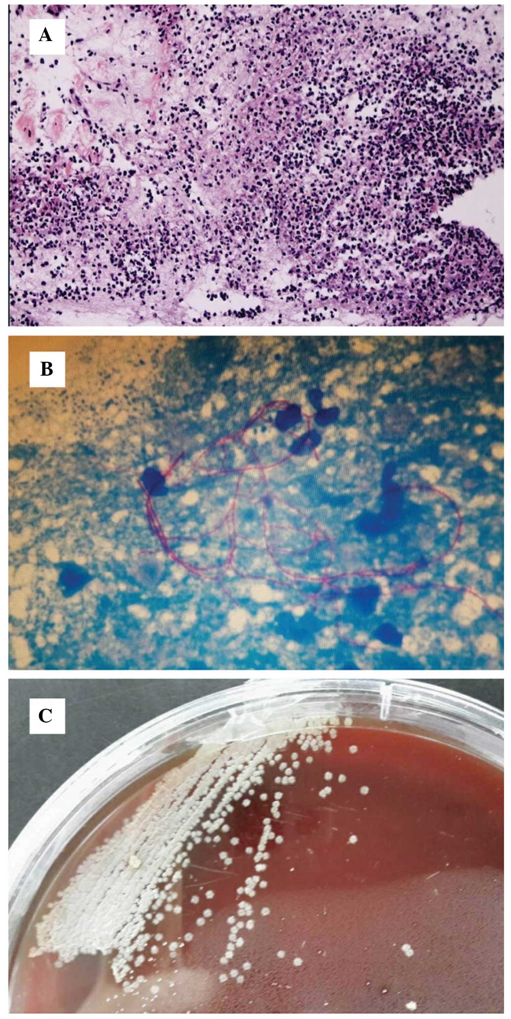
therefore, an ultrasound-guided right cervical mass biopsy was
performed. Histopathological analysis of the aspiration tissue
(Santa Cruz Biotechnology, Inc., La Jolla, TX, USA) demonstrated
granulation tissue and abscess formation (Fig. 3A). A modified acid-fast
stain-positive organism was detected with smooth and granular
colonies (Fig. 3B and C). Subculture
and microscopy demonstrated the organism was a branching
gram-positive rod; thus, Nocardia was confirmed. The
cutaneous, soft tissue lesion and lung symptoms were diagnosed as
nocardiosis.
As the patient presented with nocardiosis with the
characteristics of dissemination, further examinations were
performed. Firstly, routine and chemical testing of pleural
effusion was performed, demonstrating the effusion was pH 7.4, and
positive for Rey's reaction, which indicates exudate. The
proportion of pleural fluid was 1.025, and the WBC count was
significantly elevated at 1.8×10⁹ cells/l, with 91%
monocytes and 9% polymorphonuclear cells. Protein concentration of
the hydrothorax was 32.5 g/l, and the concentrations of glucose,
lactate dehydrogenase and adenosine deaminase were 2.1 mmol/l, 468
U/l and 27 U/l, respectively. These results confirmed a
parapneumonic effusion. Contrast-enhanced CT of the abdomen
indicated multiple low-density lesions in the right lobe of the
liver, which, with ring-enhancement, were consistent with the
imaging features of an abscess (Fig.
4). MRI scanning of the head (Magnetom Avanto 1.5T; Siemens
Healthineers) demonstrated no abnormalities. Therefore the patient
was diagnosed with disseminated nocardiosis of the subcutaneous
soft tissue, lungs and liver.
The patient was initially treated with ceftriaxone
(2 g/day) and sulfamethoxazole trimethoprim (2 tablets twice
daily). Levothyroxine sodium (25 µg/day) was administered for
subclinical hypothyroidism. Since the diagnosis of disseminated
nocardiosis was confirmed, etiological investigations and drug
sensitivity testing was conducted. Culturing of the mass biopsy
tissue showed that the patient was positive for N.
otitidiscaviarum. Using the disk diffusion method (Kirkby-Bauer
method), susceptibility testing demonstrated the following
diameters for areas of non-bacterial growth in response to various
antibiotics: Amoxicillin-clavulanic acid, 6 mm; cefepime 6 mm;
imipenem, 6 mm; cefotaxime, 6 mm; ceftriaxone, 9 mm; erythromycin,
12 mm; ampicillin, 16 mm; gentamicin, 22 mm; ciprofloxacin, 24 mm;
trimethoprim-sulfamethoxazole (TMP-SMZ), 30 mm; linezolid, 38 mm;
minocycline, 40 mm; and amikacin, 42 mm. Therefore, on the basis of
these results, combined minocycline and TMP-SMZ therapy was chosen.
Following administration for 2 months, the mass on the right-hand
side of the patient's neck markedly reduced in size (Fig. 1B and C) and the lung lesion gradually
improved.
Discussion
The PubMed database was searched and 23 cases were
retrieved with ‘Nocardia otitidiscaviarum’ as the search term from
January 1990 to August 2014 as shown in Table I (3–25). These
23 cases included 11 cases of subcutaneous and soft tissue
involvement, 5 patients with pulmonary involvement, 2 cases of
brain involvement, 1 case of pyothorax involvement, 1 case of
subcutaneous, soft tissue and lung involvement, 1 case of
subcutaneous, soft tissue and brain involvement, 1 case of
pyothorax and brain involvement, and 1 case with chest wall
abscesses, bilateral pulmonary infiltrates and intra-abdominal mass
involvement. The mean age of the patients was 56.61±18.58 years,
and there were 6 female and 17 male patients. Nine cases were
immunocompetent patients, of which 7 cases presented with cutaneous
infection; all but one of the patients with cutaneous infection had
a history of trauma. The remaining two cases presented with lung
infections with no history of trauma. A total of 14 cases were
immunosuppressed patients, including the following conditions:
Rheumatoid joints (n=3); acquired immune deficiency syndrome (AIDS;
n=2); intravenous drug use (n=1); renal transplantation (n=1);
heart transplantation (n=1); asthma (n=1); chronic obstructive
pulmonary disease (n=1); diabetes mellitus and thrombocytopenia
(n=1); sickle cell anemia and chronic renal insufficiency (n=1);
diabetes mellitus and renal transplantation (n=1); and tuberculosis
and chronic respiratory infection (n=1). Among these
immunosuppressed patients, 9 patients were treated with
corticosteroids (9/14); the exceptions were 2 cases of AIDS, 1 case
of intravenous drug abuse, 1 case of sickle cell anemia and chronic
renal insufficiency and 1 case of tuberculosis and chronic
respiratory infection. Among the 23 cases, 13 patients underwent
drug susceptibility testing, which demonstrated that 77% of cases
(10/13) were sensitive to TMP-SMX, with the remaining 3 cases being
resistant to TMP-SMX. Furthermore, 12 cases (12/13) were sensitive
to aminoglycoside and 12 cases (12/13) were resistant to
cephalosporin.
 | Table I.Studies reviewed, including patient
clinical characteristics. |
Table I.
Studies reviewed, including patient
clinical characteristics.
| Author, year | Age (years) | Gender | Underlying
disease | Sites of
infection | Treatment (drug
susceptibility) | Ref |
|---|
| Praveen et al,
2014 | 60 | Male | Chronic obstructive
pulmonary disease, trauma | Lymphocutaneous | TMP-SMX: No clinical
improvement. Amikacin + linezolid for 4 weeks: Clinical
improvement. Oral linezolid for 8 weeks (susceptible to amikacin,
minocycline and linezolid. | (3) |
| Ramamoorthi et
al, 2011 | 36 | Female | Health no trauma | Lung | Cotrimaxazole for 6
months. | (4) |
| Chen et al,
2011 | 51 | Male | Health trauma | Subcutaneous, soft
tissue | TMP-SMX and
repeated debridement. | (5) |
| Betran et
al, 2010 | 57 | Male | DM and
thrombocytopenia treated with corticosteroid | Cavitary
pneumonia | TMP-SMX
(susceptible to amikacin, gentamycin and TMP-SMX. | (6) |
| Pelaez et
al, 2009 | 85 | Female | Hypertension
coronary disease, chronic obstructive pulmonary disease | Lung, pyothorax,
brain | TMP-SMX and
imipenem for 10 days: Tonic-clonic convulsions. Oral linezolid for
1 week: Anemia and thrombocytopenia. | (7) |
| Thoms et al,
2007 | 55 | Male | Health no
trauma | Cutaneous nodules
and abscesses | Amikacin and
imipenem for 4 weeks. TMP-SMX for 1 year (according to
susceptibility testing). | (8) |
| Sharma et
al, 2007 | 36 | Female | Sickle cell anemia
and end-stage renal disease, no trauma | Lung | Intravenous TMP-SMX
for 2 weeks. Amikacin and oral gaitifloxacin for 2 weeks. Oral
gaitifloxacin for 7 months (sensitive to amikacin, ciprofloxacin,
gaitifloxacin, clarithromycin, gentamicin, kanamycin, tobramycin,
sulfisoxazole and linezolid). | (9) |
| Fabre et al,
2005 | 70 | Male | Rheumatoid
arthritis treated with infliximab and prednisone, trauma | Subcutaneous, soft
tissue ulcer | Ofloxacin and
clindamycin for 3 months. | (10) |
| Yoshida et
al, 2004 | 69 | Male | Rheumatoid
vasculitis with prednisone | Pyothorax | Imipen, minocyclin
and TMP-SMX: Clinical improvement. Levofloxacin and gentamicin
sulfate (susceptibility to levofloxacin and gentamicin sulfate,
TMP-SMX and erythromycin intermediate). | (11) |
| Hemmersbach et
al, 2004 | 44 | Male | DM, previous
tuberculosis renal transplant (3 years prior) | Brain,
subcutaneous, soft tissue | TMP-SMX
(susceptibility to TMP-SMX, gentamicin, ofloxacin, ciprofloxacin
and doxycycline. Resistance to cefotaxime, ceftriaxone, imipenem
and tobramycin, amikacin was not tested). | (12) |
| Dikensoy et
al, 2004 | 65 | Male | Immune
competent | Lung | Amikacin and
TMP-SMX for 20 days: Duodenal ulcer perforation. TMP-SMX for 4
months (sensitive to TMP-SMX, amikacine and tobramycin only). | (13) |
| Jennifer et
al, 2002 | 77 | Male | Rheumatoid
arthritis with prednisoline trauma | Subcutaneous, soft
tissue | Minocycline for 2
weeks, clarithromycin for 6 months (intermediate sensitivity to
minocyline, sensitive to clarithromycin). | (14) |
| Wada et al,
2002 | 69 | Female | Health trauma | Subcutaneous, soft
tissue | TMP-SMX for 6
months. | (15) |
| Duran et al,
2001 | 21 | Male | Intravenous drug
abuser | Brain | Ceftriaxone for 20
days: Lesion increased. Cefotaxamine and metronidazole, imipenem
and TMP-SMX for 45 days: Improvement, oral TMP-SMX and 6 months
(resistance to penicillin, second and third generation
cephalosporins, erythromycin and vancomycin, sensitive to
aminoglycoside, TMP-SMX, ciprofloxacin, intermediate susceptibility
to imipenem and minocyte). | (16) |
| Hartmann et
al, 2000 | 50 | Female | Renal
transplantation with prednisoline | Brain | Meopenem and
rifampicin for 6 weeks. Oral ciprofloxacin and rifampicin for 2
months. | (17) |
| Taniguchi et
al, 1998 | 76 | Male | Tuberculosis
chronic respiratory infection | Lung | Imipenem and oral
minocycline: Responded poorly. TMP-SMX: Improvement (susceptible to
minocycline, TMP-SMX, tobramycin, clindamycin, kanamycin and
netilmycin). | (18) |
| Sandre et
al, 1997 | 59 | Male | AIDS | Chest wall
abscesses, bilateral pulmonary infiltrates intra-abdominal
mass | TMP-SMX and
amikacin and surgical debridement. | (19) |
| Mereghetti et
al, 1997 | 31 | Male | Health trauma | Subcutaneous, soft
tissue | TMP-SMX and
imipenem for 3 week. TMP-SMX for 3 weeks (resistant to penicillins,
cephalosporins, quinolones, erythromycin, clindamycin, tetracycline
chloramphenicol, fosfomycin, vancomycin, tobramycin and gentamicin;
sensitive to TMP-SMX, imipenem, amikacin and kanamycin). | (20) |
| Suzuki et
al, 1995 | 78 | Female | Therapy with
prednisolone for bronchial asthma unknown | Subcutaneous | Minocycline,
doxycycline and ofloxacin. | (21) |
| Clark et al,
1995 | 86 | Male | Health trauma | Subcutaneous, soft
tissue | TMP-SMX for 10
weeks (susceptibility to aminoglycoside, TMP-SMX, ceftraxone and
imipenem). | (22) |
| Castelli et
al, 1994 | 31 | Male | Intravenous drug
abuser AIDS trauma | Subcutaneous, soft
tissue | TMP-SMX. | (23) |
| Yang et al,
1993 | 36 | Male | Unknown | Subcutaneous, soft
tissue | TMP-SMX for 5
months. | (24) |
| Simmons et
al, 1992 | 60 | Male | Heart
transplant | Lung, subcutaneous,
soft tissue | TMP-SMX for 10
days: No response. Ciprofloxacin for 24 days: No improvement.
Imipenem/cilastatin and TMP-SMX and doxycycline for 14 days:
Improvement. Imipenem/cilastatin and TMP-SMX and doxycycline for 14
days. Doxycycline for another 2 months (susceptibility to
ciprofloxacin, amikacin, tetracycline and imipenem; resistant to
TMP-SMX, methoxazole, cefriaxone, cefoxitin and
amoxicillin/clavulanate. | (25) |
According to the site of infection, human
nocardiosis can be clinically classified into nocardial mycetoma or
pulmonary, extrapulmonary, central nervous system, cutaneous or
subcutaneous nocardiosis (3).
The present case is a patient who initially
presented with a persistent cough (lasting 50 days) followed by a
right cervical mass with no history of trauma. The diagnosis was
primary lung infection caused by N. otitidiscaviarum. The
patient had participated in carpentry work for 3 years and often
had contact with soil and sawdust, and the infection may have been
introduced into the respiratory tract via the inspiration of soil
and/or sawdust.
Pulmonary nocardiosis is often associated with
nocardia empyema, and it has been reported that N.
otitidiscaviarum is capable of inducing empyema (11). Analysis of the right pleural effusion
in the present case demonstrated that it was an exudate dominated
by monocytes (pH 7.4) with 2.1 mmol/l glucose. These results
indicated that the pleural fluid was parapneumonic without
empyema.
It has previously been reported that Nocardia
spp. are capable of causing disseminated disease that can readily
enter the bloodstream and spread throughout the body, including the
skin, lung, central nervous system and abdominal organs (3). Therefore, contrast-enhanced CT was
performed, which detected a liver abscess, and cranial MRI
demonstrated no structural disease. These results confirmed the
diagnosis of disseminated nocardiosis of the subcutaneous soft
tissue, lungs and liver.
N. otitidiscaviarum was first isolated in
1924 by Snijders (26). Gordon and
Mihm were the first to define the biochemical criterion for its
identification and distinguish it from other Nocardia
species (27). Although the name
Nocardia caviae was originally proposed, the organism is
currently known as N. otitidiscaviarum, as defined in
Bergey's Manual of Systematic Bacteriology (28). Although primary nocardiosis may be
induced by infection with any Nocardia spp., N.
brasiliensis is isolated from the majority of cases (~80%)
(29). In a study conducted in the
USA, only 2.9% (10/347) of infections due to Nocardia were
identified as N. otitidiscaviarum (30). In Germany, Nocardia spp. were
isolated in 131 patients at a national reference laboratory between
1979 and 1991, with only 8 patients having N.
otitidiscaviarum infections (31). Therefore N. otitidiscaviarum
causes fewer infections than other species of Nocardia. This
low incidence has been attributed to reduced pathogenicity or its
lower prevalence in soil (32).
Tan et al (33) retrospectively reviewed the laboratory
records of the Bacteriology Laboratory at National Taiwan
University Hospital between January 1998 and June 2008 and
discovered that the major types of Nocardia infection were
cutaneous infection (56.6%), pulmonary infection (33.6%) and
disseminated infection (7.1%). Therefore, disseminated nocardiosis
is a rare disease. It has also been demonstrated that disseminated
systemic infection arises in the respiratory tract and is usually
associated with N. asteroides, in both healthy and
immunocompromised hosts, although immunocompromised patients have a
greater tendency to develop pulmonary or disseminated disease
(34). The present patient was a
healthy host with disseminated nocardiosis caused by N.
otitidiscaviarum, which is rare. To the best of our knowledge,
the present case report is the first to describe a case of
disseminated nocardiosis of the subcutaneous soft tissue, lungs and
liver, caused by N. otitidiscaviarum in an immunocompetent
host.
The present report is also the first to summarize
the literature concerning the clinical characteristics of N.
otitidiscaviarum from 1990 to 2014, to the best of our
knowledge. The present literature review demonstrated that
subcutaneous and soft tissue were the most common sites of
infection with N. otitidiscaviarum (14/23 cases). Amongst
the 23 cases retrieved, 39% of the patients were immunocompetent
(9/23). The majority of cases of primary cutaneous nocardiosis in
healthy patients had a history of local trauma (5/9 cases). Three
immunocompetent patients presented with primary lung infection with
no history of trauma, and may have become infected by exposure to
soil, although N. otitidiscaviarum has a lower prevalence in
soil, as compared with other Nocardia spp. These 3 cases
involved only the lungs, and were not disseminated infections;
therefore, the present patient is the only case of disseminated
infection caused by N. otitidiscaviarum in a healthy patient
without a history of local trauma, to date.
Glucocorticoids are well characterized and widely
used for immune suppression, however, glucocorticoid use is also
one of the risk factors for invasive nocardiosis (1,11). In
the present review, corticosteroid administration was considered to
be a major contributors to the impaired immunity of the patients
(9/23 cases).
Nocardia spp. infection can be observed and
confirmed by subculture and positive microscopic detection of a
branching gram-positive rod and modified acid-fast stain. It is
clinically imperative that the species be identified, as N.
farcinica is resistant to third generation cephalosporins and
tobramycin, and N. brasiliensis is resistant to minocycline
(35). Although there is no uniform
standard or recommended application method from the National
Committee for Standardization of Clinical Trials, the majority of
previous studies have used the diameter of a zone of inhibition to
determine antimicrobial susceptibility (36). It has previously been reported that
the sensitivity of N. otitidiscaviarum to β-lactam
antibiotics is poor (37), which is
consistent with the results of the present case. According to the
economic conditions of the patient, minocycline combined with
TMP-SMZ was recommended.
As with other types of nocardial infections, the
optimal duration of antibiotic therapy for N.
otitidiscaviarum infections remains unknown. Brown-Elliott
et al (29) previously
reported that relapse was rare in cases treated for 4–6 months,
although prolonged antibiotic chemotherapy may be required for
complete eradication. Furthermore, it has been reported that
systemic symptoms disappear after ≥6 weeks, and that
immunosuppressed patients should be treated for ≥6 months and
patients with central nervous system involvement should sustain
treatment for 12 months (9). In
nocardiosis cases with CNS involvement, treatment with a
combination of antibiotics, such as amikacin and minocycline, is
recommended for ≥2 months (13).
Although treatment with sulfonamides has been effective in the
majority of cases, susceptibility testing for individual patients
should guide definitive therapy, particularly for N.
otitidiscaviarum infections, which may be resistant to these
agents (10,11,19). The
present patient received minocycline and TMP-SMZ for 4 months and
was considered to be cured.
In conclusion, the present study is the first report
of disseminated N. otitidiscaviarum infection in an
immunocompetent patient without a history of trauma. Clinical and
microbiological manifestations of nocardiosis vary for different
Nocardia spp.; therefore accurate identification of the
species is crucial for effective diagnosis and treatment.
Susceptibility testing should guide definitive therapy,
particularly for N. otitidiscaviarum infections, which may
be resistant to β-lactam antibiotics. Further experience in the
treatment of N. otitidiscaviarum infections is required in
order to assess drug susceptibility and clinical outcomes.
References
|
1
|
Faghri J, Bourbour S, Moghim S, Meidani M,
Safaei HG, Hosseini N, Esfahani BN, Fazeli H and Sedighi M:
Comparison of three phenotypic and deoxyribonucleic acid extraction
methods for isolation and identification of Nocardia spp. Adv
Biomed Res. 3:151–154. 2014. View Article : Google Scholar : PubMed/NCBI
|
|
2
|
Yaşar Z, Acat M, Onaran H, Ozgü MA, Fener
N, Talay F and Cetinkaya E: An unusual case of pulmonary
nocardiosis in immunocompetent patient. Case Rep Pulmonol.
2014:9634822014.PubMed/NCBI
|
|
3
|
Shahapur PR, Peerapur BV, Shahapur RP,
Honnutagi RM and Biradar MS: Lymphocutaneous nocardiosis caused by
Nocardia otitidiscaviarum. A case report and review of literature.
J Nat Sci Biol Med. 5:197–201. 2014. View Article : Google Scholar : PubMed/NCBI
|
|
4
|
Ramamoorthi K, Pruthvi BC, Rao NR, Belle J
and Chawla K: Pulmonary nocardiosis due to Nocardia
otitidiscaviarum in an immunocompetent host - a rare case report.
Asian Pac J Trop Med. 4:414–416. 2011. View Article : Google Scholar : PubMed/NCBI
|
|
5
|
Chen B, Zhu LY, Xuan X, Wu LJ, Zhou TL,
Zhang XQ and Li BX: Isolation of both Pseudozyma aphidis and
Nocardia otitidiscaviarum from a mycetoma on the leg. Int J
Dermatol. 50:714–719. 2011. View Article : Google Scholar : PubMed/NCBI
|
|
6
|
Betrán A, Villuendas MC, Rezusta A, Moles
B, Rubio MC, Revillo MJ, Boiron P, Bello S and Rodríguez-Nava V:
Cavitary pneumonia caused by Nocardia otitidiscaviarum. Braz J
Microbiol. 41:329–332. 2010. View Article : Google Scholar : PubMed/NCBI
|
|
7
|
Pelaez AI, Garcia-Suarez MM, Manteca A,
Melon O, Aranaz C, Cimadevilla R, Mendez FJ and Vazquez F: A fatal
case of Nocardia otitidiscaviarum pulmonary infection and brain
abscess: Taxonomic characterization by molecular techniques. Ann
Clin Microbiol Antimicrob. 8:11–16. 2009. View Article : Google Scholar : PubMed/NCBI
|
|
8
|
Thoms KM, Zimmermann O, Schupp P, Thoms S
and Emmert S: Nocardia otitidiscaviarum: Cause of long-term
cutaneous abscesses on the leg of an immunocompetent man. Arch
Dermatol. 143:1086–1087. 2007. View Article : Google Scholar : PubMed/NCBI
|
|
9
|
Sharma M, Gilbert BC, Benz RL and Santoro
J: Disseminated Nocardia otitidiscaviarum infection in a woman with
sickle cell anemia and end-stage renal disease. Am J Med Sci.
333:372–375. 2007. View Article : Google Scholar : PubMed/NCBI
|
|
10
|
Fabre S, Gibert C, Lechiche C, Jorgensen C
and Sany J: Primary cutaneous Nocardia otitidiscaviarum infection
in a patient with rheumatoid arthritis treated with infliximab. J
Rheumatol. 32:2432–2433. 2005.PubMed/NCBI
|
|
11
|
Yoshida K, Bandoh S, Fujita J, Tokuda M,
Negayama K and Ishida T: Pyothorax caused by Nocardia
otitidiscaviarum in a patient with rheumatoid vasculitis. Intern
Med. 43:615–619. 2004. View Article : Google Scholar : PubMed/NCBI
|
|
12
|
Hemmersbach-Miller M, Martel AC, Benítez
AB and Sosa AO: Brain abscess due to Nocardia otitidiscaviarum:
Report of a case and review. Scand J Infect Dis. 36:381–384. 2004.
View Article : Google Scholar : PubMed/NCBI
|
|
13
|
Dikensoy O, Filiz A, Bayram N, Balci I,
Zer Y, Celik G and Ekinci E: First report of pulmonary Nocardia
otitidiscaviarum infection in an immunocompetent patient from
Turkey. Int J Clin Pract. 58:210–213. 2004. View Article : Google Scholar : PubMed/NCBI
|
|
14
|
Alberts JH and Boyd AS: Nocardia
otitidiscaviarum: An unusual Nocardia species causing a primary
lymphocutaneous infectious process in a mildly immunosuppressed
patient. Skinmed. 1:62–64. 2002. View Article : Google Scholar : PubMed/NCBI
|
|
15
|
Wada A, Matsuda S, Kubota H, Miura H and
Iwamoto Y: Primary lymphocutaneous nocardiosis caused by Nocardia
otitidiscaviarum. Hand Surg. 7:285–287. 2002. View Article : Google Scholar : PubMed/NCBI
|
|
16
|
Durán E, López L, Martínez A, Comuñas F,
Boiron P and Rubio MC: Primary brain abscess with Nocardia
otitidiscaviarum in an intravenous drug abuser. J Med Microbiol.
50:101–103. 2001.PubMed/NCBI
|
|
17
|
Hartmann A, Halvorsen CE, Jenssen T,
Bjørneklett A, Brekke IB, Bakke SJ, Hirschberg H, Tønjum T and
Gaustad P: Intracerebral abscess caused by Nocardia
otitidiscaviarum in a renal transplant patient cured by evacuation
plus antibiotic therapy. Nephron. 86:79–83. 2000. View Article : Google Scholar : PubMed/NCBI
|
|
18
|
Taniguchi H, Mukae H, Ashitani J, Ihi T,
Sakamoto A, Kohno S and Matsukura S: Pulmonary Nocardia
otitidiscaviarum infection in a patient with chronic respiratory
infection. Intern Med. 37:872–876. 1998. View Article : Google Scholar : PubMed/NCBI
|
|
19
|
Sandre RM and Summerbell RC: Disseminated
Nocardia otitidiscaviarum in a patient with AIDS. Can J Infect Dis.
8:347–350. 1997. View Article : Google Scholar : PubMed/NCBI
|
|
20
|
Mereghetti L, van der Mee-Marquet N,
Dubost AF and Boiron P: Nocardia otitidiscaviarum infection of a
traumatic skin wound. Eur J Clin Microbiol Infect Dis. 16:383–384.
1997. View Article : Google Scholar : PubMed/NCBI
|
|
21
|
Suzuki Y, Toyama K, Utsugi K, Yazawa K,
Mikami Y, Fujita M and Shinkai H: Primary lymphocutaneous
nocardiosis due to Nocardia otitidiscaviarum: The first case report
from Japan. J Dermatol. 22:344–347. 1995. View Article : Google Scholar : PubMed/NCBI
|
|
22
|
Clark NM, Braun DK, Pasternak A and
Chenoweth CE: Primary cutaneous Nocardia otitidiscaviarum
infection: case report and review. Clin Infect Dis. 20:1266–1270.
1995. View Article : Google Scholar : PubMed/NCBI
|
|
23
|
Castelli L, Zlotnik H, Ponti R and Vidotto
V: First reported Nocardia otitidiscaviarum infection in an AIDS
patient in Italy. Mycopathologia. 126:131–136. 1994. View Article : Google Scholar : PubMed/NCBI
|
|
24
|
Yang LJ, Chan HL, Chen WJ and Kuo TT:
Lymphocutaneous nocardiosis caused by Nocardia caviae: The first
case report from Asia. J Am Acad Dermatol. 29:639–41. 1993.
View Article : Google Scholar : PubMed/NCBI
|
|
25
|
Simmons BP, Gelfand MS and Roberts GD:
Nocardia otitidiscaviarum (caviae) infection in a heart transplant
patient presented as having a thigh abscess (Madura thigh). J Heart
Lung Transplant. 11:824–826. 1992.PubMed/NCBI
|
|
26
|
Snijders EP: Verslag van het
wetenschappenllijk gdeelte der vergaderingen van der afdelling
Sumatras oostkurst. Geneesk Tijdschr Ned Indie. 64:75–77. 1924.(In
Dutch).
|
|
27
|
Ferreira NP and Tracey RP: Numerical
taxonomy of cholesterol-degrading soil bacteria. J Appl Microbiol.
57:429–446. 2008.
|
|
28
|
Goodfellow M, Kämpfer P, Busse HJ,
Trujillo ME, Suzuki KI, Ludwig W and Whitman WB: The
ActinobacteriaBergeys Manual of Systematic Bacteriology. 5. 2nd.
Springer; New York, NY: 2012, View Article : Google Scholar
|
|
29
|
Gomez-Flores A, Welsh O, Said-Fernández S,
Lozano-Garza G, Tavar-Alejandro RE and Vera-Cabrera L: In Vitro and
In Vivo Activities of Antimicrobials against Nocardia brasiliensis.
Antimicrob Agents Chemother. 48:832–837. 2004. View Article : Google Scholar : PubMed/NCBI
|
|
30
|
Brown-Elliott BA, Brown JM, Conville PS
and Wallace RJ Jr: Clinical and laboratory features of the Nocardia
spp. based on current molecular taxonomy. Clin Microbiol Rev.
19:259–282. 2006. View Article : Google Scholar : PubMed/NCBI
|
|
31
|
Schaal KP and Lee HJ: Actinomycete
infections in humans: A review. Gene. 115:201–211. 1992. View Article : Google Scholar : PubMed/NCBI
|
|
32
|
Duran E, Lopez L, Martinez A, Comuñas F,
Boiron P and Rubio MC: Primary brain abscess with Nocardia
otitidiscaviarum in an intravenous drug abuser. J Med Microbiol.
50:101–103. 2001.PubMed/NCBI
|
|
33
|
Tan CK, Lai CC and Lin SH: Clinical and
microbiological characteristics of Nocardiosis including those
caused by emerging Nocardia species in Taiwan, 1998–2008. Clin
Microbiol Infect. 16:966–972. 2010. View Article : Google Scholar : PubMed/NCBI
|
|
34
|
Mari B, Montón C and Mariscal D: Pulmonary
nocardiosis: Clinical experience in ten cases. Respiration.
68:382–388. 2001. View Article : Google Scholar : PubMed/NCBI
|
|
35
|
Miralles GD: Disseminated Nocardia
farcinica infection in an AIDS patient. Eur J Clin Microbiol Infect
Dis. 13:497–500. 1994. View Article : Google Scholar : PubMed/NCBI
|
|
36
|
McNeil MM and Brown JM: The medically
important aerobic actinomycetes: Epidemiology and microbiology.
Clin Micmbiol Rev. 7:357–417. 1994. View Article : Google Scholar
|
|
37
|
Zhang W, Peng GJ, Liu J, Hu H and Liang H:
Identification of rare Nocardia otitidiscaviarum and its drug
sensitivity analysis. Int J Lab Med. 35:1320–1325. 2014.
|