Introduction
Ginseng is a traditional Chinese herbal medicine and
is well-known as a medicinal plant throughout the world. Ginseng
has been used for centuries for the treatment of numerous diseases,
including cancer, hepatitis and immune deficiency diseases
(1–3). To date, several components of Ginseng
with broad medical functions have been identified (4–6). Ginseng
polysaccharides (GPS) have been demonstrated to have
immunomodulatory functions, such as the activation of macrophages
and natural killer (NK) cells (7,8). In
China, GPS has become a commercial product for the treatment of
various types of infections and cancer, as it is thought to
increase host immune defense and immune surveillance (9,10).
Previous studies have reported that GPS may promote the
cytotoxicity of NK cells in mice with neoplasms (7,11). The
clinical studies demonstrated that the activity of NK cells in the
peripheral blood is markedly enhanced during the course of
radiation therapy when combined with GPS. In addition, the
side-effects caused by radiation therapy are reduced (12,13).
However, the mechanism underlying these effects remains to be
elucidated.
The present study treated immunosuppressed mice with
GPS and then observed the activity and distribution of NK cells in
the blood prior to and following GPS treatment. In addition, the
expression levels of perforin, granzyme and IFN-γ mRNA in the NK
cells were also detected. The aim of this study was to investigate
the molecular mechanism underlying the NK cell activation by GPS,
and to provide a basis for the study of the immunoregulatory
activity of GPS.
Materials and methods
Preparation of GPS solution
GPS (purity >90%) was purchased from Shaanxi
Sinuote Biotech Co., Ltd. (Xi'an, China), and dissolved in
phosphate-buffered saline (PBS).
Animals
Male BALB/c mice (n=40; weight, 18–22 g; age, 4
weeks old) were obtained from Liaoning Chengda Biotechnology Co.,
Ltd. (Dalian, China). During the experimental period, the mice were
housed in a room maintained under a 12 h-light/dark cycle at 24°C.
Mice were provided with ad libitum access to standard
laboratory pellet chow and fresh water.
The mice were randomly assigned to 4 groups with 10
mice/group as follows: Normal control group (A), cyclophosphamide
(Cy) model group (B), Cy + low-dose GPS group (C), Cy + high-dose
GPS group (D). The mice of groups B, C and D were injected
intraperitoneally with 50 mg/kg Cy (Shanxi Pude Pharmaceutical Co.,
Ltd., China) on days 1–3 once/day. On the 4th day, the C and D
group mice were treated with 200 and 400 mg/kg GPS by gavage for 10
days, whereas the A and B group mice were given PBS by gavage. On
the 14th day, the mice were sacrificed by cervical dislocation, and
peripheral blood cells were obtained via heart puncture. In
addition, the spleen was collected for analysis. All experimental
procedures were conducted according to the guidelines provided by
the ethical committee of experimental animal care at Liaoning
University of Traditional Chinese Medicine (Shenyang, China).
NK cell preparation
Cells from the spleen were pooled and single-cell
suspensions were prepared as follows: The spleen was thoroughly
washed with ice-cold PBS to remove red blood cells and then
immersed in PBS minced quickly, using a pair of sharp scissors to
~1-mm3 pieces. These were placed in a 40 µm cell
strainer containing spleen pieces into a 60 mm Petri dish and add a
few milliliters of fresh medium. Using the barrel from a 2 ml
syringe, the spleens were pressed through the strainer. This was
continued until only a small amount of fibrous tissue remained in
the strainer. Using fresh medium and a Pasteur pipet, the Petri
dish was rinsed, as was the cell strainer, to ensure that all cells
have been recovered. Purified splenic NK cell populations were
further isolated using magnetic-activated cell sorting (MACS)
magnetic bead separation technology. Briefly, anti-NK cell DX5
MicroBeads (1:100) were used according to the manufacturer's
protocol (Miltenyi Biotec GmbH, Bergisch Gladbach, Germany) using
the positive selection program ‘PosselD’ on the autoMACS Pro
Separator (Miltenyi Biotec). The purity of the cells was routinely
tested by fluorescence-activated cell sorting (FACS) and ranged
from 87–91%.
Quantification of NK cell
cytotoxicity
The isolated NK cells (1×10⁵ cells) were
placed in each well of a 96-well plate in 50 µl RPMI-1640 medium
(Gibco; Thermo Fisher Scientific, Inc., Waltham, MA, USA), and then
co-cultured with K562 (target) cells (The Type Culture Collection
of the Chinese Academy of Sciences, Shanghai, China) at an
effector-to-target ratio of 4:1 for 4 h at 37°C.
The plates were centrifuged at speed 1500 × g
at 4°C for 5 min, and 100 µl supernatant was transferred to new
96-well plates. Subsequently, a lactate dehydrogenase-substrate
mixture (100 µl; cat. no. 11644793001; Roche Diagnostics,
Risch-Rotkreuz, Switzerland) containing tetrazolium salt INT was
added to the supernatants. Following incubation for 30 min in the
dark at room temperature, the absorbance was measured using a
Multiskan MK3 (Thermo Fisher Scientific, Inc.) at a wavelength of
492 nm against a reference wavelength of 600 nm. Lysis percentage
was calculated using the following equation:
Specificlysis(%)=ODexperiment–ODspontaneousODmaximum–ODspontaneous×100%
Western blot analysis
The cells were washed three times with PBS and then
lysed in radioimmunoprecipitation assay buffer in the presence of
proteinase inhibitor cocktail (Sigma-Aldrich, St. Louis., MO, USA;
and was formulated as follows: 150 mM NaCl, 1.0% IGEPAL CA-630,
0.5% sodium deoxycholate, 0.1% SDS, and 50 mM Tris, pH 8.0). The
protein concentration was determined using a bicinchoninic acid
assay (Beijing Biosynthesis Biotechnology Co., Ltd., Beijing,
China). Aliquots (25 mg) were separated by 10% SDS-PAGE and
transferred to nitrocellulose membranes. The membranes were probed
with primary antibodies (Abcam, Cambridge, UK, dilution: 1:1,000)
targeting perforin (cat. no. ab180773) and granzyme (cat. no.
ab53097) at 4°C for 12 h, washed extensively with 0.1% Tween-20 in
PBS, and incubated with secondary antibody conjugated to
horseradish peroxidase (1:10,000; cat. no. SA00001-2; Proteintech
Group, Inc. Wuhan, China) and secondary antibody fluorescein
isothiocyanate-conjugated DX5 (cat. no. MA5-16648; dilution, 1:100;
Pierce Biotechnology; Thermo Fisher Scientific, Inc.) at room
temperature for 1 h. The intensity of the protein fragments was
visualized using an X-ray image film processor (Kodak, Rochester,
NY, USA). Bands from experiments were quantified by densitometry
using Image J software (imagej.nih.gov/ij/), and results were normalized to
Actin expression in each sample .
Flow cytometric analysis
Peripheral blood (100 µl) was collected into
ethylenediaminetetraacetic acid-coated tubes. Red blood cells were
first lysed with FACS lysing solution (Beyotime Institute of
Biotechnology, Jiangsu, China) and then washed twice with PBS
containing 2% fetal bovine serum. The leukocytes were then
incubated with fluorescein isothiocyanate-conjugated anti-mouse DX5
(CD49b; Pierce Biotechnology; Thermo Fisher Scientific, Inc.) for
30 min at room temperature. A FACScan flow cytometer (BD
Biosciences, San Jose, CA, USA) was used and the data were analyzed
using FlowJo software (FlowJoV10, Flexera Software Inc., Itasca,
IL, USA).
Reverse transcription-quantitative
polymerase chain reaction (RT-qPCR)
RT-qPCR was performed using SYBR-Green PCR
Master-Mix (Applied Biosystems, Foster City, CA, USA) according to
the manufacturer's protocol. Total RNA was extracted from the
isolated NK cells using TRIzol reagent (Invitrogen; Thermo Fisher
Scientific, Inc.). The purified total RNA was quantified by a
Nano-Drop ND 1000 spectrophotometer (Nano-Drop Technologies,
Wilmington, DE, USA). The fluorescent signals were detected using
an ABI7700 Real-Time Sequence-Detection system (Applied
Biosystems). The PCR conditions were as follows: Initial
denaturation at 95°C for 5 min followed by 40 cycles of PCR
reaction; denaturation at 95°C for 20 sec; annealing at 58°C for 30
sec and elongation at 72°C for 30 sec. qPCR amplification was
performed with 10 µl final reaction mixture consisting of 0.5 µl of
reverse transcription reaction mixture, 0.1 µM of each sense and
antisense primer and 1x PCR mixture of SYBR-Green PCR Master-Mix
(Applied Biosystems; Thermo Fisher Scientific, Inc.). The primers
(listed in Table I) were synthesized
by Invitrogen. All tests were run in triplicate. The values were
normalized to GAPDH. Each group consisted of 3 parallel
wells. Negative control (DNase/RNase-Free Water, Beijing Solarbio
Science & Technology Co., Ltd., Beijing, China). There was no
RT control. The2−ΔΔCq method was used to calculate
relative expression quantify. (14)
 | Table I.Primer sequences. |
Table I.
Primer sequences.
| Gene | Primer sequences |
|---|
| IFN-γ | Forward,
5′-CCTTTGGACCCTCTGACTT-3′ |
|
| Reverse,
5′-GGACCTGTGGGTTGTTGAC-3′ |
| GAPDH | Forward,
5′-CCTCAAGATCATCAGCAAT-3′ |
|
| Reverse,
5′-CCATCCACAGTCTTCTGGGT-3′ |
Statistical analysis
The results are presented as means ± standard
deviation. A one-way analysis of variance (ANOVA) was used to
compare multiple quantitative variables, followed by Student's
t-test. P<0.05 was considered to indicate a statistically
significant result. SPSS statistical software (SPSS, Inc., Chicago,
IL, USA) was used for statistical analyses.
Results
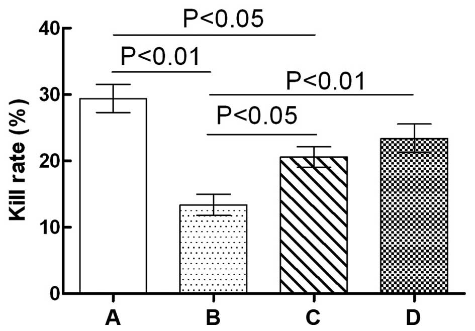
Effects of GPS on NK cell cytotoxicity
in immunosuppressed mice
The effects of GPS on NK cell cytotoxicity in
immunosuppressed mice are shown in Fig.
1. NK cell cytotoxicity in the Cy model group mice was
significantly lower compared with that of the normal control group
(P<0.01). Compared with the Cy model group, NK cell cytotoxicity
in the Cy + low-dose GPS group was significantly increased
(P<0.05); similar results were observed in the Cy + high-dose
GPS group (P<0.01). Compared with the normal control group, NK
cell cytotoxicity in the Cy + low-dose GPS group was significantly
reduced (P<0.05), although no significant difference was
observed between the Cy + high-dose GPS group and the normal
control group (P>0.05). Results showed that GPS could increase
NK cell cytotoxicity in immunosuppressed mice.
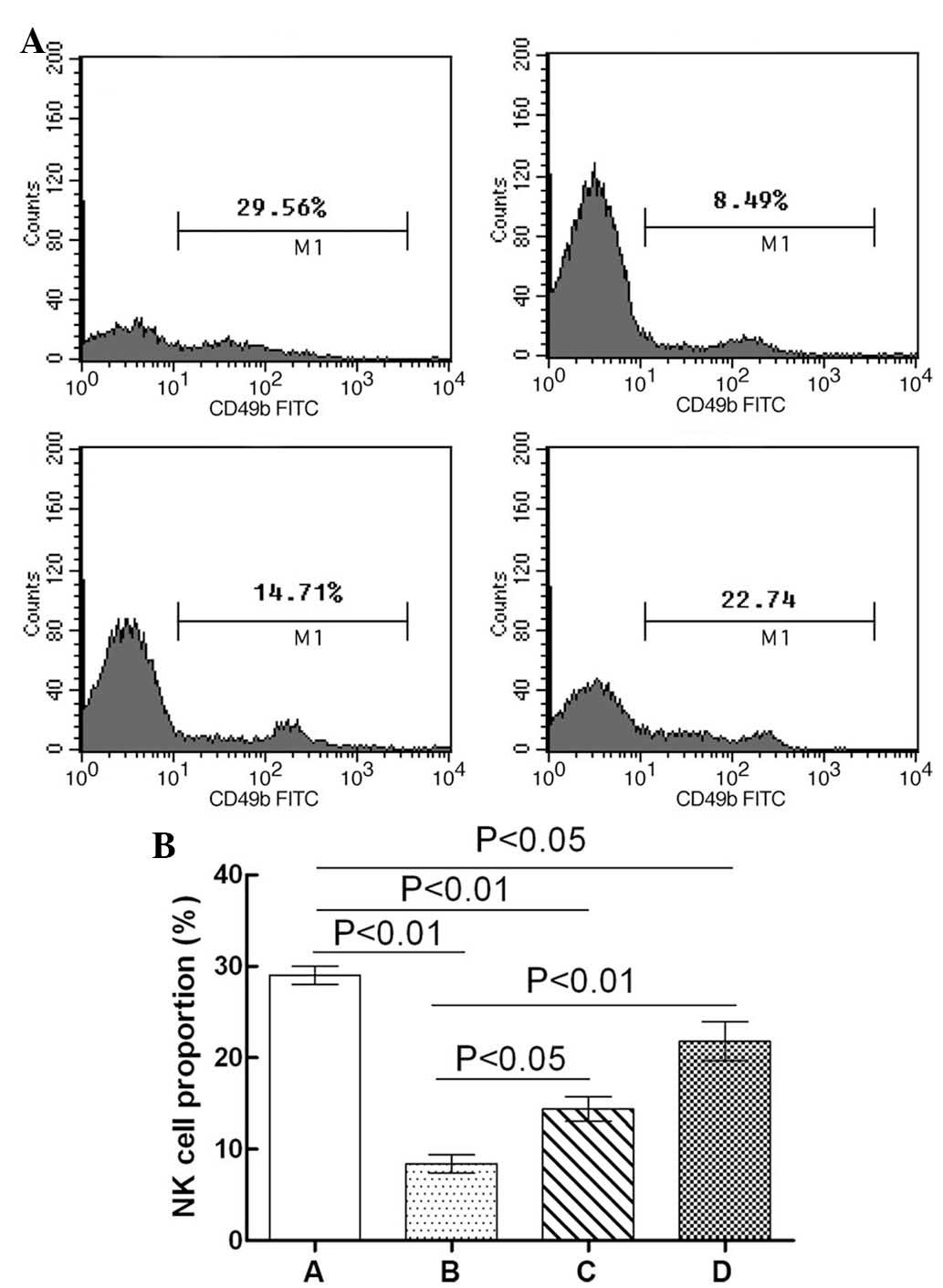
Effects of GPS on NK cell distribution
in the whole blood of immunosuppressed mice
The effects of GPS on NK cell distribution in the
whole blood of immunosuppressed mice are shown in Fig. 2. The proportion of NK cells in the
whole blood of the Cy model group mice was significantly lower
compared with the normal control group (P<0.01). Compared with
the Cy model group, the NK cell proportion in the whole blood of
the Cy + low-dose GPS mice was significantly increased
(P<0.05),; similar results were observed in the Cy + high-dose
GPS group (P<0.01). Compared with the normal control group, the
proportion of NK cells in the whole blood of the Cy + low-dose GPS
group was significantly reduced (P<0.01), and similar results
were observed for the Cy + high-dose GPS group (P<0.05). Results
showed that GPS could increase NK cell quantity in immunosuppressed
mice.
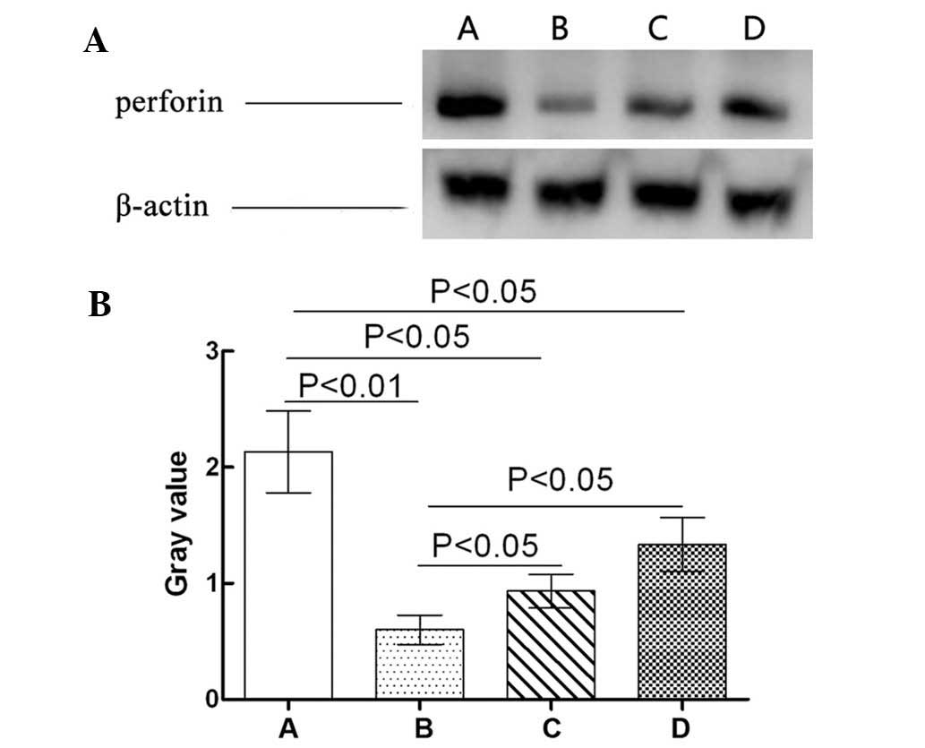
Effect of GPS on the expression levels
of perforin in the NK cells of immunosuppressed mice
The effects of GPS on the expression levels of
perforin in the NK cells of immunosuppressed mice are shown in
Fig. 3. Perforin expression levels
in the Cy model group were significantly downregulated compared
with those in the normal control group (P<0.01). Compared with
the Cy model group, perforin expression levels in the Cy + low-dose
GPS group and the Cy + high-dose GPS group were significantly
upregulated (P<0.05). However, the expression levels of perforin
in the NK cells of the Cy + low-dose GPS and Cy + high-dose GPS
groups were significantly lower compared with the normal control
group (P<0.05). Results showed that GPS was able to increase
expression levels of perforin of NK cells in immunosuppressed
mice.
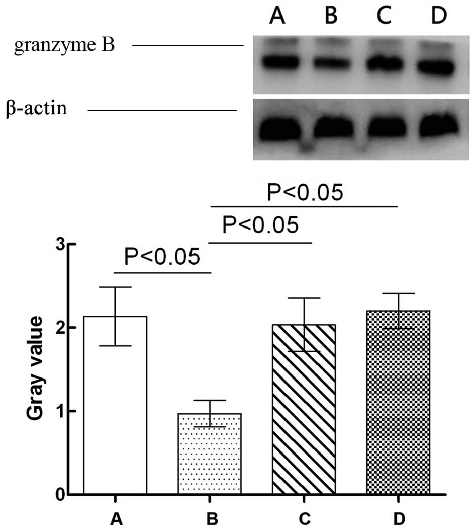
Effect of GPS on the expression levels
of granzyme B in the NK cells of immunosuppressed mice
The effects of GPS on the expression levels of
granzyme B in the NK cells of immunosuppressed mice are showed in
Fig. 4. Granzyme B expression levels
in the NK cells of the Cy model group were significantly
downregulated compared with those in the normal control grope
(P<0.05). Compared with the Cy model group, granzyme B
expression levels in the NK cells of Cy + low-dose and high-dose
GPS groups was significantly upregulated (P<0.05). Compared with
the normal control group however, the expression levels of granzyme
B in the NK cells of the Cy + low-dose and high-dose GPS mice did
not show any significant difference (P>0.05). Results indicated
that GPS could increase expression levels of granzyme B in the NK
cells in immunosuppressed mice.
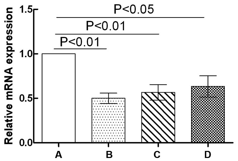
Effect of GPS on the mRNA expression
levels of IFN-γ in the NK cells of immunosuppressed mice
The effects of GPS on the mRNA expression levels of
IFN-γ in the NK cells of immunosuppressed mice are shown in
Fig. 5. Compared with the normal
control group, the mRNA expression levels of IFN-γ in the NK cells
of the Cy model group mice were significantly downregulated
(P<0.01). The mRNA expression levels of IFN-γ in the NK cells of
both the Cy + low-dose GPS group (P<0.01) and the Cy + high-dose
GPS group (P<0.05) were also significantly downregulated
compared with the normal control group. Compared with the Cy model
group, the mRNA expression levels of IFN-γ in the NK cells of both
the Cy + low-dose GPS group and the Cy + high-dose GPS group showed
no significant difference (P>0.05). Results indicated that the
effects of GPS on mRNA expression levels of IFN-γ of the NK cells
in immunosuppressed mice were not significant.
Discussion
NK cells are effector cells of the innate immune
system and act as the first line of defense against tumors or viral
infection (15,16). While killing target cells, NK cells
do not have to be sensitized by antigens and have no major
histocompatibility complex restriction (17). NK cells are able to cause the lysis
and apoptosis of target cells by releasing perforins and granzymes
(18,19). In addition, NK cells exhibit
anti-tumor and anti-viral activity by releasing various types of
cytokines, such as IFN-γ (20–22).
Investigators have demonstrated that GPS promotes NK cell
cytotoxicity and anti-tumor activity, but few studies have
investigated the mechanisms underlying these effects (23,24).
In the present study, compared with the normal
control group, the cytotoxicity and proportion of NK cells in the
blood, and the expressing levels of perforin, granzyme and IFN-γ
mRNA in the immunosuppressed model group were significantly
reduced, demonstrating that the immunosuppressed mouse model was
successfully created. Compared with the Cy model group, the
cytotoxicity and proportion of NK cells in the blood of both the Cy
+ low-dose GPS group and the Cy + high-dose GPS group were
significantly increased, suggesting that GPS was able to improve NK
cell cytotoxicity in the immunosuppressed mice and promote NK cells
differentiation. Compared with the Cy model group, the expression
levels of perforin and granzyme in both the Cy + low-dose GPS group
and the Cy + high-dose GPS group were significantly increased. This
increase in expression levels was dependent on the dosage of GPS,
and was higher in the high-dose GPS group. These results suggested
that the mechanism underlying the increase in NK cell cytotoxicity
induced by GPS may be associated with the ability of GPS to promote
the expression and release of perforins and granzymes. Furthermore,
compared with the Cy model group, the mRNA expression levels of
IFN-γ in the NK cells of the Cy + low-dose GPS group and the Cy +
high-dose GPS group showed no significant difference, indicating
that the effects of GPS on the expression levels of IFN-γ mRNA in
immunosuppressive mice were not obvious. However, the effect of GPS
on the levels of IFN-γ protein expression require further
study.
In conclusion, the present study demonstrated that
GPS is able to promote NK cell cytotoxicity in immunosuppressed
mice by increasing the proportion of NK cells in the blood and by
inducing the increased expression of perforins and granzymes. Thus,
the present study investigated the molecular mechanism underlying
the activation of NK cells activation GPS, and indicated that GPS
has a wide application in the treatment of cancer and
immunodeficiency diseases.
Acknowledgements
The present study was supported by the National
Natural Science Foundation of China (grant no. 81303079).
Glossary
Abbreviations
Abbreviations:
|
GPS
|
ginseng polysaccharide
|
|
cy
|
cyclophosphamide
|
|
NK
|
natural killer
|
|
MACS
|
magnetic-activated cell sorting
|
|
FACS
|
fluorescence-activated cell
sorting
|
|
IFN
|
interferon
|
References
|
1
|
Kwak JH, Park JY, Lee D, Kwak JY, Park EH,
Kim KH, Park HJ, Kim HY, Jang HJ, Ham J, et al: Inhibitory effects
of ginseng sapogenins on the proliferation of triple negative
breast cancer MDA-MB-231 cells. Bioorg Med Chem Lett. 24:5409–5412.
2014. View Article : Google Scholar : PubMed/NCBI
|
|
2
|
Lee MH, Lee BH, Lee S and Choi C:
Reduction of hepatitis A virus on FRhK-4 cells treated with Korean
red ginseng extract and ginsenosides. J Food Sci. 78:M1412–M1415.
2013. View Article : Google Scholar : PubMed/NCBI
|
|
3
|
Jiao L, Zhang X, Li B, Liu Z, Wang M and
Liu S: Anti-tumour and immunomodulatory activities of
oligosaccharides isolated from Panax ginseng C.A. Meyer. Int J Biol
Macromol. 65:229–233. 2014. View Article : Google Scholar : PubMed/NCBI
|
|
4
|
Li C, Cai J, Geng J, Li Y, Wang Z and Li
R: Purification, characterization and anticancer activity of a
polysaccharide from Panax ginseng. Int J Biol Macromol. 51:968–973.
2012. View Article : Google Scholar : PubMed/NCBI
|
|
5
|
Ban JY, Kang SW, Lee JS, Chung JH, Ko YG
and Choi HS: Korean red ginseng protects against neuronal damage
induced by transient focal ischemia in rats. Exp Ther Med.
3:693–698. 2012.PubMed/NCBI
|
|
6
|
Yang JW and Kim SS: Ginsenoside Rc
promotes anti-adipogenic activity on 3T3-L1 adipocytes by
down-regulating C/EBPα and PPARγ. Molecules. 20:1293–1303. 2015.
View Article : Google Scholar : PubMed/NCBI
|
|
7
|
Ni W, Zhang X, Wang B, Chen Y, Han H, Fan
Y, Zhou Y and Tai G: Antitumor activities and immunomodulatory
effects of ginseng neutral polysaccharides in combination with
5-fluorouracil. J Med Food. 13:270–277. 2010. View Article : Google Scholar : PubMed/NCBI
|
|
8
|
Wang J, Zuo G, Li J, Guan T, Li C, Jiang
R, Xie B, Lin X, Li F, Wang Y and Chen D: Induction of tumoricidal
activity in mouse peritoneal macrophages by ginseng polysaccharide.
Int J Biol Macromol. 46:389–395. 2010. View Article : Google Scholar : PubMed/NCBI
|
|
9
|
Cai JP, Wu YJ, Li C, Feng MY, Shi QT, Li
R, Wang ZY and Geng JS: Panax ginseng polysaccharide suppresses
metastasis via modulating Twist expression in gastriccancer. Int J
Biol Macromol. 57:22–25. 2013. View Article : Google Scholar : PubMed/NCBI
|
|
10
|
Cheng H, Li S, Fan Y, Gao X, Hao M, Wang
J, Zhang X, Tai G and Zhou Y: Comparative studies of the
antiproliferative effects of ginseng polysaccharides on HT-29 human
colon cancer cells. Med Oncol. 28:175–181. 2011. View Article : Google Scholar : PubMed/NCBI
|
|
11
|
Yamaoka Y, Kawakita T, Kaneko M and Nomoto
K: A polysaccharide fraction of Zizyphi fructus in augmenting
natural killer activity by oral administration. Biol Pharm Bull.
19:936–939. 1996. View Article : Google Scholar : PubMed/NCBI
|
|
12
|
Xie FY, Zeng ZF and Huang HY: Clinical
observation on nasopharyngeal carcinoma treated with combined
therapy of radiotherapy and ginseng polysaccharide injection.
Zhongguo Zhong Xi Yi Jie He Za Zhi. 21:332–334. 2001.(In Chinese).
PubMed/NCBI
|
|
13
|
Cho YJ, Son HJ and Kim KS: A 14-week
randomized, placebo-controlled, double-blind clinical trial to
evaluate the efficacy and safety of ginseng polysaccharide (Y-75).
J Transl Med. 12:2832014. View Article : Google Scholar : PubMed/NCBI
|
|
14
|
Livak KJ and Schmittgen TD: Analysis of
relative gene expression data using real-time quantitative PCR and
the 2-ΔΔCt method. Methods. 25:402–408. 2001. View Article : Google Scholar : PubMed/NCBI
|
|
15
|
Watzl C: How to trigger a killer:
Modulation of natural killer cell reactivity on many levels. Adv
Immunol. 124:137–170. 2014. View Article : Google Scholar : PubMed/NCBI
|
|
16
|
Eddy JL, Krukowski K, Janusek L and
Mathews HL: Glucocorticoids regulate natural killer cell function
epigenetically. Cell Immunol. 290:120–130. 2014. View Article : Google Scholar : PubMed/NCBI
|
|
17
|
Cox JH: Evaluation of natural killer cell
activity. Methods Mol Med. 17:383–389. 1999.PubMed/NCBI
|
|
18
|
Jiang W, Zhang C, Tian Z and Zhang J:
hIFN-α gene modification augments human natural killer cell line
anti-human hepatocellular carcinoma function. Gene Ther.
20:1062–1069. 2013. View Article : Google Scholar : PubMed/NCBI
|
|
19
|
Li Q, Kobayashi M, Wakayama Y, Inagaki H,
Katsumata M, Hirata Y, Hirata K, Shimizu T, Kawada T, Park BJ, et
al: Effect of phytoncide from trees on human natural killer cell
function. Int J Immunopathol Pharmacol. 22:951–959. 2009.PubMed/NCBI
|
|
20
|
Ahlenstiel G: The natural killer cell
response to HCV infection. Immune Netw. 13:168–176. 2013.
View Article : Google Scholar : PubMed/NCBI
|
|
21
|
Campbell KS and Hasegawa J: Natural killer
cell biology: An update and future directions. J Allergy Clin
Immunol. 132:536–544. 2013. View Article : Google Scholar : PubMed/NCBI
|
|
22
|
Lin SJ, Yan DC, Lee WI, Kuo ML, Hsiao HS
and Lee PY: Effect of azithromycin on natural killer cell function.
Int Immunopharmacol. 13:8–14. 2012. View Article : Google Scholar : PubMed/NCBI
|
|
23
|
Zhou X, Shi H, Jiang G, Zhou Y and Xu J:
Antitumor activities of ginseng polysaccharide in C57BL/6 mice with
Lewis lung carcinoma. Tumour Biol. 35:12561–12566. 2014. View Article : Google Scholar : PubMed/NCBI
|
|
24
|
Jiao L, Zhang X, Li B, Liu Z, Wang M and
Liu S: Anti-tumour and immunomodulatory activities of
oligosaccharides isolated from Panax ginseng C.A. Meyer. Int J Biol
Macromol. 65:229–233. 2014. View Article : Google Scholar : PubMed/NCBI
|