Introduction
Severe acute pancreatitis (SAP), an acute
inflammatory condition of the pancreas, is considered to be a
paradigm of sterile inflammation leading to systemic multiple organ
dysfunction syndrome (MODS) and death. Acute renal injury (ARI) is
one of the main complications of SAP and significantly increases
the mortality rate of patients with AP (66.6 vs. 14.5%) (1). However, the underlying mechanisms of
ARI occurring in patients with SAP have remained to be clarified.
Increasing evidence has indicated that pro-inflammatory cytokines,
including tumor necrosis factor-α (TNF-α) and interleukin (IL)-6,
have an important role in the pathological mechanisms of SAP and
SAP-associated organ failure (2–4).
Therefore, inhibiting the transcription and translation of
mediators to reduce the secretion of pro-inflammatory factors may
ameliorate inflammation and renal failure in SAP.
Signal transducers and activators of transcription
(STATs), a protein family comprised of seven members (STAT1, −2,
−3, −4, −5a, −5b and −6), generally transduce signals from
activated receptors or intracellular kinases to the nucleus, thus
activating and regulating gene transcription (5). The Janus kinase 2 (JAK2)/STAT3 pathway
is well known to be involved in the immune response of numerous
cytokines, including TNF-α and IL-6 (6). In addition, evidence derived from
numerous clinical and experimental studies suggests the involvement
of the JAK2/STAT3 pathway in pancreatitis (7–9) or renal
diseases (10–12).
Curcumin (diferuloylmethane), the active
phytochemical component of turmeric (a spice used mostly in Asia)
has been isolated from the rhizome of the Curcuma longa
plant. Curcumin has a myriad of pharmacological effects, including
anti-inflammatory (13,14), anti-bacterial (15), anti-oxidative (16) and renal-protective activities
(17,18). A previous study suggested that
curcumin has a therapeutic role in a rat model of SAP (19). Another study has reported that
curcumin inhibited renal inflammation in cisplatin-induced
nephrotoxicity in mice (20) and
suppressed JAK2/STAT3 signaling to ameliorate renal endothelial
dysfunction in fructose-fed rats (21). All of the above suggests the
potential use of curcumin in the treatment of acute renal failure
following SAP. Thus, the present study investigated the effect of
curcumin on acute renal failure arising from SAP and to explore the
molecular mechanism.
Materials and methods
Chemicals and reagents
Curcumin and hematoxylin and eosin were purchased
from Sigma-Aldrich (Merck KGaA, Darmstadt, Germany). ELISA kits for
assaying TNF-α and IL-6 were obtained from R&D (Minneapolis,
MN, USA). Assay kits for amylase (AMY), creatinine (Cr) and blood
urea nitrogen (BUN) were purchased from Jiancheng Biotech (Nanjing,
China). TRIzol reagent was obtained from Invitrogen (Thermo Fisher
Scientific, Inc., Waltham, MA, USA) and Moloney murine leukemia
virus (M-MLV) reverse transcriptase was from Promega, Corp.
(Madison, WI, USA). Antibodies against JAK2 (cat no. 3230),
phosphorylated (p)-JAK2 (cat no. 3776), STAT3 (cat no. 4904),
p-STAT3 (cat no. 9145) and suppressor of cytokine signaling 3
(SOCS3; cat no. 2923) were purchased from Cell Signaling
Technology, Inc. (Danvers, MA, USA) and horseradish peroxidase
(HRP)-conjugated GAPDH antibody (cat no. sc-2577) was purchased
from Santa Cruz Biotechnology (Dallas, TX, USA).
Animals
A total of 30 male Sprague-Dawley rats (age, 8–10
weeks; weight, 200–250 g) were obtained from Shanghai Laboratory
Animals Co., Ltd. (Shanghai, China). All animals were housed under
standardized conditions with a 12-h light/dark cycle with free
access to standard laboratory chow and water. All protocols and
procedures used in the study complied the US National Institute of
Health Guidelines for the Care and Use of Laboratory Animals and
were approved by the Institutional Animal Care and Utilization
Committee of Fujian Medical University (Fuzhou, China).
Experimental models and groups
The rats were divided into three subgroups: Sham
controls, SAP rats treated with saline and SAP rats treated with
curcumin (100 mg/kg). Rats were fasted 12 h prior to the operation.
The three groups were dived into 3, 12 and 24 h time-point groups
(each sub-group contained 3 rats). After anesthesia with 10%
chloraldurat (0.3 ml/100 g, Sigma Aldrich; Merck KGaA, Darmstadt,
Germany), the SAP model was induced by a standard
pressure-controlled infusion of a freshly prepared 5% sodium
taurocholate (Sigma Aldrich; Merck KGaA) solution (0.1 ml/100 g)
into the bile-pancreatic duct under laparotomy as previously
described (22). Rats in the sham
control group underwent the same operative procedure without the
injection of 5% sodium taurocholate in the pancreatic duct. In the
SAP + Cur group, rats were intraperitoneally injected with curcumin
solution (100 mg/kg) 30 min prior to the establishment of the SAP
model. After surgery, all animals were subcutaneously injected with
saline (0.2 ml/kg) in order to replenish the missing fluid.
Blood and tissue preparation
The rats were sacrificed at designated time-points
after the induction of pancreatitis. The blood samples were
obtained via intracardiac puncture and then centrifuged to obtain
the serum to be stored at −20°C until assay. Pancreas and kidney
tissues were dissected quickly on ice, and parts of tissues were
immediately fixed for hematoxylin and eosin staining analysis,
while others were frozen in liquid nitrogen and stored at −80°C for
later analysis.
Hematoxylin and eosin staining
analysis
After blood collection, rat pancreas and kidney
tissues were removed, immediately fixed and preserved in 70%
ethanol. Subsequent to embedding in paraffin, specimens were cut in
4-µm-thick sections and mounted on
3-aminopropyltriethoxysilane-coated glass slides. The sections were
de-paraffinized in xylene, re-hydrated in decreasing concentrations
of ethanol in water and stained with hematoxylin and eosin reagent.
Examination of sections was performed with a light microscope
(Olympus Ltd., Tokyo, Japan).
Serum AMY, BUN and Cr assays
Serum levels of AMY and Cr as well as BUN were
measured using standard diagnostic kits following the
manufacturer's instructions.
Serum TNF-α and IL-6 levels
ELISA kits were used to detect serum TNF-α and IL-6
levels according to the manufacturer's instructions.
Reverse-transcription quantitative
polymerase chain reaction (RT-qPCR) analysis
SOCS3 mRNA in the kidney tissue was detected by
RT-qPCR analysis. In brief, total RNA was extracted from kidney
tissues with TRIzol reagent according to the manufacturer's
instructions, and was reverse-transcribed into complementary DNA
using an oligo (dT) primer (SunShine Biotechnology, Nanjing, China)
and M-MLV reverse transcriptase. The 20 µl PCR reaction mixture was
prepared and contained 2-µl cDNA, 10 µl Power SYBR-Green PCR
Supermix (Bio-Rad Laboratories, Inc., Hercules, CA, USA) and 1 µl
of each primer and H2O-DEPC to 20 µl. RT-qPCR analysis were
performed using SYBR-Green I dye (Bio-Rad Laboratories, Inc.) under
the following reaction conditions: 1 min at 95°C followed by 40
cycles of 15 sec at 95°C, 1 min at 60°C. The following primers were
used: SOCS3 forward, 5′-TTCGCCCTTAGCGTGAAGATGG-3′ and reverse,
5′-TAGTGCTCCAGCAGCTCGAAGA-3′; GAPDH forward,
5′-CTTTGGTATCGTGGAAGGACTC-3′ and reverse,
5′-GTAGAGGCAGGGATGATGTTCT-3′. The comparative Cq
(2−∆∆Cq) method was used to analyze the relative gene
expression levels, as previously described (23). GAPDH was used as an internal positive
control.
Western blot analysis
The expression of JAK2, STAT3 and SOCS3 protein as
well as the levels of p-JAK2 and p-STAT3 in kidney tissues were
measured by western blot analysis. Protein extracts were obtained
by homogenizing samples in a cell lysis buffer (10 mM Tris-HCl, 1
mM EDTA and 250 mM sucrose, pH 7.4, containing 15 µg/ml aprotinin,
5 µg/ml leupeptin, 0.1 mM phenylmethanesulfonyl fluoride, 1 mM NaF
and 1 mM Na3VO4), followed by centrifugation
at 12,000 × g for 15 min. The protein concentration was determined
using a bicinchoninic acid kit (Thermo Fisher Scientific, Inc.). A
total of 30 µg protein per lane was separated by 10% SDS-PAGE,
transferred onto polyvinylidene fluoride membranes (Millipore,
Billerica, MA, USA), blocked with 5% skim milk and incubated with
primary antibodies (p-JAK2: 1:1,000, JAK2: 1:1,000, p-STAT3,
1:1,000, SOCS3: 1:1,000, GAPDH: 1:500) at 4°C overnight. Then the
blots were incubated with the corresponding secondary antibody
(HRP-conjugated whole-goat anti-rabbit IgG, 1:10,000; cat no.
074-1506; Kirkegaard & Prerry Lab Inc., Gaithersburg, MD, USA)
for 2 h at room temperature. Finally, immunoreactive bands were
visualized via enhanced chemiluminescence (Cell Signaling
Technology, Inc., Danvers, MA, USA) and the membranes were then
immediately exposed to autoradiographic film (Eastman Kodak,
Rochester, NY, USA).
Statistical analysis
Results were expressed as the mean ± standard
deviation. Statistical analysis was performed using one-way
analysis of variance followed by a post-hoc test (Fisher's least
significant differences test) as appropriate with the statistical
analysis system GraphPad Prism 6 (GraphPad Software, Inc., La
Jolla, CA, USA). P<0.05 was considered to indicate a
statistically significant difference.
Results
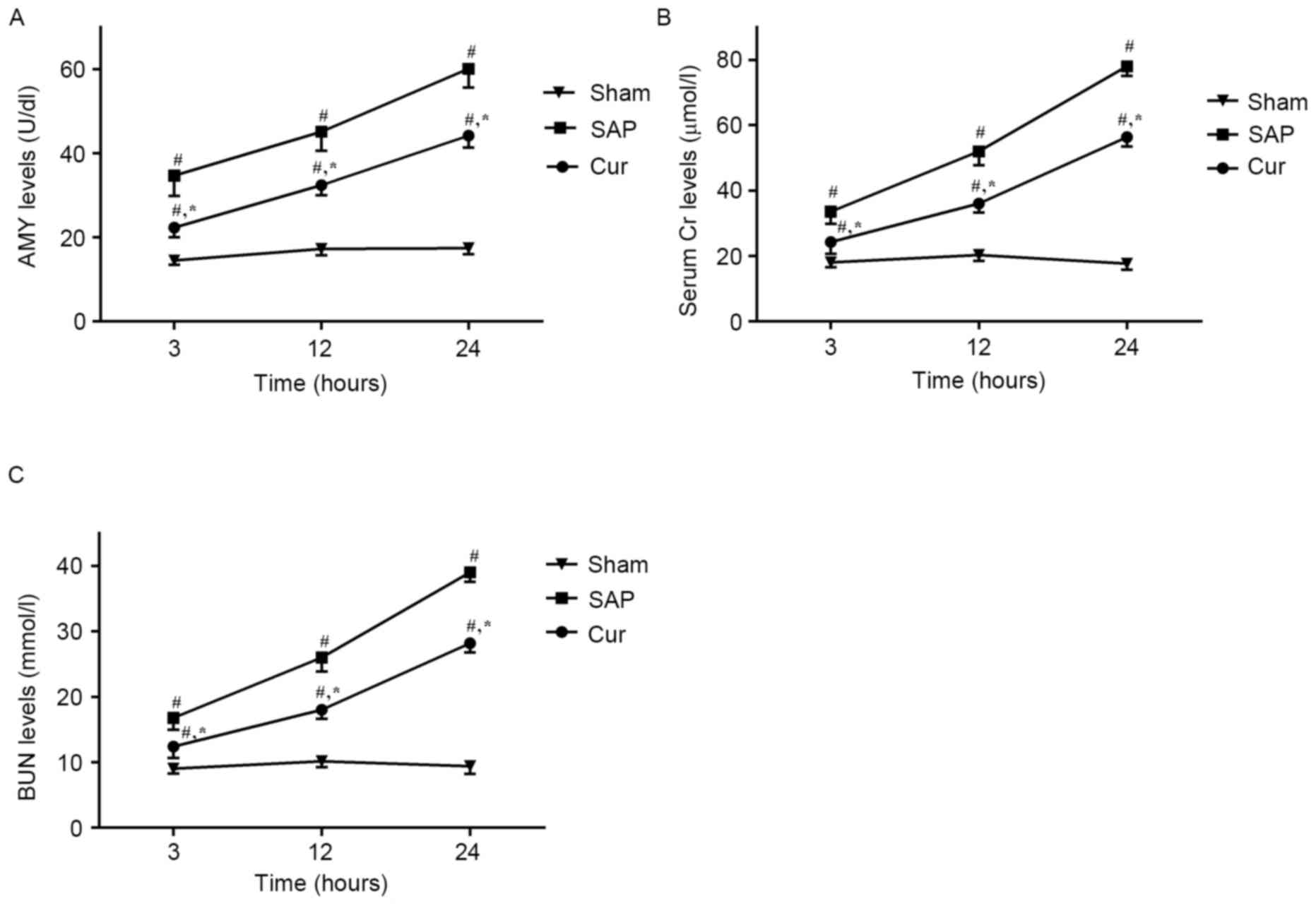
Curcumin attenuates SAP-induced
increases in serum AMY, Cr and BUN
As shown in Fig. 1A,
serum AMY levels were significantly increased in the SAP group at
all time-points in comparison with those in the Sham group.
However, in the Cur group, these increases in serum AMY levels were
significantly inhibited. Moreover, the SAP group showed elevated
levels of serum Cr (Fig. 1B) and BUN
(Fig. 1C) compared with those in the
Sham group. Treatment with curcumin obviously decreased the levels
of serum Cr and BUN at the respective time-points.
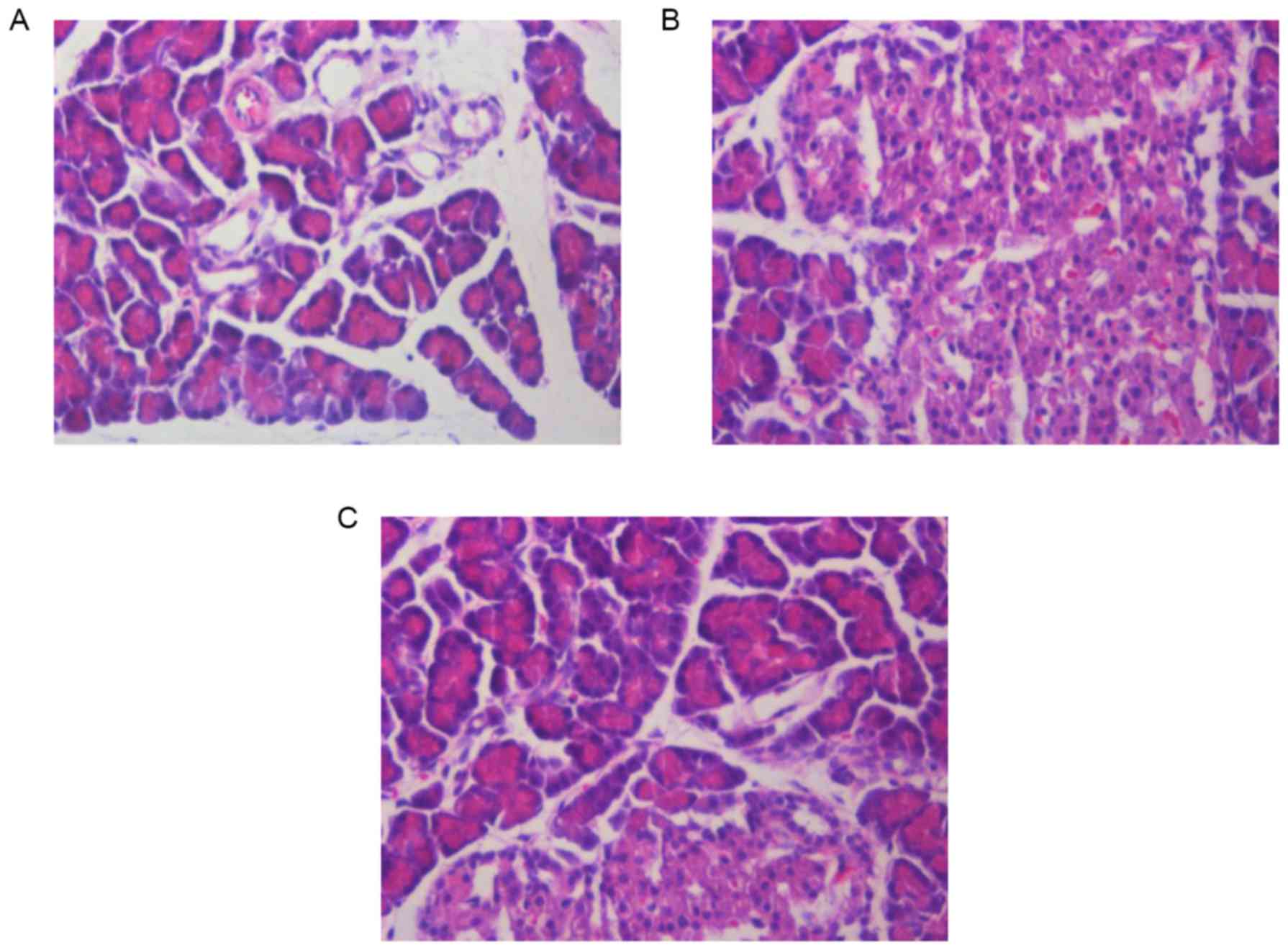
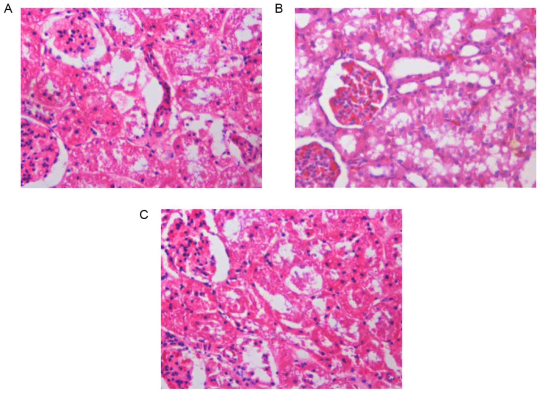
Curcumin pre-treatment reduces
histopathological changes in the pancreas and kidneys of SAP
rats
Pancreatic and renal histological changes were
assessed using hematoxylin and eosin staining. As shown in Fig. 2, obvious edema, inflammatory cell
infiltration and necrosis were observed in the pancreases of SAP
rats at 12 h (Fig. 2B). Compared to
the SAP group, the pancreatic histological injuries were
significantly ameliorated by pre-treatment with curcumin (Fig. 2C). Furthermore, Fig 3 shows that, SAP rats at 12 h
demonstrated glomerular and tubular damage as well as inflammatory
cell infiltration, suggesting renal injury (Fig. 3B). However, rats pre-treated with
curcumin demonstrated reduced histological features in the kidney
in comparison with those in the SAP rats (Fig. 3C).
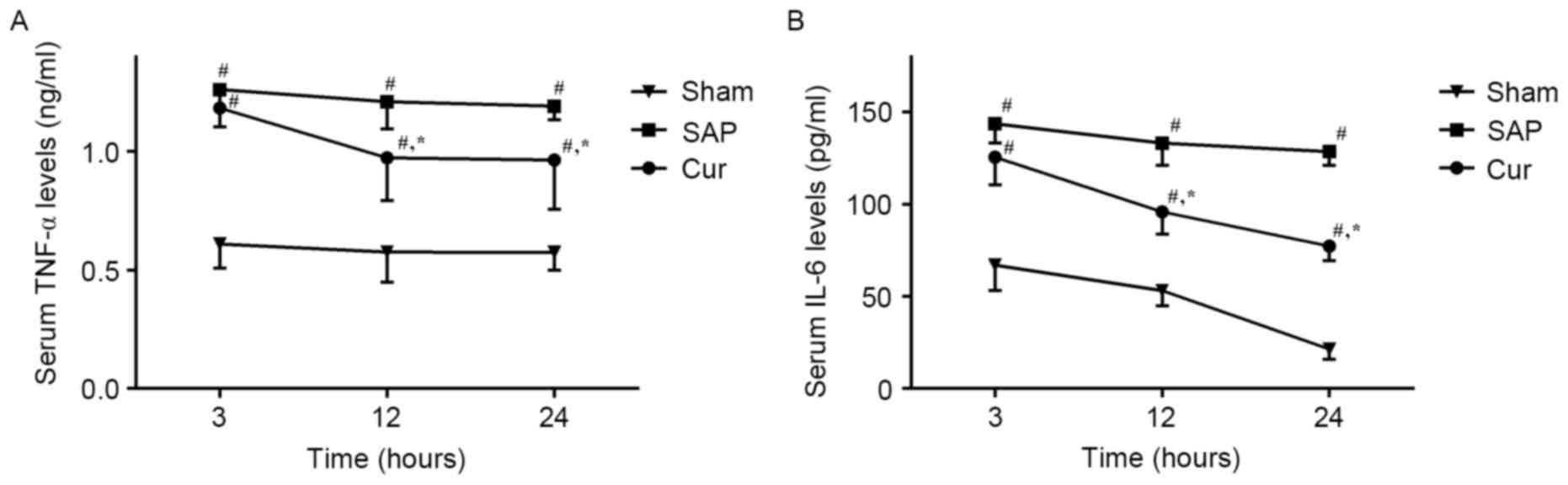
Curcumin reduces SAP-induced increases
of serum TNF-α and IL-6 levels in SAP rats
The effect of curcumin on the serum levels of
inflammatory cytokines, TNF-α and IL-6, were analyzed using ELISA
kits. As shown in Fig. 4, a
substantial increase in TNF-α and IL-6 levels was observed in the
SAP group. However, pre-treatment with curcumin significantly
decreased serum levels of TNF-α and IL-6 in this animal model.
Curcumin suppresses the JAK2/STAT3
signaling pathway in SAP rats
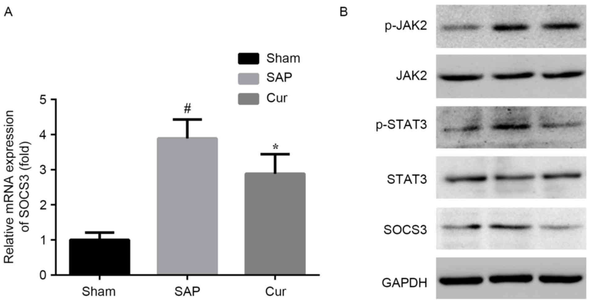
Elevated mRNA and protein levels of SOCS3 were
observed in the kidneys of SAP rats. These alterations in SAP rats
were attenuated by pre-treatment with curcumin at the mRNA and
protein level (Fig. 5A and B). To
further determine JAK and STAT3 signaling in the kidneys of rats at
12 h after induction of SAP, western blot analysis was applied.
Compared to the Sham group, the SAP group showed elevated renal
protein levels of p-JAK2 and p-STAT3. Curcumin distinctly inhibited
the activation of JAK2 and STAT3 in this model (Fig. 5B).
Discussion
The present study demonstrated that a) renal injury
caused by SAP was significantly improved by pre-treatment with
curcumin, b) curcumin greatly decreased the secretion of
inflammatory cytokines and c) the mechanism by which curcumin
ameliorates renal injury includes the reduction of inflammation and
suppression of JAK2/STAT3 pathway activation.
A previous study confirmed the protective effect of
curcumin in a rat model of SAP (19). In the present study, hyperamylasemia
and pancreatic pathological evidence were observed in the SAP model
group, which were obviously improved by pre-treatment with
curcumin. Moreover, the levels of BUN and serum creatinine were
increased, alongside obvious pathological damage, which was
indicative of renal injury in this SAP model. However,
pre-treatment with curcumin significantly ameliorated the
pathological changes in the kidneys of SAP rats and also decreased
BUN and serum Cr levels in rats with renal injury associated with
SAP.
At present, the pathogenesis of SAP-induced renal
injury remains elusive. It has been generally recognized that the
excess release of cytokines and inflammatory mediators has a
pivotal role in SAP and SAP-associated renal injury (24). Cytokines such as IL-6 and TNF-α exert
a major effect on the outcome of SAP, particularly by triggering
the systemic inflammatory response and multisystem organ failure,
which is mostly responsible for the associated mortality (25). In the present study, increased serum
levels of IL-6 and TNF-α were observed in the SAP group, which were
greatly decreased by treatment with curcumin. These results
combined with the histological and biochemical changes in the
kidneys of SAP rats indicated that curcumin attenuated renal injury
in SAP rats, at least in part via the inhibition of inflammatory
cytokines.
The JAK/STAT pathway is a pleiotropic cascade
essential to cytokine and growth hormone receptor signaling
(26). An increasing body of
evidence has suggested the involvement of the JAK/STAT pathway in
renal disease (27–29). A previous study indicated that the
activation of JAK/STAT3 signaling induces the expression of IL-1β
in cerulean-stimulated pancreatic acinar cells (30). Of note, studies have shown that
curcumin, as an inhibitor of the JAK2/STAT3 pathway, exerts marked
effects in several experimental models and human diseases (21,31). The
present study found that the phosphorylation of JAK2 and STAT3 was
significantly increased in the kidneys of SAP rats, which was
inhibited by pre-treatment with curcumin, suggesting that the
inflammatory response triggered by SAP was effectively inhibited by
curcumin via interfering with JAK2/STAT3 pathway activation.
However, few studies have investigated the role of JAK2/STAT3
signaling in renal injury following SAP. The present study provided
in vivo evidence that the JAK2/STAT3 pathway is activated in
renal injury following SAP, and that the renoprotective effect of
curcumin in SAP is associated with the inhibition of this
activation.
In conclusion, the present study demonstrated the
renoprotective effect of curcumin in a rat model of SAP. The
underlying mechanism of its effect is in part the inhibition of
JAK2/STAT3 pathway activation to reduce the inflammation cascade
and inflammatory cytokine secretion. Therefore, the findings of the
present study revealed the potential use of curcumin for the
prevention and treatment of SAP and the associated renal
injury.
Acknowledgements
The present study was supported by a grant from the
Geriatric Key Clinical Specialty Discipline Construction Programs
of China and Fujian Province (2013).
References
|
1
|
Li H, Qian Z, Liu Z, Liu X, Han X and Kang
H: Risk factors and outcome of acute renal failure in patients with
severe acute pancreatitis. J Crit Care. 25:225–229. 2010.
View Article : Google Scholar : PubMed/NCBI
|
|
2
|
Papachristou GI, Clermont G, Sharma A,
Yadav D and Whitcomb DC: Risk and markers of severe acute
pancreatitis. Gastroenterol Clin North Am. 36:277–296, viii. 2007.
View Article : Google Scholar : PubMed/NCBI
|
|
3
|
Zhang XP, Wang L and Zhou YF: The
pathogenic mechanism of severe acute pancreatitis complicated with
renal injury: A review of current knowledge. Digest Dis Sci. 53:pp.
297–306. 2008; View Article : Google Scholar : PubMed/NCBI
|
|
4
|
Zhu AJ, Shi JS and Sun XJ: Organ failure
associated with severe acute pancreatitis. World J Gastroentero.
9:2570–2573. 2003.
|
|
5
|
Rawlings JS, Rosler KM and Harrison DA:
The JAK/STAT signaling pathway. J Cell Sci. 117:1281–1283. 2004.
View Article : Google Scholar : PubMed/NCBI
|
|
6
|
Yang X, He G, Hao Y, Chen C, Li M, Wang Y,
Zhang G and Yu Z: The role of the JAK2-STAT3 pathway in
pro-inflammatory responses of EMF-stimulated N9 microglial cells. J
Neuroinflammation. 7:542010. View Article : Google Scholar : PubMed/NCBI
|
|
7
|
Yu JH and Kim H: Role of janus
kinase/signal transducers and activators of transcription in the
pathogenesis of pancreatitis and pancreatic cancer. Gut Liver.
6:417–422. 2012. View Article : Google Scholar : PubMed/NCBI
|
|
8
|
Vona-Davis LC, Frankenberry KA, Waheed U,
Peterson E and McFadden DW: Expression of STAT3 and SOCS3 in
pancreatic acinar cells 1,2. J Surg Res. 127:14–20. 2005.
View Article : Google Scholar : PubMed/NCBI
|
|
9
|
Ju KD, Lim JW, Kim KH and Kim H: Potential
role of NADPH oxidase-mediated activation of Jak2/Stat3 and
mitogen-activated protein kinases and expression of TGF-β1 in the
pathophysiology of acute pancreatitis. Inflamm Res. 60:791–800.
2011. View Article : Google Scholar : PubMed/NCBI
|
|
10
|
Horiguchi A and Asano T, Kuroda K, Sato A,
Asakuma J, Ito K, Hayakawa M, Sumitomo M and Asano T: STAT3
inhibitor WP1066 as a novel therapeutic agent for renal cell
carcinoma. Brit J Cancer. 102:1592–1599. 2010. View Article : Google Scholar : PubMed/NCBI
|
|
11
|
Brands MW, Banes-Berceli AK, Inscho EW,
Al-Azawi H, Allen AJ and Labazi H: Interleukin 6 knockout prevents
angiotensin II hypertension: role of renal vasoconstriction and
janus kinase 2/signal transducer and activator of transcription 3
activation. Hypertension. 56:879–884. 2010. View Article : Google Scholar : PubMed/NCBI
|
|
12
|
Pang M, Ma L, Gong R, Tolbert E, Mao H,
Ponnusamy M, Chin YE, Yan H, Dworkin LD and Zhuang S: A novel STAT3
inhibitor, S3I-201, attenuates renal interstitial fibroblast
activation and interstitial fibrosis in obstructive nephropathy.
Kidney Int. 78:257–268. 2010. View Article : Google Scholar : PubMed/NCBI
|
|
13
|
Aggarwal BB and Harikumar KB: Potential
therapeutic effects of curcumin, the anti-inflammatory agent,
against neurodegenerative, cardiovascular, pulmonary, metabolic,
autoimmune, and neoplastic diseases. Int J Biochem Cell Biol.
41:40–59. 2009. View Article : Google Scholar : PubMed/NCBI
|
|
14
|
Wang ME, Chen YC, Chen IS, Hsieh SC, Chen
SS and Chiu CH: Curcumin protects against thioacetamide-induced
hepatic fibrosis by attenuating the inflammatory response and
inducing apoptosis of damaged hepatocytes. J Nutr Biochem.
23:1352–1366. 2012. View Article : Google Scholar : PubMed/NCBI
|
|
15
|
Mun SH, Joung DK, Kim YS, Kang OH, Kim SB,
Seo YS, Kim YC, Lee DS, Shin DW, Kweon KT and Kwon DY: Synergistic
antibacterial effect of curcumin against methicillin-resistant
Staphylococcus aureus. Phytomedicine. 20:714–718. 2013. View Article : Google Scholar : PubMed/NCBI
|
|
16
|
Kim BH, Lee ES, Choi R, Nawaboot J, Lee
MY, Lee EY, Kim HS and Chung CH: Protective effects of curcumin on
renal oxidative stress and lipid metabolism in a rat model of Type
2 diabetic nephropathy. Yonsei Med J. 57:664–673. 2016. View Article : Google Scholar : PubMed/NCBI
|
|
17
|
Trujillo J, Chirino YI, Molina-Jijón E,
Andérica-Romero AC, Tapia E and Pedraza-Chaverrí J: Renoprotective
effect of the antioxidant curcumin: Recent findings. Redox Biol.
1:448–456. 2013. View Article : Google Scholar : PubMed/NCBI
|
|
18
|
Karahan MA, Yalcin S, Aydogan H,
Büyükfirat E, Kücük A, Kocarslan S, Yüce HH, Taskın A and Aksoy N:
Curcumin and dexmedetomidine prevents oxidative stress and renal
injury in hind limb ischemia/reperfusion injury in a rat model. Ren
Fail. 38:693–698. 2016. View Article : Google Scholar : PubMed/NCBI
|
|
19
|
Zhong K: Curcumin mediates a protective
effect via TLR-4/NF-κB signaling pathway in rat model of severe
acute pancreatitis. Cell Biochem Biophys. 73:175–180. 2015.
View Article : Google Scholar : PubMed/NCBI
|
|
20
|
Ueki M, Ueno M, Morishita J and Maekawa N:
Curcumin ameliorates cisplatin-induced nephrotoxicity by inhibiting
renal inflammation in mice. J Biosci Bioeng. 115:547–551. 2013.
View Article : Google Scholar : PubMed/NCBI
|
|
21
|
Zhang DM, Li YC, Xu D, Ding XQ and Kong
LD: Protection of curcumin against fructose-induced hyperuricaemia
and renal endothelial dysfunction involves NO-mediated JAK-STAT
signalling in rats. Food Chem. 134:2184–2193. 2012. View Article : Google Scholar : PubMed/NCBI
|
|
22
|
Zhang XH, Li ML, Wang B, Guo MX and Zhu
RM: Caspase-1 inhibition alleviates acute renal injury in rats with
severe acute pancreatitis. World J Gastroenterol. 20:10457–10463.
2014. View Article : Google Scholar : PubMed/NCBI
|
|
23
|
Livak KJ and Schmittgen TD: Analysis of
relative gene expression data using real-time quantitative PCR and
the 2(−Delta Delta C(T)) Method. Methods. 25:402–408. 2001.
View Article : Google Scholar : PubMed/NCBI
|
|
24
|
Makhija R and Kingsnorth AN: Cytokine
storm in acute pancreatitis. J Hepatobiliary Pancreat Surg.
9:401–410. 2002. View Article : Google Scholar : PubMed/NCBI
|
|
25
|
Yu J, Deng W, Wang W, Ding Y, Jin H, Chen
C, Chen X, Xiong X and Xu S: Inhibition of poly (ADP-ribose)
polymerase attenuates acute kidney injury in sodium
taurocholate-induced acute pancreatitis in rats. Pancreas.
41:1299–1305. 2012. View Article : Google Scholar : PubMed/NCBI
|
|
26
|
Chuang PY and He JC: JAK/STAT signaling in
renal diseases. Kidney Int. 78:231–234. 2010. View Article : Google Scholar : PubMed/NCBI
|
|
27
|
Kuratsune M, Masaki T, Hirai T,
Kiribayashi K, Yokoyama Y, Arakawa T, Yorioka N and Kohno N: Signal
transducer and activator of transcription 3 involvement in the
development of renal interstitial fibrosis after unilateral
ureteral obstruction. Nephrology (Carlton). 12:565–571. 2007.
View Article : Google Scholar : PubMed/NCBI
|
|
28
|
Berthier CC, Zhang H, Schin M, Henger A,
Nelson RG, Yee B, Boucherot A, Neusser MA, Cohen CD, Carter-Su C,
et al: Enhanced expression of janus kinase-signal transducer and
activator of transcription pathway members in human diabetic
nephropathy. Diabetes. 58:469–477. 2009. View Article : Google Scholar : PubMed/NCBI
|
|
29
|
Lu TC, Wang ZH, Feng X, Chuang PY, Fang W,
Shen Y, Levy DE, Xiong H, Chen N and He JC: Knockdown of Stat3
activity in vivo prevents diabetic glomerulopathy. Kidney Int.
76:63–71. 2009. View Article : Google Scholar : PubMed/NCBI
|
|
30
|
Yu JH, Kim KH and Kim H: Suppression of
IL-1beta expression by the Jak 2 inhibitor AG490 in
cerulein-stimulated pancreatic acinar cells. Biochem Pharmacol.
72:1555–1562. 2006. View Article : Google Scholar : PubMed/NCBI
|
|
31
|
Duan W, Yang Y, Yan J, Yu S, Liu J, Zhou
J, Zhang J, Jin Z and Yi D: The effects of curcumin post-treatment
against myocardial ischemia and reperfusion by activation of the
JAK2/STAT3 signaling pathway. Basic Res Cardiol. 107:2632012.
View Article : Google Scholar : PubMed/NCBI
|