Introduction
Chronic kidney disease (CKD) is a major healthcare
burden and the 18th leading cause of disease-related
mortality worldwide, with an estimated prevalence of 8–16%
worldwide (1,2). Renal interstitial fibrosis (RIF) is the
major pathological basis for many types of CKD, and the inhibition
of RIF has been considered as an effective therapeutic strategy to
protect the kidney (3). RIF is
generally characterized by an excessive accumulation and deposition
of extracellular matrix (ECM) that progressively leads to the
degradation of functional nephrons. This ECM is predominantly
produced by α-smooth muscle actin (α-SMA)-expressing
myofibroblasts, which may originate from renal epithelial tubules
through epithelial-to-mesenchymal transition (EMT) (4). EMT is able to induce a downregulation
of E-cadherin in epithelial cells, which leads to a loss of
adhesion and polarity in these cells (5). In the process of EMT, the expression of
myofibroblast cell marker α-SMA is increased in tubular epithelial
cells, whereas epithelial marker E-cadherin expression is decreased
(6).
Transforming growth factor-β1 (TGF-β1) has generally
been considered as an essential factor in RIF (7) and it is recognized that TGF-β1 and its
downstream mediator, connective tissue growth factor (CTGF), are
associated with most forms of CKD. EMT and fibrosis are promoted by
the expression of TGF-β1, whereas they are prevented by the
inhibition of TGF-β1 by various methods (8). CTGF is also known to be an important
matrix-cellular regulatory factor, which participates in
angiogenesis, and appears to be a central mediator of EMT and
fibrosis (9).
In addition, fibrosis is able to induce ischemia and
hypoxia, and hypoxia-inducible factor-1α (HIF-1α) is a
transcription factor associated with cellular responses to hypoxia;
recent reports have suggested that HIF-1α has an important role in
stimulating EMT in vivo (10,11).
Furthermore, vascular endothelial growth factor (VEGF) is a
downstream factor of HIF-1α, which promotes endothelial cell
proliferation, induces microvascular hyperpermeability and is
associated with interstitial matrix remodeling (12,13).
The fruit of Gardenia jasminoides Ellis
(Rubiaceae) has been used in Traditional Chinese Medicine for the
treatment of fever, jaundice, headache, edema, hypertension, and
hepatic disorders. Gardenia extract contains multiple active
chemical components, and a previous study by the present authors
indicated that the ethanol extract of gardenia fruits (EEG)
primarily contains eight constituents (14). Previous studies have indicated that
these components have various pharmacological properties, including
anti-inflammatory effects (15–17) and
the ability to attenuate oxidative stress (18); notably, inflammation and oxidative
stress are critical pathogenic factors in fibrogenesis. Gardenia
has previously been reported to exert antifibrotic effects in liver
fibrosis (19) However, to the best
of our knowledge, there are currently no published studies
regarding the antifibrotic effect of EEG in renal tissues.
Unilateral ureteral obstruction (UUO) is the most
frequently used experimental model for the investigation of the
mechanisms of RIF and for the evaluation of therapeutic methods to
alleviate fibrosis. Therefore, the effects of EEG on UUO-induced
RIF were studied in vivo. Furthermore, potential underlying
mechanisms were also explored. The present results demonstrated
that EEG was able to inhibit the induction of EMT by decreasing
TGF-β1, CTGF, HIF-1a, and increasing VEGF expression.
Materials and methods
Plant materials and reagents
The following antibodies were used for
immunohistochemistry and western blotting: Rabbit polyclonal
antibodies against HIF-1α, VEGF, TGF-β1, CTGF, β-actin, goat anti
rabbit IgG-Biotin (cat. nos: PB2045, BA0407, BA0290, BA0752, BA2305
and BA1003, respectively; all Wuhan Boster Biological Technology,
Ltd., Wuhan, China), E-cadherin and α-SMA (cat. nos: 20874 and
14395; Wuhan Sanying Biotechnology, Wuhan, China). An SABC kit
(cat. no.: SA1022; Wuhan Boster Biological Technology, Ltd.), NAG
ELISA kit (cat. no.: RA20482, Shanghai Qiaodu Biomart, Shanghai,
China), bicinchoninic acid assay protein quantification kit and
enhanced chemiluminescence plus kit (cat. nos: P0010, P0018;
Beyotime Institute of Biotechnology, Haimen, China) were also
purchased. A Masson Trichrome staining kit (cat. no.: DC0033;
Beijing Leagene Biotechnology Co., Ltd. Beijing, China), bovine
serum albumin (BSA; Sangon Biotech, Shanghai, China),
radioimmunoprecipitation assay (RIPA) lysis buffer (Shanghai
Solarbio Biotechnology Co., Ltd. Shanghai, China) and chloral
hydrate (Shanghai Sinopharm group, Shanghai, China) were used in
the present study. The dried gardenia fruits were purchased from
Yuanchunlin Pharmacy (Zhuhai, China) in January 2014 and identified
by Yang Chen. Voucher specimens were deposited at the Department of
Pharmaceutical Sciences, Zunyi Medical University Zhuhai Campus
(Zhuhai, China). Reference gardenoside, 6β-hydroxy geniposide,
geniposidic acid, geniposide, crocin-1, crocin-2, crocin-3 and
crocin-4, were isolated from EEG by high-speed counter-current
chromatography in a previous study by the present authors (14). The structures of these compounds are
displayed in Fig. 1.
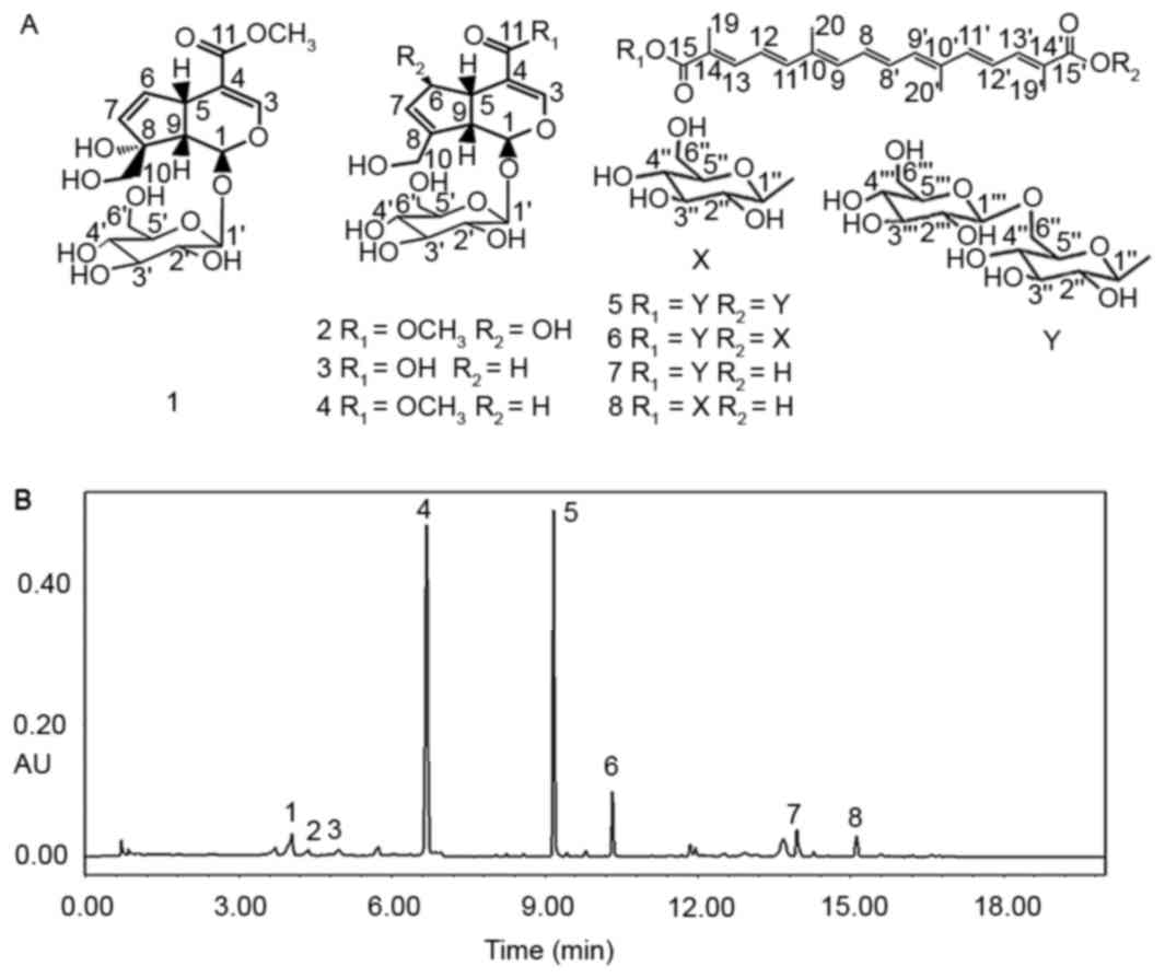 | Figure 1.Structures and UPLC chromatogram of
EEG. (A) Structures of compounds 1–8. (B) UPLC chromatogram of EEG.
The detection wavelength was as follows: 0–7 min, 238 nm; 7–20 min,
440 nm. UPLC, ultra performance liquid chromatography; EEG, ethanol
extract of gardenia fruits; 1, gardenoside; 2, 6β-hydroxy
geniposide; 3, geniposidic acid; 4, geniposide; 5, crocin-1; 6,
crocin-2; 7, crocin-3; 8, crocin-4. |
Preparation of crude extracts and
ultra performance liquid chromatography (UPLC) analysis
Chemical extraction and ultra performance liquid
chromatography analysis were performed according to our previous
study (14). Briefly, the dried
gardenia fruits (2 kg) were ground to coarse powder and extracted
with ethanol-water (40%) via cold percolation (2×2 l) at 25°C for
24 h. The alcohol extract was concentrated by rotary evaporation,
and the sample was stored at 4°C prior to animal experiments. The
mobile phase was composed of acetonitrile (A) and pure water (B),
that was programmed as follows: From 0–4.5 min, 10–18% A; 4.5–6.0
min, linear gradient increase from 18–28% A; 6.0–12.0 min, linear
gradient increase from 28–38% A; 12.0–15.0 min, linear gradient
increase from 38–50% A; 15.0–20.0 min, linear gradient increase
from 50–10% A. The flow rate was 0.3 ml/min and the detection
wavelength was as follows: 0–7 min, 238 nm; 7–20 min, 440 nm.
Experimental groups and protocol
A total of 30 male 8-week-old Sprague-Dawley rats,
weighing 180–220 g, were provided by the Animal Experimental Center
of Guangdong Province (Guangzhou, China). Rats had free access to
food and water at a temperature of 22±1°C, a relative humidity of
55±2%, and under a 12 h light/dark cycle. The rats were randomly
assigned into three groups (n=10 each): i) Sham-operation plus
vehicle (sham group); ii) UUO plus vehicle (UUO group); and iii)
UUO plus EEG (EEG group). Briefly, after anesthesia an
intraperitoneal injection of 10% chloral hydrate (300 mg/kg), the
left kidney and ureter were exposed via a flank incision. Then the
left ureter was ligated at two points and cut between the
ligatures. Sham group animals underwent an identical procedure
without ligation. Rats were gavaged with EEG at a dose of 200
mg/kg/day in the EEG group, whereas rats in the sham and UUO groups
were gavaged with the same volume of vehicle (distilled water) once
daily. All rats were sacrificed 14 days after UUO surgery, the left
kidneys were excised, and harvested for immunochemistry and western
blotting. Blood and urine samples were harvested for biochemical
analysis. The present methodology was approved by the Ethics
Committee of Zunyi Medical University.
Physiological parameters
N-acetyl-β-d-glucosaminidase (NAG), which is a
marker of renal tubular damage was measured in urine samples using
an ELISA kit according to the manufacturer's protocol. Serum
creatinine (Cr) and blood urea nitrogen (BUN) levels were measured
by a Roche automatic biochemical analysis.
Histopathologic examination
kidney tissues were fixed in 10% neutral formalin
solution for 24 h at room temperature, embedded in paraffin, and
sectioned into 4 µm sections that were stained with
hematoxylin-eosin (H&E) and Masson's trichrome stains using a
Masson Trichrome staining kit according to the manufacturer's
protocol. A total of 10 non-consecutive high-power fields of each
renal section were examined by light microscopy. The H&E
staining was used to provide a semi-quantitative score on the
degree of tubulointerstitial lesions (20), and Masson staining was used to
calculate the renal interstitial fibrosis area (21).
Immunohistochemical analysis
Renal sections (4 µm) were cut from
paraffin-embedded tissues. Sections were blocked with 5% BSA at
room temperature for 20 min. Slides were incubated overnight at 4°C
with the following primary antibodies: HIF-1α (1:100), VEGF
(1:100), E-cad (1:50), α-SMA (1:50), TGF-β1 (1:50) and CTGF (1:50).
Following washing with PBS (3 times), slide sections were incubated
at room temperature with the secondary antibody (1:100) for 20 min.
The immune complexes were then detected by the diaminobenzidine
substrate. Finally, the brown reaction product was observed via
light microscopy (magnification, ×400). Furthermore, 10
non-consecutive visual fields were randomly selected in the renal
section. The integrated optical density (IOD) total and mean
density (IOD/area) of each visual field were determined using
Image-Pro Plus software 6.0 (Media Cybernetics, Inc., Rockville,
MD, USA).
Western blotting
In the kidney tissue-based assay, kidney tissue
samples from each group were homogenized with lysis buffer plus 1
mM PMSF and protease inhibitor cocktail. Proteins were treated with
RIPA lysis buffer containing a cocktail of protease inhibitors (2
µg/ml aprotinin, 1 mM phenylmethyl sulfonylfuoride, and 10 µg/ml
leupeptin). Lysates were incubated for 30 min at 4°C and
centrifuged at 12,000 × g for 10 min at room temperature.
Supernatant protein concentration was then determined using to a
bicinchoninic acid protein quantification kit. Protein samples (20
µg) were separated by 10% SDS-PAGE and transferred to a PVDF
membrane using a wet transferring method. The membranes were
blocked with 5% BSA for 1 h at 4°C and incubated overnight at 4°C
with primary antibodies against HIF-1α (1:100), VEGF (1:100), E-cad
(1:100), α-SMA (1:100), TGF-β1 (1:100), CTGF (1:100) and β-actin
proteins (1:100). Membranes were subsequently incubated for 2 h at
room temperature with horseradish peroxidase-conjugated antibodies
(anti-rabbit IgG; 1:1,000). Protein bands were visualized using an
electrochemiluminescence kit and densitometric analysis of the
western blot results was performed with Image J version 1.48
software (National Institutes of Health, Bethesda, MD, USA).
Statistical analysis
Experimental data were analyzed using SPSS 13.0
(SPSS, Inc., Chicago, IL, USA) and all data are presented as the
mean ± standard deviation. Student's t-test or one-way analysis of
variance was used in statistical analysis of the data. P<0.05
was considered to indicate a statistically significant
difference.
Results
UPLC analysis of gardenia extract
Under the chromatographic conditions used in the
present study, eight compounds were well-resolved from EEG with
baseline separation, namely gardenoside, 6β-hydroxy geniposide,
geniposidic acid, geniposide, crocin-1, crocin-2, crocin-3 and
crocin-4 (Fig. 1). Identification of
the compounds was confirmed by direct comparison of UV spectra and
the retention time of each analyte with those obtained from the
references. UPLC analysis of gardenia alcoholic extract indicates
that iridoid glycosides and crocetin derivatives are the primary
components of this herb. In addition, gardenoside, 6β-hydroxy
geniposide, geniposidic acid, geniposide, crocin-1, crocin-2,
crocin-3 and crocin-4 were determined to account for 1.721, 0.384,
0.362, 26.041, 9.841, 1.365, 0.771 and 0.785 g/100 g ethanol
extract of gardenia, respectively.
Physiological parameters
As presented in Table
I, Compared with the sham group, NAG was significantly
increased in the UUO group, whereas this was significantly
decreased in the EEG group (both P<0.05). No significant
differences were observed in Scr and BUN levels among the three
groups.
 | Table I.Physiological parameters of renal
interstitial fibrosis model rats. |
Table I.
Physiological parameters of renal
interstitial fibrosis model rats.
| Group | Rats (n) | Scr (µmol/l) | BUN (mmol/l) | NAG (U/l) |
|---|
| Sham | 10 | 38.96±7.86 | 6.09±0.97 | 9.10±3.55 |
| UUO | 10 | 42.41±4.64 | 8.48±0.99 |
27.12±9.89a |
| EEG | 10 | 40.25±4.01 | 6.93±0.55 |
12.35±4.76b |
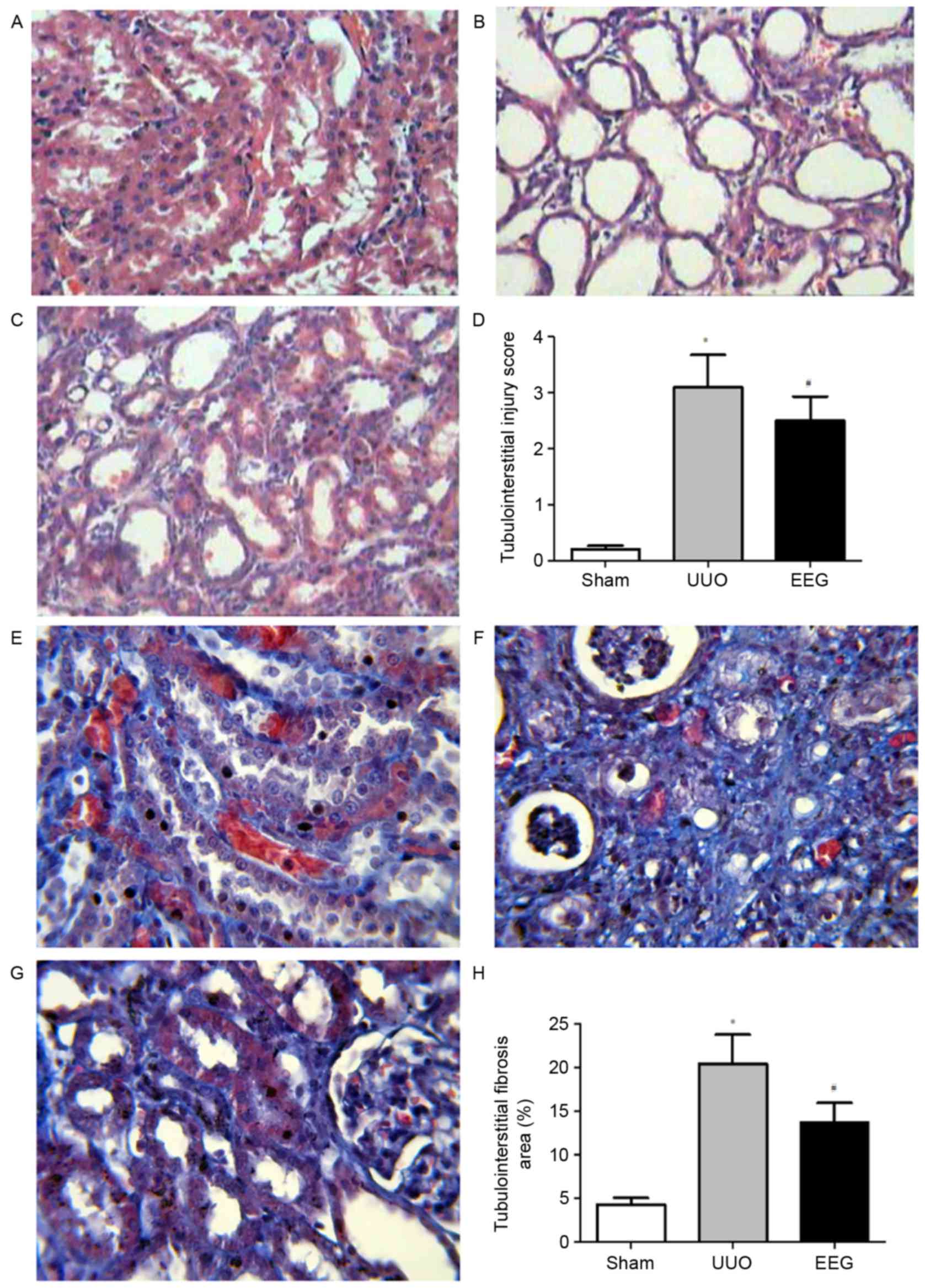
EEG ameliorates UUO-induced renal
histologic changes
H&E staining (Fig.
2) demonstrated that kidney histology was normal in the sham
group (Fig. 2A), whereas rats in the
UUO group developed severe tubulointerstitial damage at 14 days
following UUO operation (Fig. 2B).
However, the EEG group exhibited an improved histological
appearance, with attenuated inflammatory cellular infiltration, and
reduced tubular expansion and atrophy (Fig. 2C). Significantly decreased the
tubulointerstitial injury scores of obstructed rats (P<0.05;
Fig. 2D).
Effect of EEG on UUO-induced RIF
Masson staining indicated marked tubulointerstitial
fibrosis in UUO group (Fig. 2F).
Compared with the sham group (Fig.
2E), the percentage area of the fibrous area in the UUO group
was significantly higher (P<0.05), whereas this was
significantly ameliorated by EEG treatment (P<0.05; Fig. 2G and H).
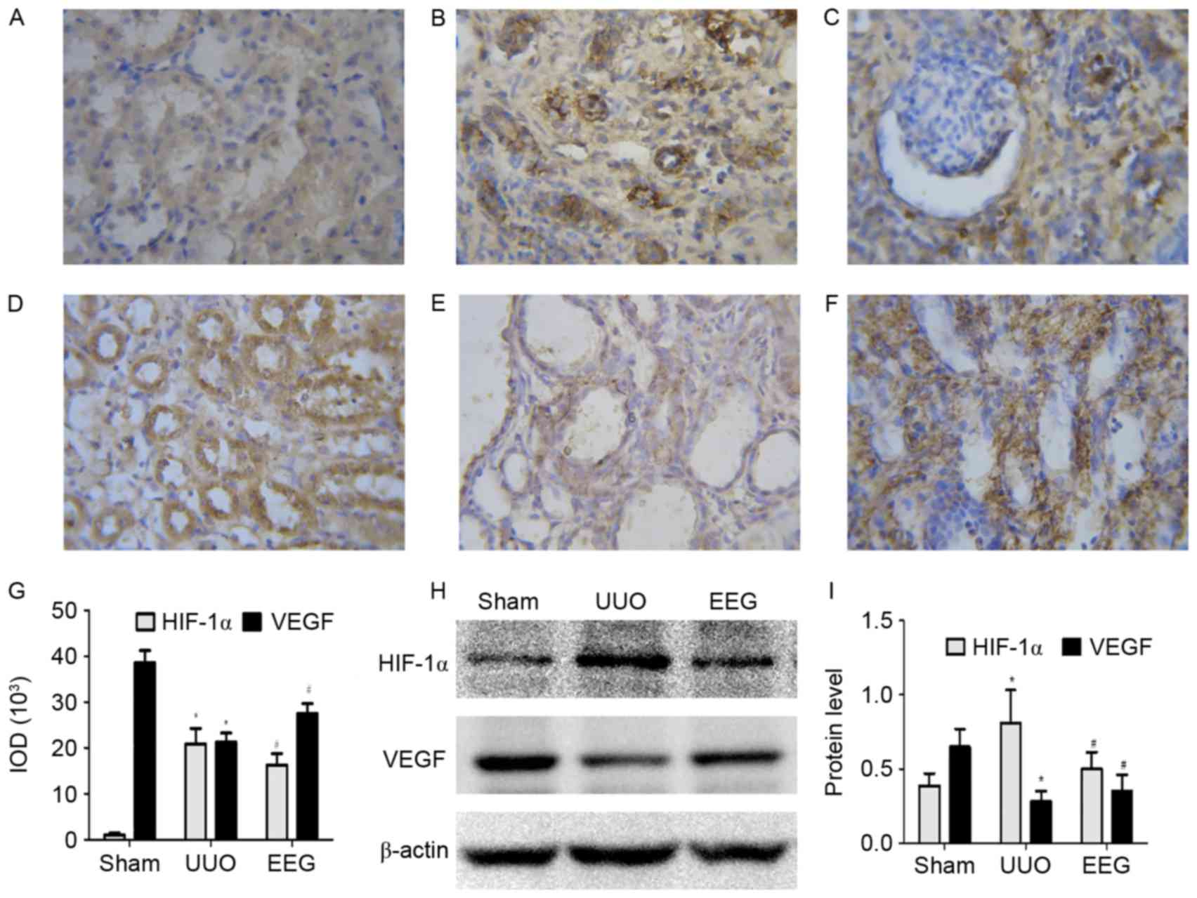
Immunostaining and western blot
analysis of HIF-1α and VEGF
Immunostaining and western blot analysis of HIF-1α
and VEGF are presented in Fig. 3.
Compared with the sham group, HIF-1α expression was markedly
increased in fibrotic areas and tubular epithelial cells in the UUO
group (Fig. 3A and B). Compared with
the UUO group, HIF-1α staining was markedly decreased in tubules in
the EEG group (Fig. 3C). Western
blot analysis revealed that expression of HIF-1α protein was
significantly reduced in the EEG group compared with the UUO group
(P<0.05; Fig. 3H and I), which
was consistent with the immunohistochemistry (P<0.05; Fig. 3G). This suggests that expression of
HIF-1α protein was reduced by EEG treatment.
VEGF was most prominent in tubular epithelial cells
in the sham group (Fig. 3D). In the
UUO group (Fig. 3E), the expression
of VEGF was significantly decreased compared with the sham group
(P<0.05), whereas the level of VEGF protein in the EEG group
(Fig. 3E) was significantly
increased compared with UUO group (P<0.05; Fig. 3F and G). The level of VEGF protein
detected by western blotting exhibited trends similar to those
observed in immunohistochemistry (Fig.
3H and I). These results suggest that the level of VEGF protein
was enhanced in the EEG group.
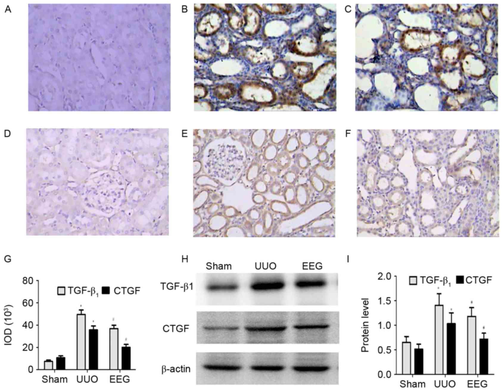
Immunostaining and western blot
analysis of TGF-β1 and CTGF
Immunostaining and western blot analysis of TGF-β1
and CTGF are presented in Fig. 4. As
shown in Fig. 4A-C and G, there was
a significant increase in TGF-β1 expression in the interstitial
cells and the tubular epithelial cells, and interstitial fibrotic
regions of kidneys in the UUO group compared with the sham group
(P<0.05); however, this increase was significantly inhibited by
EEG (P<0.05). Similar observations were made in western blot
analysis (Fig. 4H and I).
In the sham group, faint CTGF staining was detected
in the interstitial cells (Fig. 4D)
which was significantly increased in the UUO group (P<0.05;
Fig. 4E and G). The expression of
CTGF protein in the EEG group was significantly reduced compared
with the UUO group (Fig. 4F).
Similar observations were made in western blot analysis (Fig. 4H and I).
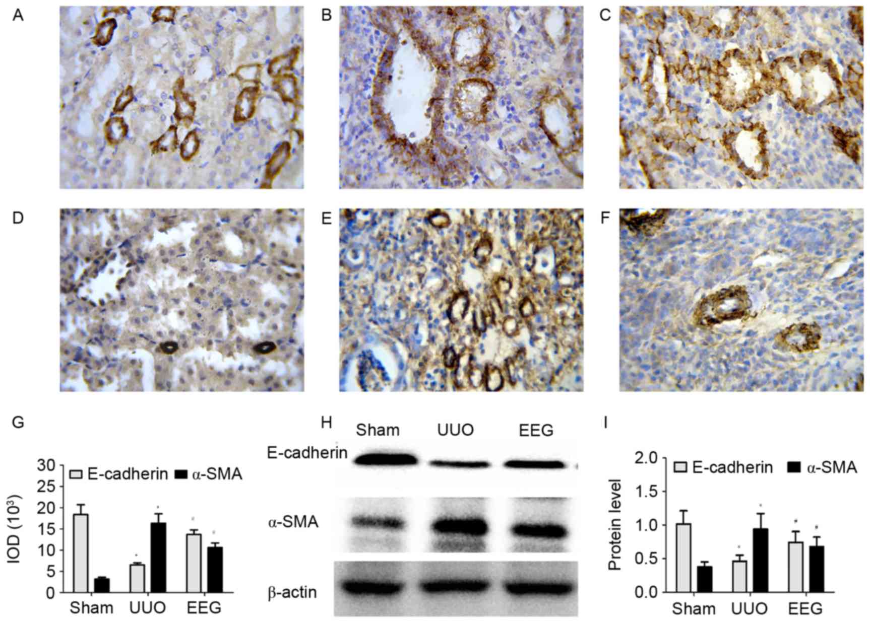
Immunostaining and western blot
analysis of E-cadherin and α-SMA
Immunostaining and western blot analysis of
E-cadherin and α-SMA are presented in Fig. 5. In the sham group, staining of
E-cadherin was predominantly observed in the cytoplasmic membrane
and the tubuloepithelial cells (Fig.
5A). There was a significant reduction in the level of
E-cadherin in the UUO group compared with sham group (P<0.05;
Fig. 5B and G); however, the level
of E-cadherin in the EEG group was significantly increased compared
with the UUO group (P<0.05; Fig. 5C
and G). Similar observations were made in western blot analysis
(Fig. 5H and I).
In the sham group, faint α-SMA staining was observed
(Fig. 5D. There was a significant
increase expression of α-SMA in the UUO group compared with the
sham group (P<0.05; Fig. 5E and
G). EEG treatment significantly reduced this α-SMA expression
(P<0.05; Fig. 5F and G). Similar
observations were made in western blot analysis (Fig. 5H and I).
Discussion
Extraction with ethanol was used to obtain an
extract of gardenia fruit that contained eight constituents
including crocetin, and geniposide and genipin, which are marked
anti-infective drugs. A recent study has discovered that crocetin
treatment may protect against burn-induced small intestinal injury
by inhibiting oxidative stress and inflammatory response (22). Furthermore, crocetin may alleviate
cardiac, skin and lung fibrosis due to suppression of reactive
oxygen species-dependent signaling pathways or a reduction in
endothelin-1 (23,24). It has also been reported that
geniposide is able to suppress the release of pro-inflammatory
cytokines tumor necrosis factor-α, interleukin-6 and interleukin-1β
in vitro and in vivo, and is also able to block the
phosphorylation of nuclear factor-κB, p65 and p38, as well as
inhibiting the expression of extracellular signal-related kinases
(ERKs) and c-Jun N-terminal kinases in
lipopolysaccharide-stimulated primary mouse macrophages (16). Furthermore, geniposide is able to
inhibit TGF-β1-induced EMT in hepatic fibrosis by depressing the
TGF-β/Smad and ERK-mitogen-activated protein kinase signaling
pathways (25).
Recent studies have demonstrated that EEG is
protective against liver fibrosis (19,26,27);
however little focus has been given to the kidney. UUO rat is a
mature experimental model for RIF in rats (22), which produces tubulointerstitial
inflammation and fibrosis. In the present study, no impaired renal
function was identified from BUN and Scr in UUO rats. These results
were caused by an increase in unobstructed kidney functions, which
compensated for the dysfunctions of obstructed-kidney. NAG in the
UUO group was significantly raised when compared with the sham
group, and EEG treatment significantly reduced this level.
In terms of pathology, the renal interstitium
widened, and the tubules expanded in the UUO group. EEG was able to
alleviate tubulointerstitial fibrosis induced by UUO. These results
suggest that the UUO model for RIF was successfully established in
the present study, and indicated that EEG is capable of depressing
the progression of RIF.
It is widely accepted that EMT is an important
mechanism associated with RIF (3).
In EMT, expression of the epithelial cell marker E-cadherin and
acquired mesenchymal features, such as expression of α-SMA, are
reduced in tubular epithelial cells. The present study demonstrated
that EEG treatment significantly decreased α-SMA and increased
E-cadherin, indicating that EEG is able to inhibit the transition
between epithelium and mesenchyme.
It has been demonstrated that TGF-β1 is a
profibrotic cytokine in the development of RIF, and it has been
identified as the most potent inducer of EMT (8). CTGF is an important downstream mediator
in the various profibrotic actions of TGF-β1, which are associated
with the degree of RIF and directly promote EMT (9,28). In
the present study, the level of CTGF and TGF-β1 was
significantly increased in the UUO group, whereas the expression of
CTGF and TGF-β1 in the EEG group were significantly
decreased compared with the UUO group. These results suggested that
EEG decreased the expression of TGF-β1 in UUO, and in turn reduced
CTGF. Therefore, EEG may inhibit EMT induction by UUO via TGF-β1
and CTGF.
Hypoxia has been recognized as a crucial
microenvironmental factor in the progression of tissue fibrosis
(29). Under hypoxic condition,
HIF-1α dimerizes with HIF-1β and this heterodimer HIF-1
translocates to the nucleus, where the activation of target genes
is mediated by binding to hypoxia-response elements (30). HIF-1α is a sensitive hypoxia
indicator, which may be a crucial factor in EMT (31,32).
HIF-1α in the UUO group was significantly higher than that in the
sham group, and following EEG treatment, the expression of HIF-1α
was significantly lower than that of the UUO group, suggesting that
hypoxia was ameliorated in the EEG group. A previous study
suggested that hypoxia and signaling through HIF-1α contribute to
the development of interstitial fibrosis via the induction of
ECM-modifying and the modulation of EMT (32). The present results indicate that EEG
inhibited EMT through HIF-1α in addition to TGF-β1 and CTGF.
VEGF is a survival factor for vascular endothelial
cells and serves an important role in the homeostasis of the
vascular endothelium, which is induced by hypoxia and
transcriptionally activated by HIF-1 (33). A reduction of VEGF was observed in
the UUO group. The decrease of VEGF was coincident with severe
interstitial fibrosis. This data may be contrary to the result of
elevated HIF-1α in this model. Several studies have suggested that
transcription factor Sp1 and the oncogene Ras are transcriptional
activators of VEGF (34,35). Consequently, the reduction of VEGF
levels in UUO may be the result of multiple factors in addition to
hypoxia and HIF-1α expression. The level of VEGF in the EEG group
was significantly increased compared with the UUO group. It was
previously reported that VEGF enhanced glomerular capillary repair
and prevented the progression of kidney diseases (36). VEGF is greatly expressed in tubular
epithelial cells, and renal tubular epithelial cells are an
important cellular resource of VEGF. There may be an intrinsic
renoprotective reason that maintains the epithelial phenotype of
tubular cells, and loss of these factors may contribute to EMT
progression. VEGF may be a candidate for this type of factor as
reduced expression of VEGF has been observed in RIF (37,38).
Furthermore, it has been demonstrated previously that VEGF
inhibited EMT through its influence on TGF-β1 and CTGF in UUO mice
(39). Therefore, it may be
speculated that EEG treatment decreased the expressions of HIF-1α
and increased VEGF, inhibiting fibrosis and EMT through decreasing
TGF-β1 and CTGF.
In conclusion, the present study demonstrated that
EEG had a protective effect on UUO rats, and may be able to
alleviate renal fibrosis and regulate EMT via decreasing the
expression of HIF-1α, CTGF, and TGF-β1, and elevating
VEGF protein levels. These results provide rationale for the
further study of the mechanisms of EEG and identify a potential
novel therapeutic method for the treatment of RIF.
Acknowledgments
The present study was supported by the Joint Fund of
Science and Technology Department of Guizhou Province [grant no.
J(2010)2], the Project Natural Science Foundation of China (grant
no. 21562051) and the project of Science and Technology Agency of
Guizhou Province [grant no. J(2015) 2157].
References
|
1
|
Jha V, Garcia-Garcia G, Iseki K, Li Z,
Naicker S, Plattner B, Saran R, Wang AY and Yang CW: Chronic kidney
disease: Global dimension and perspectives. Lancet. 382:260–272.
2013. View Article : Google Scholar : PubMed/NCBI
|
|
2
|
Lozano R, Naghavi M, Foreman K, Lim S,
Shibuya K, Aboyans V, Abraham J, Adair T, Aggarwal R, Ahn SY, et
al: Global and regional mortality from 235 causes of death for 20
age groups in 1990 and 2010: A systematic analysis for the global
burden of disease study 2010. Lancet. 380:2095–2128. 2012.
View Article : Google Scholar : PubMed/NCBI
|
|
3
|
Sun YB, Qu X, Caruana G and Li J: The
origin of renal fibroblasts/myofibroblasts and the signals that
trigger fibrosis. Differentiation. 92:102–107. 2016. View Article : Google Scholar : PubMed/NCBI
|
|
4
|
Duffield JS, Lupher M, Thannickal VJ and
Wynn TA: Host responses in tissue repair and fibrosis. Annu Rev
Pathol. 8:241–276. 2013. View Article : Google Scholar : PubMed/NCBI
|
|
5
|
Bertinat R, Silva P, Mann E, Li X, Nualart
F and Yáñez AJ: In vivo sodium tungstate treatment prevents
E-cadherin loss induced by diabetic serum in HK-2 cell line. J Cell
Physiol. 230:2437–2446. 2015. View Article : Google Scholar : PubMed/NCBI
|
|
6
|
Wang JY, Gao YB, Zhang N, Zou DW, Wang P,
Zhu ZY, Li JY, Zhou SN, Wang SC, Wang YY and Yang JK: miR-21
overexpression enhances TGF-β1-induced epithelial-to-mesenchymal
transition by target smad7 and aggravates renal damage in diabetic
nephropathy. Mol Cell Endocrinol. 392:163–172. 2014. View Article : Google Scholar : PubMed/NCBI
|
|
7
|
Farris AB and Colvin RB: Renal
interstitial fibrosis: Mechanisms and evaluation. Curr Opin Nephrol
Hypertens. 21:289–300. 2012. View Article : Google Scholar : PubMed/NCBI
|
|
8
|
López-Hernández FJ and López-Novoa JM:
Role of TGF-β in chronic kidney disease: An integration of tubular,
glomerular and vascular effects. Cell Tissue Res. 347:141–154.
2012. View Article : Google Scholar : PubMed/NCBI
|
|
9
|
Phanish MK, Winn SK and Dockrell ME:
Connective tissue growth factor-(CTGF, CCN2)-a marker, mediator and
therapeutic target for renal fibrosis. Nephron Exp Nephrol.
114:e83–e92. 2010. View Article : Google Scholar : PubMed/NCBI
|
|
10
|
Cammarata PR, Neelam S and Brooks MM:
Inhibition of hypoxia inducible factor-1 α downregulates the
expression of epithelial to mesenchymal transition early marker
proteins without undermining cell survival in hypoxic lens
epithelial cells. Mol Vis. 21:1024–1035. 2015.PubMed/NCBI
|
|
11
|
Sun Y, Wang H, Liu M, Lin F and Hua J:
Resveratrol abrogates the effects of hypoxia on cell proliferation,
invasion and EMT in osteosarcoma cells through downregulation of
the HIF-1α protein. Mol Med Rep. 11:1975–1981. 2015.PubMed/NCBI
|
|
12
|
Hakroush S, Moeller MJ, Theilig F,
Kaissling B, Sijmonsma TP, Jugold M, Akeson AL, Traykova-Brauch M,
Hosser H, Hähnel B, et al: Effects of increased renal tubular
vascular endothelial growth factor (VEGF) on fibrosis, cyst
formation, and glomerular disease. Am J Pathol. 175:1883–1895.
2009. View Article : Google Scholar : PubMed/NCBI
|
|
13
|
Sis B, Husain S, Chang J, Halloran P and
Osasan S: Decreased renal VEGF-A signaling as a mechanism for
kidney transplant fibrosis and failure. Am J Transplant. 13 Suppl
5:S1102013.
|
|
14
|
Wang YR, Chen Y, Deng L, Cai S, Liu J, Li
W, Du L, Cui G, Xu X, Lu T, et al: Systematic separation and
purification of iridoid glycosides and crocetin derivatives from
Gardenia jasminoides Ellis by high-speed counter-current
chromatography. Phytochem Anal. 26:202–208. 2015. View Article : Google Scholar : PubMed/NCBI
|
|
15
|
Hong YJ and Yang KS: Anti-inflammatory
activities of crocetin derivatives from processed Gardenia
jasminoides. Arch Pharm Res. 36:933–940. 2013. View Article : Google Scholar : PubMed/NCBI
|
|
16
|
Fu YH, Liu B, Liu J, Liu Z, Liang D, Li F,
Li D, Cao Y, Zhang X, Zhang N and Yang Z: Geniposide, from Gardenia
jasminoides Ellis, inhibits the inflammatory response in the
primary mouse macrophages and mouse models. Int Immunopharmacol.
14:792–798. 2012. View Article : Google Scholar : PubMed/NCBI
|
|
17
|
Koo HJ, Lim KH, Jung HJ and Park EH:
Anti-inflammatory evaluation of gardenia extract, geniposide and
genipin. J Ethnopharmacol. 103:496–500. 2006. View Article : Google Scholar : PubMed/NCBI
|
|
18
|
Chang KH, Chen WL, Wu YR, Lin TH, Wu YC,
Chao CY, Lin JY, Lee LC, Chen YC, Lee-Chen GJ and Chen CM: Aqueous
extract of Gardenia jasminoides targeting oxidative stress to
reduce polyQ aggregation in cell models of spinocerebellar ataxia
3. Neuropharmacology. 81:166–175. 2014. View Article : Google Scholar : PubMed/NCBI
|
|
19
|
Chen YH, Lan T, Li J, Qiu CH, Wu T, Gou HJ
and Lu MQ: Gardenia jasminoides attenuates hepatocellular injury
and fibrosis in bile duct-ligated rats and human hepatic stellate
cells. World J Gastroenterol. 18:7158–7165. 2012. View Article : Google Scholar : PubMed/NCBI
|
|
20
|
Mizuno S, Matsumoto K and Nakamura T:
Hepatocyte growth factor suppresses interstitial fibrosis in a
mouse model of obstructive nephropathy. Kidney Int. 59:1304–1314.
2001. View Article : Google Scholar : PubMed/NCBI
|
|
21
|
Mizuguchi Y, Miyajima A, Kosaka T, Asano
T, Asano T and Hayakawa M: Atorvastatin ameliorates renal tissue
damage in unilateral ureteral obstruction. J Urol. 172:2456–2459.
2004. View Article : Google Scholar : PubMed/NCBI
|
|
22
|
Zhou C, Bai W, Chen Q, Xu Z, Zhu X, Wen A
and Yang X: Protective effect of crocetin against burn-induced
intestinal injury. J Surg Res. 198:99–107. 2015. View Article : Google Scholar : PubMed/NCBI
|
|
23
|
Song Y, Zhu L and Li M: Antifibrotic
effects of crocetin in scleroderma fibroblasts and in
bleomycin-induced sclerotic mice. Clinics (Sao Paulo).
68:1350–1357. 2013. View Article : Google Scholar : PubMed/NCBI
|
|
24
|
Cai J, Yi FF, Bian ZY, Shen DF, Yang L,
Yan L, Tang QZ, Yang XC and Li H: Crocetin protects against cardiac
hypertrophy by blocking MEK-ERK1/2 signalling pathway. J Cell Mol
Med. 13:909–925. 2009. View Article : Google Scholar : PubMed/NCBI
|
|
25
|
Park JH, Yoon J, Lee KY and Park B:
Effects of geniposide on hepatocytes undergoing
epithelial-mesenchymal transition in hepatic fibrosis by targeting
TGFβ/Smad and ERK-MAPK signaling pathways. Biochimie. 113:26–34.
2015. View Article : Google Scholar : PubMed/NCBI
|
|
26
|
Chen P, Chen Y, Wang Y, Cai S, Deng L, Liu
J and Zhang H: Comparative evaluation of hepatoprotective
activities of geniposide, crocins and crocetin by CCl4-induced
liver injury in mice. Biomol Ther (Seoul). 24:156–162. 2016.
View Article : Google Scholar : PubMed/NCBI
|
|
27
|
Ma T, Huang C, Zong G, Zha D, Meng X, Li J
and Tang W: Hepatoprotective effects of geniposide in a rat model
of nonalcoholic steatohepatitis. J Pharm Pharmacol. 63:587–593.
2011. View Article : Google Scholar : PubMed/NCBI
|
|
28
|
Sanz-Rosa D, de las Heras N, Ortega MR, et
al: Role of CTGF on renal fibrosis in spontaneously hypertensive
rats. Effect of treatment with candesartan. J Hypertens. 23:327.
2005.
|
|
29
|
Haase VH: Pathophysiological consequences
of HIF activation: HIF as a modulator of fibrosis. Ann N Y Acad
Sci. 1177:57–65. 2009. View Article : Google Scholar : PubMed/NCBI
|
|
30
|
Nørregaard R, Bødker T, Jensen BL,
Stødkilde L, Nielsen S and Frøkiaer J: Increased renal
adrenomedullin expression in rats with ureteral obstruction. Am J
Physiol Regul Integr Comp Physiol. 296:R185–R192. 2009. View Article : Google Scholar : PubMed/NCBI
|
|
31
|
Harris AL: Hypoxia-a key regulatory factor
in tumour growth. Nat Rev Cancer. 2:38–47. 2002. View Article : Google Scholar : PubMed/NCBI
|
|
32
|
Higgins DF, Kimura K, Bernhardt WM,
Shrimanker N, Akai Y, Hohenstein B, Saito Y, Johnson RS, Kretzler
M, Cohen CD, et al: Hypoxia promotes fibrogenesis in vivo via HIF-1
stimulation of epithelial-to-mesenchymal transition. J Clin Invest.
117:3810–3820. 2007.PubMed/NCBI
|
|
33
|
Hellwig-Bürgel T, Stiehl DP, Katschinski
DM, Marxsen J, Kreft B and Jelkmann W: VEGF production by primary
human renal proximal tubular cells: Requirement of HIF-1,
PI3-kinase and MAPKK-1 signaling. Cell Physiol Biochem. 15:99–108.
2005. View Article : Google Scholar : PubMed/NCBI
|
|
34
|
Taoka R, Jinesh GG, Xue W, Safe S and
Kamat AM: CF3DODA-Me induces apoptosis, degrades Sp1, and blocks
the transformation phase of the blebbishield emergency program.
Apoptosis. 22:719–729. 2017. View Article : Google Scholar : PubMed/NCBI
|
|
35
|
Milanini-Mongiat J, Pouysségur J and Pagès
G: Identification of two Sp1 phosphorylation sites for p42/p44
mitogen-activated protein kinases: Their implication in vascular
endothelial growth factor gene transcription. J Biol Chem.
277:20631–20639. 2002. View Article : Google Scholar : PubMed/NCBI
|
|
36
|
Wahab NA and Mason RM: A critical look at
growth factors and epithelial-to-mesenchymal transition in the
adult kidney. Interrelationships between growth factors that
regulate EMT in the adult kidney. Nephron Exp Nephrol. 104:129–134.
2006. View Article : Google Scholar
|
|
37
|
Song YR, You SJ, Lee YM, Chin HJ, Chae DW,
Oh YK, Joo KW, Han JS and Na KY: Activation of hypoxia-inducible
factor attenuates renal injury in rat remnant kidney. Nephrol Dial
Transplant. 25:77–85. 2010. View Article : Google Scholar : PubMed/NCBI
|
|
38
|
Burt LE, Forbes MS, Thornhill BA, Kiley SC
and Chevalier RL: Renal vascular endothelial growth factor in
neonatal obstructive nephropathy. I. Endogenous VEGF. Am J Physiol
Renal Physiol. 292:F158–F167. 2007. View Article : Google Scholar : PubMed/NCBI
|
|
39
|
Lian YG, Zhou QG, Zhang YJ and Zheng FL:
VEGF ameliorates tubulointerstitial fibrosis in unilateral ureteral
obstruction mice via inhibition of epithelial-mesenchymal
transition. Acta Pharmacol Sin. 32:1513–1521. 2011. View Article : Google Scholar : PubMed/NCBI
|