Introduction
In Taiwan, hepatocellular carcinoma (HCC) is the
second-most common cause of cancer-associated mortality and its
incidence is increasing (1). A
number of risk factors for HCC have been reported previously,
including hepatitis B, hepatitis C, alcoholic liver disease and
non-alcoholic fatty liver disease (2–4). In
2016, the reported outcome of HCC was poor, with a high mortality
rate (35.5/100,000) in Taiwan (5).
Current targeted and systemic therapies for HCC face a number of
challenges, including drug toxicity and resistance (6). As such, the development of effective
prediction and surveillance tools are necessary to enable early
diagnosis, the prediction of clinical outcomes and personalized
adjuvant treatments (7). Recent
studies on HCC have focused on the development of high-throughput
microarrays for predicting HCC prognosis (8–10).
Several genes and proteins have been reported to be associated with
HCC prognosis, including forkhead box M1 (FOXM1) (10), cyclin-dependent kinase 1 (Cdk1)
(9), TP53BP1 and CDKN1B (8).
Typically, tumor progression occurs in two stages:
Growth and distant metastases. Circulating tumor cells spread from
the primary sites into the peripheral blood supply and arrive at
the metastasis site; this process involves many genes (11,12).
Recurrent, high-grade and distant metastases indicate invasive
circulating tumor cells, which are responsible for poor prognosis
in HCC patients (13). A
transmembrane glycoprotein-epithelial cell adhesion molecule
(EpCAM; CD326) serves an important role in the distant metastasis
of HCC (14). Ber-EP4 is a
monoclonal antibody that effectively labels EpCAM in epithelial
tissues and has been widely used to differentiate mesothelioma,
adenocarcinoma and basal and squamous cell carcinomas (15,16).
However, few studies have analyzed the association between EpCAM
expression and HCC prognosis. The aim of the present study was to
analyze tumor and normal tissue samples from Taiwanese patients
with HCC who had not previously received chemotherapy or targeted
therapy in order to determine whether EpCAM expression is an
independent clinicopathological indicator of HCC.
Patients and methods
Patients
A prospective search was performed in Changhua
Christian Hospital to identify patients with HCC diagnosed between
July 2011 and November 2013 at the Division of General Surgery,
Department of Surgery, Changhua Christian Hospital, Taiwan. The
present study was approved by the Institutional Review Board of
Changhua Christian Hospital (CCH IRB number: 120504). Informed
consent was obtained from all included patients. The inclusion
criteria were as follows: HCC diagnosis, no previous chemotherapy
or targeted therapies. The exclusion criteria were as follows: Age
<18 years, pregnancy, previous chemotherapy, targeted therapy,
or surgical intervention. All included patients received curative
surgical treatment and long-term follow-up. The demographic and
clinical data, including pathological stage, operative procedure
and surgical outcome, were recorded and patients were monitored
until death, censorship, or loss to follow-up. The Child-Pugh score
predicted the clinical outcome. Following surgical resection,
primary tumor tissues and matched adjacent normal tissues were
collected and analyzed using a tissue microarray. The follow-up
duration was defined as the period between the date of surgical
intervention to the date of last visit or death. Patients were
categorized into the low expression and high expression groups
depending on EpCAM expression. The median EpCAM expression in the
tumor group was used as the cut off value.
Immunohistochemistry (IHC) and
scoring
To detect EpCAM expression, tumor specimens were
embedded in paraffin, cut into 4-µm-thick sections and mounted on
poly-l-lysine-coated slides. Subsequently, 10 mM Tris-HCl (pH 7.4)
and 150 mM sodium chloride were used to deparaffinize and rinse
slides. Tissue was fixed with 4% paraformaldehyde for 10 min at
room temperature, permeabilized with 0.1% Triton X-100 for 10 min
at room temperature, blocked with 3% bovine serum albumin
(Sigma-Aldrich; Merck KGaA, Darmstadt, Germany) for 30 min at room
temperature and incubated with EpCAM (Ber-EP4) monoclonal
antibodies (cat. no. 61-0132-2; 1:100; Genemed Biotechnologies,
Inc., South San Francisco, CA, USA) in blocking buffer for 1 h at
room temperature. The slides were washed thrice with PBS and
antibodies were detected using the EnVision Detection Systems
Peroxidase/DAB, Rabbit/Mouse kit (Dako; Agilent Technologies, Inc.,
Santa Clara, CA, USA) and observed under a light microscope (BX50;
Olympus Corp., Tokyo, Japan) at a magnification of ×20 and ×40.
EpCAM expression was visualized in negative controls (adjacent
normal tissues) by performing the same IHC steps, excluding the
addition of the monoclonal antibodies. IHC results were evaluated
by a professional pathologist and the scoring system considered two
aspects: Staining intensity and percentage of positive cells. The
staining intensity was scored using 4 grades as described
previously (17,18): 0, no expression; 1, weak expression;
2, moderate expression; and 3, strong expression. The IHC score
ranged from 0 to 300 and was calculated using the following
formula: Staining intensity × percentage of positive labeled
cells.
Statistical analysis
The association between EpCAM expression and the
clinical and pathological parameters was analyzed using Chi-square
and paired-sample t-tests. SPSS 13.0 (SPSS, Inc., Chicago, IL, USA)
was used for all statistical analyses. Survival curves were plotted
using the Kaplan-Meier method and compared using log-rank test.
Cox's proportional hazards regression model was used to analyze the
association between the variables and survival data. P<0.05 was
considered to indicate a statistically significant difference.
Results
Patient characteristics
Patient characteristics are listed in Table I. A total of 185 patients (aged 26–81
years; mean, 62.9±10.8 years; male: Female, 73:27) were included in
the present study. The majority of patients had Child-Pugh score A
(Child-Pugh points: 4.79±1.8), with a mean tumor size of 45.9 mm.
The prevalence of hepatitis B and C was 54.6 and 33.5%,
respectively. Following surgical excision, pathology revealed
moderately differentiated tumors in most patients (n=97; 52.4%) and
well-differentiated tumors in 12 patients (6.5%), with poorly
differentiated tumors in 76 patients (41.1%). A total of 143
patients (76.4%) were determined to have HCC at clinical stage I.
The mean follow-up time was 857 days. During this period, 20
(10.8%) patients experienced tumor recurrence.
 | Table I.Clinicopathological characteristics of
patients with hepatocellular carcinoma. |
Table I.
Clinicopathological characteristics of
patients with hepatocellular carcinoma.
| Characteristics | Patient data |
|---|
| Age (years) | 62.8±10.8 |
| Sex, n (%) |
|
|
Female | 50
(27.0) |
|
Male | 135 (73.0) |
| Recurrence rate, n
(%) | 20
(10.8) |
| Survival days |
857±342.5 |
| Child-Pugh
score | 4.79±1.8 |
| Differentiation, n
(%) |
|
|
Well | 12 (6.5) |
|
Moderate | 97
(52.4) |
|
Poor | 76
(41.1) |
| Clinical stage, n
(%) |
|
| Stage
I | 143 (76.4) |
| Stage
II | 21
(11.4) |
| Stage
III | 21
(11.4) |
| Tumor size
(mm) | 45.9±37.2 |
| Hepatitis B, n
(%) | 101 (54.6) |
| Hepatitis C, n
(%) | 62
(33.5) |
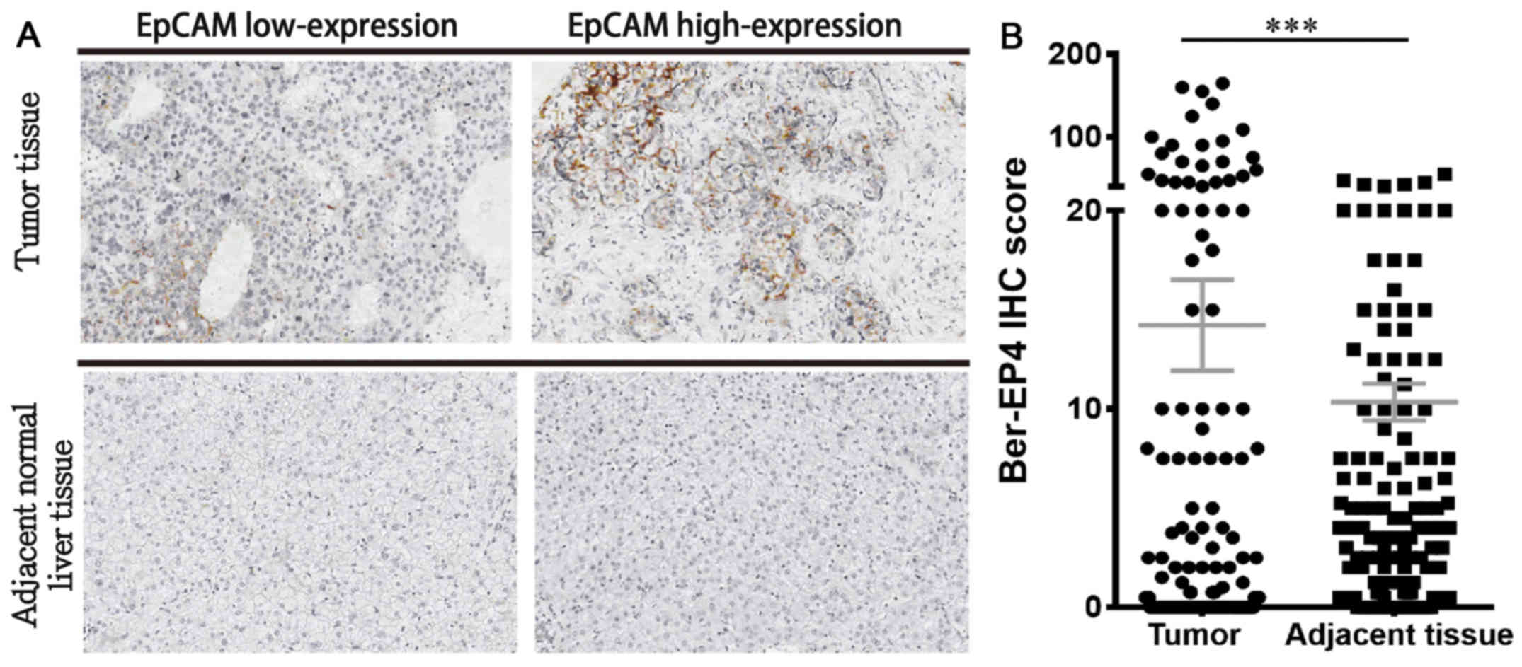
EpCAM expression
IHC staining revealed that all 185 HCC and normal
adjacent tissues were positive for EpCAM expression. EpCAM
expression was upregulated in tumor tissues compared with matched
adjacent normal liver tissues (Fig.
1A). In addition, EpCAM expression was significantly higher in
HCC tissues compared with the paired adjacent normal liver tissues
(P<0.001; Fig. 1B). The authors
of the current study hypothesized that high EpCAM expression
promotes poor clinical outcomes in the low-expression group. Among
the clinicopathological parameters assessed, the differentiation
grade was positively associated with high EpCAM expression
(P<0.05; Table II). No
significant association was observed between high EpCAM expression
and hepatitis B or C.
| Table II.Association between
clinicopathological characteristics and epithelial cell adhesion
molecule expression in patients with hepatocellular carcinoma. |
Table II.
Association between
clinicopathological characteristics and epithelial cell adhesion
molecule expression in patients with hepatocellular carcinoma.
|
| EpCAM |
|
|---|
|
|
|
|
|---|
| Variables | Low expression | High
expression | P-value |
|---|
| Age (years) | 63.2±11.6 | 62.1±9.6 | 0.292 |
| Sex, n |
|
| 0.511 |
|
Female | 27 | 23 |
|
|
Male | 65 | 70 |
|
| Recurrence, n |
|
| 0.354 |
|
Negative | 80 | 85 |
|
|
Positive | 12 | 8 |
|
| Differentiation,
n |
|
| 0.042 |
|
Well | 6 | 6 |
|
|
Moderate | 40 | 57 |
|
|
Poor | 46 | 30 |
|
| Clinical stage,
n |
|
| 0.497 |
| Stage
I, II | 84 | 80 |
|
| Stage
III, IV | 12 | 9 |
|
| Tumor size
(mm2) | 46.2±39.6 | 45.6±34.9 | 0.384 |
| Hepatitis B, n |
|
| 1.000 |
|
Negative | 42 | 42 |
|
|
Positive | 50 | 51 |
|
| Hepatitis C, n |
|
| 1.000 |
|
Negative | 61 | 62 |
|
|
Positive | 31 | 31 |
|
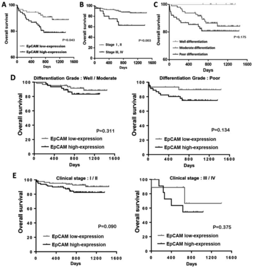
Survival analysis
Overall survival analysis revealed a significant
difference in two factors: Clinical stage and EpCAM expression
(P<0.05; Table III). The median
survival time was 928 days for patients with stage I and II tumors
and 642 days for patients with stage III or IV tumors. The median
survival time was 832 days in the group with high EpCAM expression
(survival rate, 80%) and 976 days in the group with low EpCAM
expression (survival rate, 90%).
| Table III.Univariate analysis of overall
survival for patients with hepatocellular carcinoma. |
Table III.
Univariate analysis of overall
survival for patients with hepatocellular carcinoma.
|
| Overall
survival |
|
|---|
|
|
|
|
|---|
| Variables | Median survival
days (n) | Survival (%) | Log-rank |
|---|
| Sex |
|
| 0.904 |
|
Female | 895 | 86 |
|
|
Male | 1,034 | 85.2 |
|
| Recurrence |
|
| 0.099 |
|
Negative | 935 | 83.6 |
|
|
Positive | 634 | 100 |
|
|
Differentiation |
|
| 0.175 |
|
Well | 860 | 100 |
|
|
Moderate | 918 | 86.6 |
|
|
Poor | 832 | 81.6 |
|
| Clinical stage |
|
| 0.003 |
| Stage
I, II | 928 | 87.8 |
|
| Stage
III, IV | 642 | 66.7 |
|
| Hepatitis B |
|
| 0.967 |
|
Negative | 919 | 85.7 |
|
|
Positive | 909 | 85.1 |
|
| Hepatitis C |
|
| 0.567 |
|
Negative | 915 | 84.6 |
|
|
Positive | 895 | 87.1 |
|
| Epithelial cell
adhesion molecule |
|
| 0.043 |
|
Low | 976 | 90.3 |
|
|
High | 832 | 80.4 |
|
The association between high EpCAM expression and
clinical outcomes in HCC patients was investigated (Table II). Overall survival analysis
revealed that patients with a high clinical stage and high EpCAM
expression had lower survival rates and reduced survival duration,
compared with those at clinical stages III and IV, and low EpCAM
expression. Cox regression analysis confirmed the prognostic
significance of a high clinical stage and high EpCAM expression
(Table IV). Kaplan-Meier analysis
revealed that patients with high EpCAM expression had a shorter
overall survival time compared with those with low EpCAM expression
(Fig. 2A); similar results were
noted for high clinical stage and differentiation grade in patients
with HCC (Fig. 2). These results
suggest that high EpCAM expression serves an important role in
determining the clinical outcomes of patients with HCC.
| Table IV.Univariate and multivariate analyses
of clinical characteristics in patients with hepatocellular
carcinoma. |
Table IV.
Univariate and multivariate analyses
of clinical characteristics in patients with hepatocellular
carcinoma.
|
| Univariate
analysis | Multivariate
analysis |
|---|
|
|
|
|
|---|
| Variables | HR | 95% CI | P-value | HR | 95% CI | P-value |
|---|
| Sex |
|
Female | – | – | – |
|
|
|
|
Male | 1.054 | 0.446–2.493 | 0.904 |
|
|
|
| Recurrence |
|
Negative | – | – | – |
|
|
|
|
Positive | 0.043 | 0.000–13.801 | 0.285 |
|
|
|
| Clinical stage |
| Stage
I, II | – | – | – | – | – | – |
| Stage
III, IV | 3.487 | 1.465–8.302 | 0.005 | 3.255 | 1.365–7.762 | 0.008 |
|
Differentiation |
| Well or
moderate | – | – | – |
|
|
|
|
Poor | 1.734 | 0.815–3.690 | 0.153 |
|
|
|
| EpCAM |
|
Low | – | – | – | – | – | – |
|
High | 2.238 | 1.004–4.989 | 0.049 | 2.108 | 0.943–4.712 | 0.069 |
Discussion
EpCAM is a transmembrane glycoprotein that regulates
Ca2+-independent cell-cell adhesion via several
functions, including cell migration, proliferation and
differentiation (19–21). In addition, EpCAM is involved in
c-Myc- and cyclin A/E-mediated cell cycle progression and
proliferation (22). High EpCAM
expression has been reported to be regulated by Wnt/β-catenin
signaling, which is responsible for the tumorigenic and invasive
abilities of HCC (23). High EpCAM
expression is reportedly associated with poor clinical outcomes in
breast (24), ovarian (25) and esophageal squamous cell carcinoma
(26). Furthermore, Schmelzer et
al (27) has proposed EpCAM as a
hepatic stemness marker. Together, these reports suggest that EpCAM
may be a good marker for HCC prognosis.
In the present study, EpCAM expression in primary
HCC was investigated, as well as its impact on clinical outcomes.
The results revealed that high EpCAM expression was significantly
associated with high differentiation grade. Bae et al
(28) previously reported that high
EpCAM expression was associated with high histologic grade, while
EpCAM downregulation inhibited HCC proliferation. These findings
support the results of the present study, which indicate that high
EpCAM expression is associated with high differentiation grade and
poor outcome. EpCAM immunoreactivity has been reported in 15.6–35%
of HCCs (29–31) and is associated with young age, poor
differentiation grade and high clinical stage (28,30,32,33). In
the present study, EpCAM expression was significantly associated
with differentiation grade; however, a high proportion of patients
enrolled in the present study had stage I tumors (76.4%) compared
with previous studies (28,34,35).
Furthermore, no significant association was observed between EpCAM
expression and clinical stage. However, Kaplan-Meier analysis
demonstrated that EpCAM serves an important role in patients with
tumors of high clinical stage. With regards to long-term patient
follow-up, EpCAM expression was not significant in multivariate
analysis.
The carrier rate of hepatitis B and C in Taiwan is
high and these viruses induce rapid development of HCC (36). A number of oncogenes and DNA
micromutations, including WNT, β-catenin, p53, Janus kinase, signal
transducer and activator of transcription and mitogen-activated
protein kinase-1, have been investigated in the progression and
development of hepatocarcinogenesis in patients with hepatitis B
and C (37–39). However, few studies have investigated
the novel biomarker EpCAM. Kimura et al (40) reported that high EpCAM expression is
frequently observed in patients with hepatitis B virus. In
addition, they demonstrated that EpCAM-expressing cells have high
anti-cancer drug resistance. This trend was not observed in the
present study. Furthermore, no significant difference was observed
in overall survival analysis and Kaplan-Meier analysis. This may be
due to a number of reasons; firstly, the patient sample was small
and the effects of high-expression of EpCAM may not have been
accurately detected. Secondly, a larger number of patients with
stage I and II tumors were included in the present study compared
with previous studies (28,34,41).
Previous studies have reported that EpCAM expression is more
significantly associated with clinical outcome in high-stage tumors
(28,34,41).
Thirdly, liver cirrhosis rates and Child-Pugh score were lower in
in the present study compared with previous reports (28,34,41).
Although no association was observed between high EpCAM expression
and poor clinical outcome in HCC patients with hepatitis B and C
virus infection in the present study, we believe that such
association may exist. Further studies are required to confirm the
role of high EpCAM expression in HCC. The authors of the present
study also believe that high EpCAM expression may be associated
with poor prognosis in HCC. Thus, high EpCAM expression may be a
prognostic factor of poor outcome in patients with HCC.
In conclusion, the present study suggests a
potential role of EpCAM as an important risk factor for poor
survival in HCC and EpCAM expression can be measured using routine
IHC. Further studies are required to investigate EpCAM as a
biomarker for HCC. The preliminary data herein suggests that HCC
patients with high EpCAM expression may benefit from targeted
therapy and immunotherapy. Thus, anti-EpCAM therapy is an appealing
strategy for HCC and should be explored in the future.
Acknowledgements
Not applicable.
Funding
The present study was funded by the Ministry of
Science and Technology, Taiwan (grant no. MOST
106-2314-B-442-001-MY3) and Show Chwan Memorial Hospital (grant no.
RB17004).
Availability of data and materials
The datasets used and/or analyzed during the current
study are available from the corresponding author on reasonable
request.
Authors' contributions
CJK and CJL were involved in the acquisition and
analysis of data. PYC and MYW were involved in the design of the
study and organized the manuscript.
Ethics approval and consent to
participate
The current study was approved by the Ethics
Committee of Changhua Christian Hospital (Changhua, Taiwan,
R.O.C.). All patients provided written informed consent.
Patient consent for publication
Not applicable.
Competing interests
The authors declare that they have no competing
interests.
References
|
1
|
Chen DS: Hepatocellular carcinoma in
taiwan. Hepatol Res. 2 Suppl 37:S101–S105. 2007. View Article : Google Scholar
|
|
2
|
Hung TH, Liang CM, Hsu CN, Tai WC, Tsai
KL, Ku MK, Wang JW, Tseng KL, Yuan LT, Nguang SH, et al:
Association between complicated liver cirrhosis and the risk of
hepatocellular carcinoma in taiwan. PLoS one. 12:e01818582017.
View Article : Google Scholar : PubMed/NCBI
|
|
3
|
Kim GA, Lee HC, Choe J, Kim MJ, Lee MJ,
Chang HS, Bae IY, Kim HK, An J, Shim JH, et al: Association between
non-alcoholic fatty liver disease and cancer incidence rate. J
Hepatol. 2:32294–32298. 2017.
|
|
4
|
Huang YT, Yang HI, Liu J, Lee MH, Freeman
JR and Chen CJ: Mediation analysis of hepatitis b and c in relation
to hepatocellular carcinoma risk. Epidemiology. 27:14–20. 2016.
View Article : Google Scholar : PubMed/NCBI
|
|
5
|
Altekruse SF, McGlynn KA and Reichman ME:
Hepatocellular carcinoma incidence, mortality, and survival trends
in the united states from 1975 to 2005. J Clin Oncol. 27:1485–1491.
2009. View Article : Google Scholar : PubMed/NCBI
|
|
6
|
Tazi el M, Essadi I, M'Rabti H, Touyar A
and Errihani PH: Systemic treatment and targeted therapy in
patients with advanced hepatocellular carcinoma. N Am J Med Sci.
3:167–175. 2011. View Article : Google Scholar : PubMed/NCBI
|
|
7
|
Yuan S, Wang J, Yang Y, Zhang J, Liu H,
Xiao J, Xu Q, Huang X, Xiang B, Zhu S, et al: The prediction of
clinical outcome in hepatocellular carcinoma based on a six-gene
metastasis signature. Clini Cancer Res. 23:289–297. 2017.
View Article : Google Scholar
|
|
8
|
Yu GP, Xiao QY, Shi ZQ, Tang LS, Ma XP,
Zhang LY, Chen HT, Wang WJ, Zhang PY, Ding DL, et al: Genetic
polymorphisms in apoptosis-related genes and the prognosis of
hepatocellular carcinoma. Am J Cancer Res. 5:3249–3259.
2015.PubMed/NCBI
|
|
9
|
Cai J, Li B, Zhu Y, Fang X, Zhu M, Wang M,
Liu S, Jiang X, Zheng J, Zhang X and Chen P: Prognostic biomarker
identification through integrating the gene signatures of
hepatocellular carcinoma properties. EbioMedicine. 19:18–30. 2017.
View Article : Google Scholar : PubMed/NCBI
|
|
10
|
Song BN and Chu IS: A gene expression
signature of FOXM1 predicts the prognosis of hepatocellular
carcinoma. Exp Mol Med. 50:e4182018. View Article : Google Scholar : PubMed/NCBI
|
|
11
|
Llovet JM and Bruix J: Testing molecular
therapies in hepatocellular carcinoma: The need for randomized
phase II trials. J Clin Oncol. 27:833–835. 2009. View Article : Google Scholar : PubMed/NCBI
|
|
12
|
Ringelhan M, Pfister D, O'Connor T,
Pikarsky E and Heikenwalder M: The immunology of hepatocellular
carcinoma. Nat Immunol. 19:222–232. 2018. View Article : Google Scholar : PubMed/NCBI
|
|
13
|
Zhou Y, Wang B, Wu J, Zhang C, Zhou Y,
Yang X, Zhou J, Guo W and Fan J: Association of preoperative EpCAM
circulating tumor cells and peripheral treg cell levels with early
recurrence of hepatocellular carcinoma following radical hepatic
resection. BMC Cancer. 16:5062016. View Article : Google Scholar : PubMed/NCBI
|
|
14
|
Wang MH, Sun R, Zhou XM, Zhang MY, Lu JB,
Yang Y, Zeng LS, Yang XZ, Shi L, Xiao RW, et al: Epithelial cell
adhesion molecule overexpression regulates epithelial-mesenchymal
transition, stemness and metastasis of nasopharyngeal carcinoma
cells via the PTEN/AKT/mTOR pathway. Cell Death Dis. 9:22018.
View Article : Google Scholar : PubMed/NCBI
|
|
15
|
Beer TW, Shepherd P and Theaker JM: Ber
EP4 and epithelial membrane antigen aid distinction of basal cell,
squamous cell and basosquamous carcinomas of the skin.
Histopathology. 37:218–223. 2000. View Article : Google Scholar : PubMed/NCBI
|
|
16
|
Sheibani K, Shin SS, Kezirian J and Weiss
LM: Ber-EP4 antibody as a discriminant in the differential
diagnosis of malignant mesothelioma versus adenocarcinoma. Am J
Surg Pathol. 15:779–784. 1991. View Article : Google Scholar : PubMed/NCBI
|
|
17
|
Chen YL, Chen PM, Lin PY, Hsiau YT and Chu
PY: ABCG2 overexpression confers poor outcomes in hepatocellular
carcinoma of elderly patients. Anticancer Res. 36:2983–2988.
2016.PubMed/NCBI
|
|
18
|
Yu HC, Hung MH, Chen YL, Chu PY, Wang CY,
Chao TT, Liu CY, Shiau CW and Chen KF: Erlotinib derivative
inhibits hepatocellular carcinoma by targeting CIP2A to reactivate
protein phosphatase 2A. Cell Death Dis. 5:e13592014. View Article : Google Scholar : PubMed/NCBI
|
|
19
|
Maetzel D, Denzel S, Mack B, Canis M, Went
P, Benk M, Kieu C, Papior P, Baeuerle PA, Munz M and Gires O:
Nuclear signalling by tumour-associated antigen EpCAM. Nat Cell
Biol. 11:162–171. 2009. View
Article : Google Scholar : PubMed/NCBI
|
|
20
|
Litvinov SV, van Driel W, van Rhijn CM,
Bakker HA, van Krieken H, Fleuren GJ and Warnaar SO: Expression of
Ep-CAM in cervical squamous epithelia correlates with an increased
proliferation and the disappearance of markers for terminal
differentiation. Am J Pathol. 148:865–875. 1996.PubMed/NCBI
|
|
21
|
Osta WA, Chen Y, Mikhitarian K, Mitas M,
Salem M, Hannun YA, Cole DJ and Gillanders WE: EpCAM is
overexpressed in breast cancer and is a potential target for breast
cancer gene therapy. Cancer Res. 64:5818–5824. 2004. View Article : Google Scholar : PubMed/NCBI
|
|
22
|
Chaves-Perez A, Mack B, Maetzel D,
Kremling H, Eggert C, Harréus U and Gires O: EpCAM regulates cell
cycle progression via control of cyclin D1 expression. Oncogene.
32:641–650. 2013. View Article : Google Scholar : PubMed/NCBI
|
|
23
|
Yamashita T, Budhu A, Forgues M and Wang
XW: Activation of hepatic stem cell marker EpCAM by
Wnt-beta-catenin signaling in hepatocellular carcinoma. Cancer Res.
67:10831–10839. 2007. View Article : Google Scholar : PubMed/NCBI
|
|
24
|
Spizzo G, Went P, Dirnhofer S, Obrist P,
Simon R, Spichtin H, Maurer R, Metzger U, von Castelberg B, Bart R,
et al: High Ep-CAM expression is associated with poor prognosis in
node-positive breast cancer. Breast cancer Res Treat. 86:207–213.
2004. View Article : Google Scholar : PubMed/NCBI
|
|
25
|
Spizzo G, Went P, Dirnhofer S, Obrist P,
Moch H, Baeuerle PA, Mueller-Holzner E, Marth C, Gastl G and Zeimet
AG: Overexpression of epithelial cell adhesion molecule (Ep-CAM) is
an independent prognostic marker for reduced survival of patients
with epithelial ovarian cancer. Gynecol Oncol. 103:483–488. 2006.
View Article : Google Scholar : PubMed/NCBI
|
|
26
|
Stoecklein NH, Siegmund A, Scheunemann P,
Luebke AM, Erbersdobler A, Verde PE, Eisenberger CF, Peiper M,
Rehders A, Esch JS, et al: Ep-CAM expression in squamous cell
carcinoma of the esophagus: A potential therapeutic target and
prognostic marker. BMC Cancer. 6:1652006. View Article : Google Scholar : PubMed/NCBI
|
|
27
|
Schmelzer E, Wauthier E and Reid LM: The
phenotypes of pluripotent human hepatic progenitors. Stem Cells.
24:1852–1858. 2006. View Article : Google Scholar : PubMed/NCBI
|
|
28
|
Bae JS, Noh SJ, Jang KY, Park HS, Chung
MJ, Park CK and Moon WS: Expression and role of epithelial cell
adhesion molecule in dysplastic nodule and hepatocellular
carcinoma. Int J Oncol. 41:2150–2158. 2012. View Article : Google Scholar : PubMed/NCBI
|
|
29
|
Yeh CT, Kuo CJ, Lai MW, Chen TC, Lin CY,
Yeh TS and Lee WC: CD133-positive hepatocellular carcinoma in an
area endemic for hepatitis B virus infection. BMC Cancer.
9:3242009. View Article : Google Scholar : PubMed/NCBI
|
|
30
|
Kim H, Choi GH, Na DC, Ahn EY, Kim GI, Lee
JE, Cho JY, Yoo JE, Choi JS and Park YN: Human hepatocellular
carcinomas with ‘Stemness’-related marker expression: Keratin 19
expression and a poor prognosis. Hepatology. 54:1707–1717. 2011.
View Article : Google Scholar : PubMed/NCBI
|
|
31
|
Yang XR, Xu Y, Yu B, Zhou J, Qiu SJ, Shi
GM, Zhang BH, Wu WZ, Shi YH, Wu B, et al: High expression levels of
putative hepatic stem/progenitor cell biomarkers related to tumour
angiogenesis and poor prognosis of hepatocellular carcinoma. Gut.
59:953–962. 2010. View Article : Google Scholar : PubMed/NCBI
|
|
32
|
Shan YF, Huang YL, Xie YK, Tan YH, Chen
BC, Zhou MT, Shi HQ, Yu ZP, Song QT and Zhang QY: Angiogenesis and
clinicopathologic characteristics in different hepatocellular
carcinoma subtypes defined by EpCAM and α-fetoprotein expression
status. Med Oncol. 28:1012–1016. 2011. View Article : Google Scholar : PubMed/NCBI
|
|
33
|
Yamashita T, Forgues M, Wang W, Kim JW, Ye
Q, Jia H, Budhu A, Zanetti KA, Chen Y, Qin LX, et al: EpCAM and
alpha-fetoprotein expression defines novel prognostic subtypes of
hepatocellular carcinoma. Cancer Res. 68:1451–1461. 2008.
View Article : Google Scholar : PubMed/NCBI
|
|
34
|
Sung JJ, Noh SJ, Bae JS, Park HS, Jang KY,
Chung MJ and Moon WS: Immunohistochemical expression and clinical
significance of suggested stem cell markers in hepatocellular
carcinoma. J Pathol Transl Med. 50:52–57. 2016. View Article : Google Scholar : PubMed/NCBI
|
|
35
|
Fong D, Seeber A, Terracciano L, Kasal A,
Mazzoleni G, Lehne F, Gastl G and Spizzo G: Expression of EpCAM(MF)
and EpCAM(MT) variants in human carcinomas. J Clin Pathol.
67:408–414. 2014. View Article : Google Scholar : PubMed/NCBI
|
|
36
|
Liu CJ, Chen PJ, Chen DS, Tseng TC and Kao
JH: Perspectives on dual hepatitis B and C infection in taiwan. J
Formos Med Assoc. 115:298–305. 2016. View Article : Google Scholar : PubMed/NCBI
|
|
37
|
Hussain SP, Schwank J, Staib F, Wang XW
and Harris CC: TP53 mutations and hepatocellular carcinoma:
Insights into the etiology and pathogenesis of liver cancer.
Oncogene. 26:2166–2176. 2007. View Article : Google Scholar : PubMed/NCBI
|
|
38
|
Vilchez V, Turcios L, Marti F and Gedaly
R: Targeting Wnt/β-catenin pathway in hepatocellular carcinoma
treatment. World J Gastroenterol. 22:823–832. 2016. View Article : Google Scholar : PubMed/NCBI
|
|
39
|
Shi F, Lian S, Wu P and Shen L:
Transarterial chemoembolization with or without microwave ablation
in the treatment of intermediate (BCLC B) hepatocellular carcinoma.
J Clin Oncol. 35:e156352017. View Article : Google Scholar
|
|
40
|
Kimura O, Kondo Y, Kogure T, Kakazu E,
Ninomiya M, Iwata T, Morosawa T and Shimosegawa T: Expression of
EpCAM increases in the Hepatitis B related and the
treatment-resistant hepatocellular carcinoma. BioMed Res Int.
2014:1729132014. View Article : Google Scholar : PubMed/NCBI
|
|
41
|
Seino S, Tsuchiya A, Watanabe Y, Kawata Y,
Kojima Y, Ikarashi S, Yanai H, Nakamura K, Kumaki D, Hirano M, et
al: Clinical outcome of hepatocellular carcinoma can be predicted
by the expression of hepatic progenitor cell markers and serum
tumour markers. Oncotarget. 9:21844–21860. 2018. View Article : Google Scholar : PubMed/NCBI
|