Introduction
In periapical lesions of deciduous teeth, the apex
and bifurcation sites, encompassing the cementum, periodontal
ligaments and alveolar bone, can harbor co-infections involving
anaerobic bacteria and other pathogens (1). These infections may trigger
heightened osteoclast activity or alveolar bone resorption
(2). In severe cases, infection
may extend to permanent teeth, precipitating premature loss of
primary teeth and adversely affecting the typical growth and
development of permanent teeth (3,4).
Root canal treatment is the foremost intervention for periapical
disease in deciduous teeth. This treatment not only proves
effective in alleviating pain, controlling inflammation and
encouraging lesion healing, but also supports the natural turnover
of deciduous teeth, thereby preserving the typical development of
permanent teeth (5,6). The success of root canal treatment
relies on the effective control of bacterial infections within the
root canal, a pivotal step encompassing the removal of infectious
material, root canal disinfection and subsequent filling with
absorbable materials (7,8). Presently, the manual use of stainless
steel files remains the primary instrument for root canal treatment
in pediatric deciduous teeth (9).
Nevertheless, a previous study reported that NiTi root canal
preparation devices exhibit promise for treating infections in
deciduous teeth, particularly in the mechanical preparation step
(10). Additionally, optimizing
root canal lengths has proven instrumental in enhancing the
efficiency and safety of root canal preparation, thereby
substantially reducing bacterial counts and metabolites within the
root canal (11,12). The practice of enlarging the root
canal preparation area has gained traction in clinical practice,
demonstrating its capability to mitigate postoperative responses
and achieve heightened postoperative filling rates (13). However, the intricate anatomy of
the root canal may pose challenges to accomplishing the desired
resection through mechanical preparation with a NiTi
instrument.
Antibiotics serve a crucial role in the clinical
control of root canal infections and are a valuable complement to
mechanical cleaning (14). While
effective mechanical cleaning coupled with antibiotic sealing holds
the potential for superior root canal disinfection, this
combination has not been extensively reported. Nakamura et
al (15) reported that
antibiotics, in conjunction with ultrasonic flushing, were more
effective than nonactivated irrigation in enhancing root canal
cleaning. However, Klyn et al (16) reported that there was no
significant difference in the cleaning efficacy when antibiotics
were combined with ultrasonic flushing or other debridement methods
in the apical region. Therefore, after the ultrasonic treatment
(with or without NiTi device), the removal of infectious substances
and microorganisms using appropriate intracanal medications or
rinses is a pivotal step in the treatment process for root canal
infection.
Given the diverse range of microorganisms that can
infect root canals, relying on a single antimicrobial drug may fall
short in achieving thorough disinfection. Therefore, tailored
antibiotic use combining multiple drugs into a paste, based on the
common flora identified in root canal lesions, may effectively
control inflammation and inhibit bacterial proliferation (17). Cogulu et al (18) reported that pathogenic bacteria in
children with periapical lesions are predominantly anaerobic, often
mixed with other bacteria like Clostridium sclerotinia and
Streptococcus. Clinical manifestations of these infections
include pain, discomfort upon percussion, root canal exudate and
pus formation (19). A commonly
used antibiotic combination for the treatment of these pathogens is
the metronidazole, ciprofloxacin and minocycline paste (20). This paste, composed of the
aforementioned antibiotics, not only exhibits broad-spectrum
antibacterial properties, but also demonstrates robust efficacy,
rendering it widely applied in clinical practice.
Cefixime, a third-generation cephalosporin
antibiotic, demonstrates high stability against β-lactamase
produced by gram-negative bacilli and exhibits potent
bacteriostatic activity (21). Its
efficacy against gram-negative bacilli in the oral cavity surpasses
that of both first- and second-generation cephalosporins, making it
highly suitable for treating root canal infections (22). By contrast, dexamethasone is an
adrenocorticosteroid drug renowned for its anti-inflammatory,
anti-allergic and analgesic effects and is therefore used to reduce
pain in root canal treatments (23,24).
The present study aimed to assess the clinical
efficacy of a mechanical NiTi file combined with ultrasonic
cleaning and multiple antibiotic paste through a randomized
controlled study. The incorporation of cefixime and dexamethasone,
alongside additional antibiotics, into an existing triple
antibiotic regimen will deepen understanding of treating periapical
inflammation in deciduous teeth. This study aimed to provide
further knowledge and practical guidance for enhancing treatment
outcomes and the overall quality of life for patients undergoing
root canal treatment.
Materials and methods
Clinical data collection
The present study received approval from the Medical
Ethics Committee of the Affiliated Hospital of Southwest Medical
University (approval no. 20200916002; Sichuan, China). The
recruitment of subjects commenced in October 2020 and concluded in
October 2021. All participants were enrolled from the Department of
Pediatric Stomatology at The Affiliated Stomatological Hospital of
Southwest Medical University (Sichuan, China). A total of 90
patients, each with one tooth exhibiting periapical inflammation,
were selected as the subjects for the study. During the course of
the study, the legal guardian of all children provided written
consent for participation in the study.
Study design and inclusion and
exclusion criteria
A total of 90 patients were randomly assigned to
groups A, B and C (n=30/group), in the process of root canal
preparation. The inclusion of two control groups (B and C) was
designed to compare the efficacy of the intervention. Both group A
and group B were prepared by using M3 primary tooth machine and
nickel-titanium file. In group A, ultrasonic root canal washing was
combined with multiple antibiotics sealing (metronidazole,
ciprofloxacin, minocycline, cefixime and dexamethasone) followed by
two weeks of root canal filling. In group B, ultrasonic root canal
washing was performed followed by calcium hydroxide sealing for two
weeks. In group C, root canal was prepared using stainless steel
hand K-file followed by calcium hydroxide sealing for two weeks of
root canal filling. By having two control groups, the relative
efficacy of the proposed intervention in comparison to different
treatment strategies could be assessed. This design allowed for a
more comprehensive evaluation of the intervention's efficacy in the
context of treating periapical inflammation in deciduous teeth.
The inclusion criteria for the study were as
follows: i) A definitive diagnosis of acute or chronic periapical
inflammation of deciduous teeth; ii) the affected root was in a
stable stage (patient aged 4-7 years) which is the period of time
from the formation of deciduous tooth roots to their resorption and
X-ray images demonstrate no root atresia calcification or root
resorption; iii) The FrankI treatment compliance evaluation scale
was utilized to score the degree of compatibility and all scores
were >3 points (25); and iv)
informed consent was provided by the patient's legal guardian.
The exclusion criteria for the study were as
follows: i) History of antibiotic therapy within the last 1 month;
ii) history of antibiotic allergies; iii) contraindications to root
canal treatment; iv) pulp penetration and root divergence; v)
patients who did not cooperate with treatment and exhibited poor
compliance; vi) incomplete medical records; and vii) patients with
systemic diseases.
Experimental materials
A multi-antibiotic paste, composed of metronidazole
(0.2 g/tablet, Huazhong Pharmaceutical Co., Ltd.; approval no.
H42020388), ciprofloxacin (0.25 g/ capsule; Zhejiang Jingxin
Pharmaceutical Co., Ltd.; approval no. H33020387), minocycline (0.1
g/capsule; Hanhui Pharmaceutical Co., Ltd.; approval no.
H20174081), cefixime (0.1 g/capsule; Chengdu Brilliant
Pharmaceutical Co., Ltd.; approval no. H20041661) and dexamethasone
(0.75 mg/tablet; Guangdong Nanguo Pharmaceutical Co., Ltd.;
approval no. H44024618) was prepared in equal proportions at a
ratio of 1:1:1:1:1.
Intervention
All patients underwent X-ray imaging prior to root
canal treatment. Patients underwent initial, secondary, tertiary
and follow-up visits during the treatment regime.
Initial visit
Patient diagnosis and treatment planning were
carried out and newly diagnosed cases underwent the removal of
caries, pulp opening and hole preparation. The top of the pulp
chamber was removed, and the pulp was extracted. The affected area
was rinsed, and moisture was removed by drying the area. The area
was sealed with calcium hydroxide (Ivoclar Vivadent ApexCal;
approval no. 20153172564) for 1 week.
Second visit. The sealing agent was removed,
and the root canal extracted using a 15K file (Dentsply Sirona)
lubricated with 17% EDTA (Longly Biotechnology). Root ZX root canal
measuring instrument was used to measure the root canal length of
group A and group B. Root canal preparation was performed with the
M3 baby tooth file combined with ultrasonic cleaning (Yirui M3
taper 0.06; Changzhou Yirui Machinery Processing Co., Ltd.) In
Group C, root canals were enlarged with a Gates Glidden drill
(Dentsply Sirona) and prepared by hand with stainless steel K files
(Dentsply Sirona; nos. 15, 20, 25 and 30) in combination with 17%
EDTA. The root canal was rinsed with ≥5 ml of a 0.25% sodium
hypochlorite solution and flushed with 0.9% normal saline. In Group
A, the tip of the root canal was dried using hygroscopic paper and
was subsequently filled with the multiple antibiotic paste before
being temporarily sealed using glass ionomer (Changshu Shangchi
Dental Materials Co., Ltd.; approval no. 20193171718). In Groups B
and C, the root canal was dried with tissue, filled with 23.9%
ApexCal Calcium hydroxide and temporarily sealed with glass
ionomer.
Third visit. Patients returned 2 weeks after
drug sealing and if there were no obvious issues and the local
mucosa returned to its normal state, the paste in the root canal
was washed away with purified water. The root canal lumen with
filled with vitex paste (Neo Dental Chemical Products Co., Ltd.), a
material based on glass ionomer cement, and filled with composite
resin. If there were no obvious issues 1 week after filling, a
pre-coronation examination, which includes oral examination and
X-rays, was performed to ensure the effectiveness of root canal
treatment and the stability of the filling. Root canal treatment
was performed by the same doctor.
Follow-up visits. Follow-up visits were
conducted at 6 and 12 months after root canal treatment. Clinical
symptoms of the affected tooth and the condition around the root
were recorded by X-ray. X-ray records were completed by the dental
assistant.
Evaluation of pain intensity
Following the surgery, the pain level of patients
was assessed through a questionnaire analyzing pain scores. A
WeChat (Tencent; version 8.0.40) mini-program facilitated pain
follow-ups on 1, 3 and 7 days after the treatment. The Visual
Analog Scale (VAS) was employed to gauge the severity of pain among
the treatment groups. The scale comprised a total score of 10
points, where higher scores indicated more intense pain. Pain
levels were categorized as follows: i) 0 (no pain); ii) 1-3 (mild
pain); iii) 4-6 (moderate pain); and iv) 7-10 (severe pain).
Therapeutic efficacy evaluation
The assessment of root canal treatment outcomes was
conducted at the 6 and 12-month post-treatment marks, based on a
number of criteria. The criteria for an effective treatment were as
follows: i) No noticeable loosening or swelling of the treated
tooth post-surgery; ii) healing of the original gum leakage; iii)
X-rays demonstrated either the disappearance or reduction of
shadows in the affected area; and iv) no anomalies in the inherited
permanent tooth germs. The criteria for an ineffective treatment
were as follows: i) Presence of postoperative bleeding, spontaneous
pain, occlusal pain, redness, swelling, sinus formation or tooth
loosening; and ii) X-rays demonstrated no substantial changes, an
increase in shadows in the thinning area of the apical bone or
progressive pathological resorption.
Statistical analysis
Data sorting and analysis were conducted using SPSS
software (version 23.0; IBM Corp.). The VAS scores were presented
as median ± interquartile range and were analyzed using the
Kruskal-Wallis test followed by Dunn's post hoc test. Fisher's
exact test followed by Bonferroni's correction was used to analyze
the therapeutic effect after root canal treatment. Count data were
expressed as percentages.
Results
Study criteria
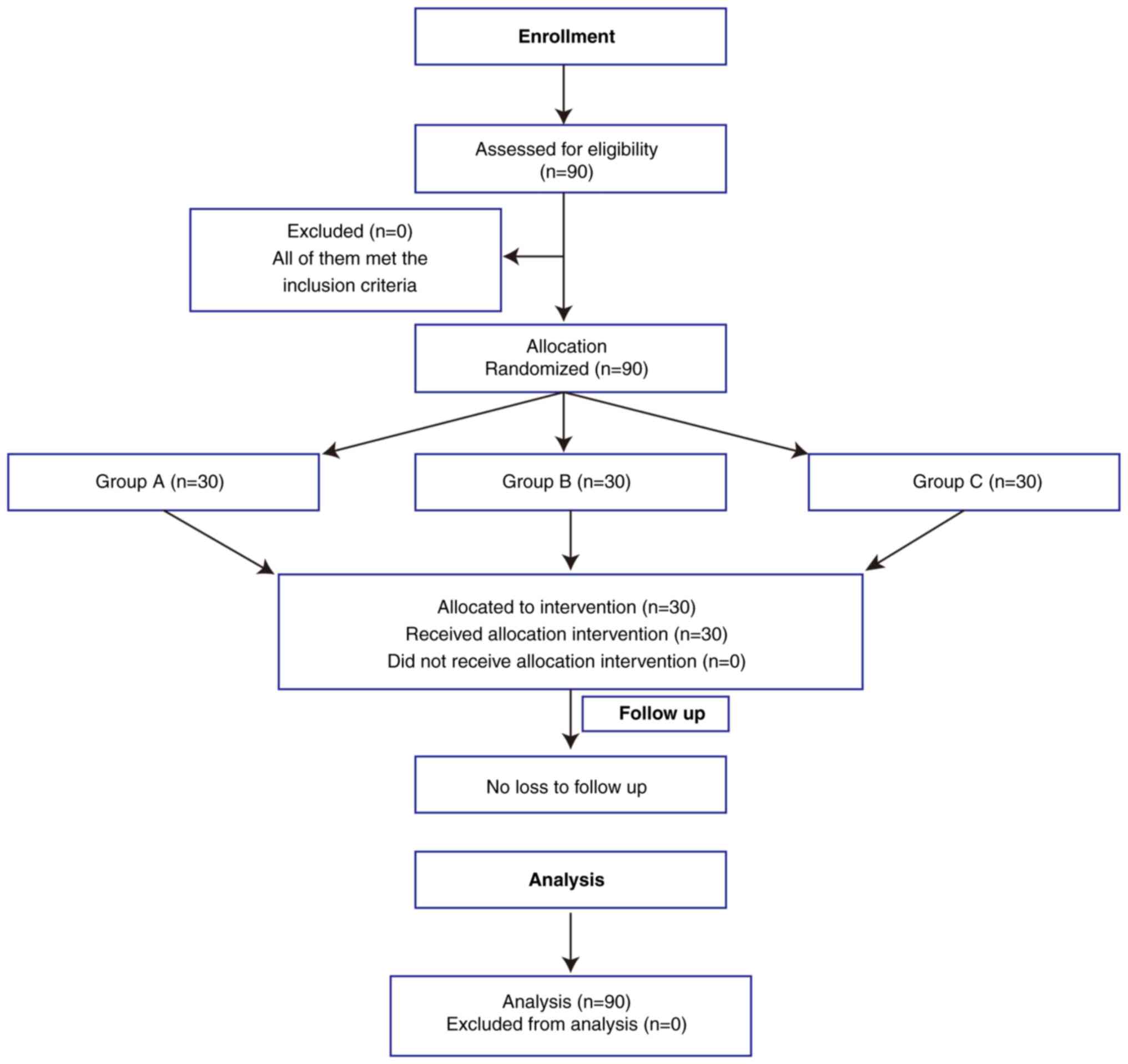
A total of 90 patients were enrolled in the present
study, each having one affected tooth with periapical periodontitis
of deciduous teeth (Fig. 1).
Evaluation of pain severity on days 1,
3 and 7 post-treatment
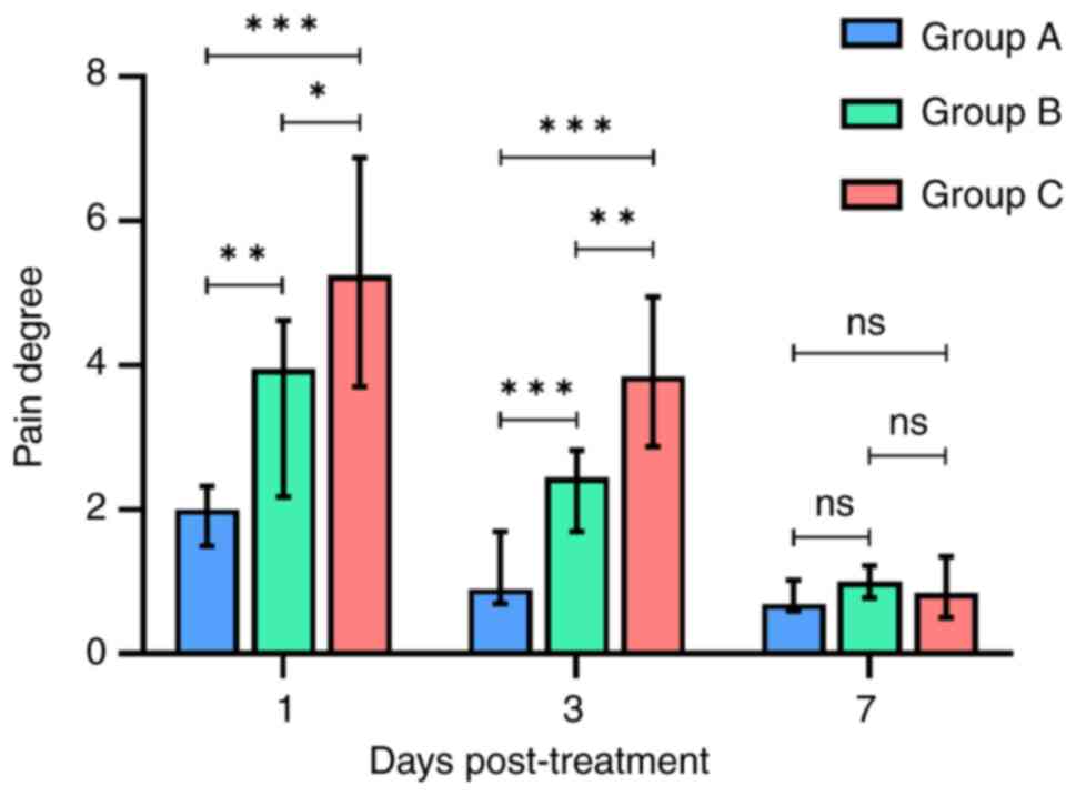
The pain response scores at 1, 3 and 7 days after
surgery were analyzed (Fig. 2;
Tables SI, SII and SIII). Notably, Group A exhibited
significantly lower pain scores compared with Groups B and C on
days 1 and 3 post-treatment (P<0.05). Group B demonstrated a
significantly lower postoperative pain scores compared with Group C
on days 1 and 3 post-treatment (P<0.05). However, by day 7
post-treatment, there was no significant difference in pain scores
among the three treatment groups (P>0.05). These findings
suggest that, during the early postoperative period, the combined
approach of the NiTi device, ultrasonic irrigation and multiple
antibiotic treatment administered to Group A may have contributed
to superior pain reduction compared with the other treatments.
Evaluation of treatment efficacy at 6
and 12 months post-treatment
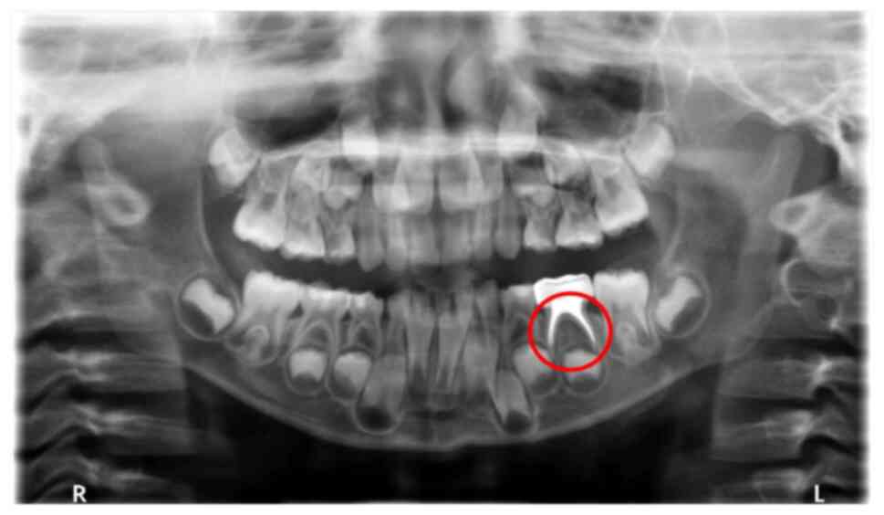
The efficacy of the treatments were evaluated 6
months after surgery. X-rays from Group B were used as examples for
efficacy evaluation. No abnormal images were detected of the root
and germ of the permanent tooth of the left lower second deciduous
molar, which indicated effective treatment 6 months following
surgical treatment (Fig. 3).
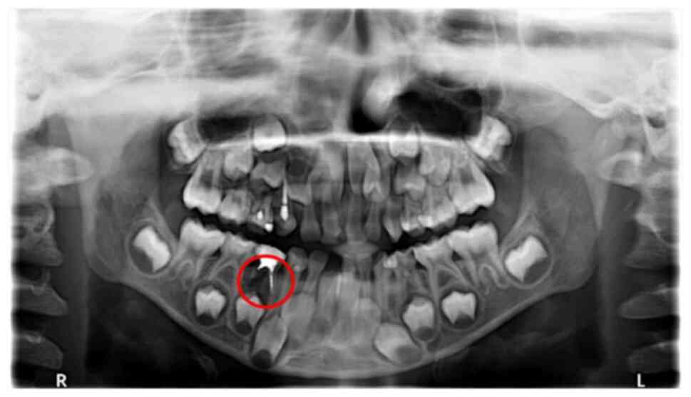
Conversely, the distal root resorption of the right lower primary
molar displayed a low periapical density shadow, thus was recorded
as an ineffective treatment (Fig.
4). The observation of treatment outcomes at 6 and 12 months
post-treatment reinforced the distinctions demonstrated between the
various treatments applied in the present study. Imaging of the
effectively treated teeth exhibited normalcy in the periapical
region, whereas ineffectively treated teeth demonstrated
significant abnormal shadows. These results highlighted the role of
research in assessing therapeutic effects in clinical applications
and may offer practical insights for the long-term management of
periapical inflammation of deciduous teeth.
At 6 months after the root canal procedures, there
was one ineffective case in Group A, two in Group B and eight in
Group C, which resulted in an effective treatment rate of 96.66,
93.33 and 73.33%, respectively (Table
I). The effective treatment rate in both Groups A and B were
significantly higher compared with that of Group C (P<0.05).
However, there was no significant difference in the effective
treatment rate between Groups A and B (P>0.05). This highlighted
the potential superior efficacy observed in Groups A and B compared
with Group C at the 6-month interval post-root canal treatment.
 | Table IComparison of therapeutic effect 6
months after root canal treatment. |
Table I
Comparison of therapeutic effect 6
months after root canal treatment.
| Treatment group | Number of teeth,
n | Effective treatment,
n (%) | Ineffective
treatment, n (%) | P-value |
|---|
| A | 30 | 29 (96.66) | 1 (3.34) | P>0.05 (vs. Group
B) |
| B | 30 | 28 (93.33) | 2 (6.67) | P=0.0489 (vs. Group
C) |
| C | 30 | 22 (73.33) | 8 (26.67) | P=0.0160 (vs. Group
A) |
In the extended observation at 12 months following
root canal treatment, each of the three treatment groups exhibited
cases of ineffective treatment (Table
II). The effective treatment rate for Group A was 93.33%, which
was significantly higher compared with that of Group C which was
63.33% (P<0.05). However, there was no significant difference in
the effective treatment rate of Group B (70.00%) and Group C.
Neither between Group B and Group A. These findings emphasized the
increased efficacy exhibited by the treatment administered to Group
A, which suggests its superiority over the treatment administered
to Group C over a 12-month period. These data provided support for
the efficacy of the treatment protocol implemented in Group A.
| Table IIComparison of therapeutic effect 12
months after root canal treatment. |
Table II
Comparison of therapeutic effect 12
months after root canal treatment.
| Treatment group | Number of teeth,
n | Effective treatment,
n (%) | Ineffective
treatment, n (%) | P-value |
|---|
| A | 30 | 28 (93.33) | 2 (6.67) | P>0.05 (vs. Group
B) |
| B | 30 | 21 (70.00) | 9 (30.00) | P>0.05 (vs. Group
C) |
| C | 30 | 19 (63.33) | 11 (36.67) | P=0.0197 (vs. Group
A) |
Discussion
Root canal treatment stands as a pivotal therapeutic
approach in managing apical infections of deciduous teeth, with the
overarching objective of facilitating the physiological shedding of
deciduous teeth (26). The pivotal
role of microorganisms within the root canal system in initiating
and perpetuating pulp and periapical ailments underscores the
criticality of effectively eliminating bacteria from the root canal
for the success of the treatment (27). The intricate anatomy of the root
canal, coupled with the deep infiltration of bacteria and debris
into dentin tubules, pose challenges to traditional treatment
methods such as mechanical NiTi preparation and ultrasonic
flushing, particularly in addressing collateral branches of the
root canal (28,29). Consequently, the present study
employed a randomized controlled design to assess the clinical
efficacy of combining mechanical NiTi file preparation with
ultrasonic cleaning, alongside the application of a multiple
antibiotic paste. This multifaceted approach aimed to overcome the
limitations of traditional techniques and enhance the overall
efficacy of root canal treatment in deciduous teeth.
The results of the present study demonstrated
differences in the pain scores reported among the three treatment
groups on days 1 and 3 post-treatment. Specifically, Group A
exhibited significantly lower pain VAS scores compared with Groups
B and C, with Group B also demonstrating lower scores compared with
Group C. By day 7, all treatment groups demonstrated a substantial
improvement in pain levels. Post-root canal preparation, pain can
stem from inadequacies in the procedure, for example, mechanical
injury caused by instrumentations (hand files or rotary) or
chemical damages due to disinfection (30). In the present study, the superior
pain outcomes in Groups A and B, compared with Group C, suggested
that a meticulous approach to root canal preparation and more
effective cleaning methods may have contributed to reduced pain
levels. The antibiotic pastes employed in the present study
displayed efficacy in eradicating Streptococcus and
Fusobacterium sclerotia, thereby mitigating swelling,
alleviating pain and sterilizing the area. Moreover, the
application of glucocorticoids, specifically dexamethasone, exerted
inhibitory effects on the inflammatory process, minimizing early
inflammatory symptoms such as redness, swelling, heat and pain. The
amalgamation of these factors significantly alleviated
postoperative pain in Group A compared with Group C. These results
suggested that the combined utilization of NiTi mechanical
preparations, ultrasonic irrigation and multi-faceted antibiotic
therapy presented substantial advantages in reducing pain post-root
canal treatment in deciduous teeth. This multifaceted approach
potentially holds promise in enhancing the treatment compliance of
pediatric patients.
The outcomes of the present study demonstrated
notable differences in the effective treatment rates between Group
A and Group B. At the 6-month mark post-root canal treatment,
coupled with the application of tightly closed metal preformed
crowns, Group A and Group B exhibited effective rates of 96.66 and
93.33%, respectively. Effective treatment rates showed a
significant disparity at the 12-month mark, with effective rates of
93.33 and 70.00% for Groups A and B, respectively. The superior
response rate in Group A compared with Group B could potentially be
attributed to the multi-antibiotic treatment administered within 2
weeks after root canal preparation. This intervention served a
crucial role in controlling the microorganisms within the root
canal, thereby facilitating the recovery of inflammation in the
periapical tissue. These findings highlighted the efficacy of the
antibiotic regimen administered to Group A, substantiating its role
in promoting the resolution of periapical inflammation.
In the present study, the multi-antibiotic protocol
adopted in Group A comprised metronidazole, ciprofloxacin,
minocycline, cefixime and dexamethasone. Each component in this
regimen served a specific purpose to collectively enhance the
therapeutic outcome. Metronidazole, classified as a nitroimidazole
organic compound, exhibits a broad-spectrum antibacterial effect,
particularly against anaerobic bacteria (31). Ciprofloxacin, a fluoroquinolone,
exerts potent antimicrobial activity against gram-negative bacteria
by primarily inhibiting DNA replication (32). Minocycline, belonging to the
tetracycline class, disrupts bacterial protein synthesis,
contributing to its antimicrobial effects (33). Cefixime, a cephalosporin
antibiotic, demonstrates robust antibacterial efficacy against
streptococcal sclerotinia and Fusobacterium (21). Lastly, dexamethasone, a
glucocorticoid, possesses anti-inflammatory, anti-allergic and
analgesic properties (24).
By contrast, the application of calcium hydroxide as
a sealant in Group B, while biologically active and capable of
disrupting bacterial biofilms, creating an alkaline environment and
neutralizing acidic substances, exhibits antibacterial efficacy
inferior to that of certain types of antibiotic pastes (34). Consequently, Group A demonstrated a
superior treatment outcome, which was potentially due to the potent
antimicrobial properties of its multi-antibiotic regimen.
Additionally, the treatment efficiency of Group B surpassed that of
Group C. This result may be attributed to the combined use of NiTi
mechanical preparation and ultrasonic irrigation, which could
facilitate the effective removal of infectious materials and smear
layers within the root canal (14). This synergistic approach enhanced
the overall treatment efficiency, potentially highlighting the
importance of employing multiple treatment strategies for optimal
therapeutic results.
Further research should focus on proposing potential
mechanisms for the observed effects reported in the present study.
Furthermore, a more detailed discussion of limitations, along with
suggestions for future research to address these limitations, is
encouraged. For example, the use of a single antibiotic compared
with the use of mixed antibiotics in the present study to optimize
the ratio of the components of the antibiotics mixture. While the
findings of the present study may have scientific and clinical
significance, specifying how these findings could be practically
applied in clinical settings and delineating steps for using NiTi
devices and antibiotics in root canal treatment are important for
the broader implementation of treatment strategies to patients.
The findings of the present study demonstrated that
a significant pain reduction was achieved through the synergistic
approach of NiTi mechanical preparation, ultrasonic irrigation and
multi-antibiotic occlusion during root canal treatment of deciduous
teeth. This combined methodology not only effectively mitigated
postoperative pain but also enhanced overall treatment efficiency.
The positive outcomes observed with the application of NiTi files,
ultrasonic irrigation and multi-antibiotic closure in the
management of periapical inflammation in primary teeth may signify
its potential for broader clinical adoption in the future.
The present study analyzed the efficacy of the NiTi
file, ultrasonic irrigation and multi-antibiotic closure approach
in the clinical treatment of primary teeth with periapical
inflammation. This method potentially holds promise for wider
implementation and may contribute to the future advancement of
contemporary clinical practices. Furthermore, the use of multiple
antibiotic pastes as a treatment option for root canal closure in
deciduous teeth offers an efficient treatment for patients in
clinical settings.
Supplementary Material
Comparison of the pain degree on day 1
after root canal surgery among different treatment groups.
Comparison of the pain degree on day 3
after root canal surgery among different treatment groups.
Comparison of the pain degree on day 7
after root canal surgery among different treatment groups.
Acknowledgements
Not applicable.
Funding
Funding: This work was financially supported by the Southwest
Medical University Youth Program (grant no. 2021ZKQN025) and
Project of Stomatological Institute of Southwest Medical University
(grant no. 2021XJYJS01).
Availability of data and materials
The data generated in the present study may be
requested from the corresponding author.
Authors' contributions
ZZ and GF conceived the study, designed the study
and drafted the original manuscript. ZZ analyzed the data and
conducted the experiments. GF critically revised the manuscript and
provided constructive feedback. ZZ and GF confirm the authenticity
of all the raw data. All authors have read and approved the final
version of the manuscript.
Ethics approval and consent to
participate
All procedures performed in the study involving
human participants were approved by The Medical Ethics Committee of
The Affiliated Hospital of Southwest Medical University (approval
no. 20200916002; Sichuan, China). The legal guardians of all
patients provided written informed consent for participation in the
study.
Patient consent for publication
Not applicable.
Competing interests
The authors declare that they have no competing
interests.
References
|
1
|
Nair PN: Pathogenesis of apical
periodontitis and the causes of endodontic failures. Crit Rev Oral
Biol Med. 15:348–381. 2004.PubMed/NCBI View Article : Google Scholar
|
|
2
|
Andreasen FM: Transient apical breakdown
and its relation to color and sensibility changes after luxation
injuries to teeth. Endod Dent Traumatol. 2:9–19. 1986.PubMed/NCBI View Article : Google Scholar
|
|
3
|
Liu D, Peng X, Wang S, Han Q, Li B, Zhou
X, Ren B, Xu HHK, Weir MD, Li M, et al: A novel antibacterial
resin-based root canal sealer modified by Dimethylaminododecyl
Methacrylate. Sci Rep. 9(10632)2019.PubMed/NCBI View Article : Google Scholar
|
|
4
|
Chum JD, Lim DJZ, Sheriff SO, Pulikkotil
SJ, Suresh A and Davamani F: In vitro evaluation of octenidine as
an antimicrobial agent against Staphylococcus epidermidis in
disinfecting the root canal system. Restor Dent Endod.
44(e8)2019.PubMed/NCBI View Article : Google Scholar
|
|
5
|
Zehnder M: Root canal irrigants. J Endod.
32:389–398. 2006.PubMed/NCBI View Article : Google Scholar
|
|
6
|
Chen Y, Li H, Li M, Yang L, Sun Q and Chen
K: Analysis of survival and factors associated with failure of
primary tooth pulpectomies performed under general anaesthesia in
children from South China. Int J Paediatr Dent. 30:225–233.
2020.PubMed/NCBI View Article : Google Scholar
|
|
7
|
Nagarathna C, Vishwanathan S,
Krishnamurthy NH and Bhat PK: Primary molar pulpectomy using two
different obturation techniques: A clinical study. Contemp Clin
Dent. 9:231–236. 2018.PubMed/NCBI View Article : Google Scholar
|
|
8
|
Jiang J, Sun J, Huang Z, Bi Z, Yu G, Yang
J and Wang Y: The state of the art and future trends of root canal
files from the perspective of patent analysis: A study design.
Biomed Eng Online. 21(90)2022.PubMed/NCBI View Article : Google Scholar
|
|
9
|
Al-Obaida MI, Alzuwayer AA, Alanazi SS and
Balhaddad AA: In Vitro analysis of the fatigue resistance of four
single file canal preparation instruments. Materials (Basel).
15(688)2022.PubMed/NCBI View Article : Google Scholar
|
|
10
|
Topçuoğlu G, Topçuoğlu HS, Delikan E,
Aydınbelge M and Dogan S: Postoperative pain after root canal
preparation with hand and rotary files in primary molar teeth.
Pediatr Dent. 39:192–196. 2017.PubMed/NCBI
|
|
11
|
Singh A, Purohit BM and Mittal P:
Periodontal predicaments and associated risk factors among patients
with schizophrenia. Neurology, Psychiatry and Brain Research.
32:36–41. 2019.
|
|
12
|
Divya DV, Ghanashyam PM, Naga RA, Venkata
SR, Pavani RS and Santosh Kumar KVK: Triple antibiotic paste versus
propolis: A clinical quest for the reliable treatment of periapical
lesions in primary molars. Saudi Dental Journal. 9:34–39. 2019.
|
|
13
|
Wu D, Gao J, Hu X, Xiao Z, Huang Z, Zhang
L, Chen X and He Y: Evaluation algorithm of root canal shape based
on steklov spectrum analysis. Comput Math Methods Med.
2019(4830914)2019.PubMed/NCBI View Article : Google Scholar
|
|
14
|
Wang C, Xu P, Li X, Zheng Y and Song Z:
Research progress of stimulus-responsive antibacterial materials
for bone infection. Front Bioeng Biotechnol.
10(1069932)2022.PubMed/NCBI View Article : Google Scholar
|
|
15
|
Nakamura VC, Pinheiro ET, Prado LC,
Silveira AC, Carvalho APL, Mayer MPA and Gavini G: Effect of
ultrasonic activation on the reduction of bacteria and endotoxins
in root canals: A randomized clinical trial. Int Endod J 51 Suppl.
1:e12–e22. 2018.PubMed/NCBI View Article : Google Scholar
|
|
16
|
Klyn SL, Kirkpatrick TC and Rutledge RE:
In Vitro comparisons of debris removal of the EndoActivatorTM
System, the F File, Ultrasonic Irrigation, and NaOCl irrigation
alone after hand-rotary instrumentation in human mandibular molars.
J Endod. 36:1367–1371. 2010.PubMed/NCBI View Article : Google Scholar
|
|
17
|
Narayanan LL and Vaishnavi C: Endodontic
microbiology. J Conserv Dent. 13:233–239. 2010.PubMed/NCBI View Article : Google Scholar
|
|
18
|
Cogulu D, Uzel A, Oncag O and Eronat C:
PCR-based identification of selected pathogens associated with
endodontic infections in deciduous and permanent teeth. Oral Surg
Oral Med Oral Pathol Oral Radiol Endod. 106:443–449.
2008.PubMed/NCBI View Article : Google Scholar
|
|
19
|
Pak JG and White SN: Pain prevalence and
severity before, during, and after root canal treatment: A
systematic review. J Endod. 37:429–438. 2011.PubMed/NCBI View Article : Google Scholar
|
|
20
|
Fischer NG, Münchow EA, Tamerler C,
Bottino MC and Aparicio C: Harnessing biomolecules for bioinspired
dental biomaterials. J Mater Chem B. 8:8713–8747. 2020.PubMed/NCBI View Article : Google Scholar
|
|
21
|
Manzoori JL, Amjadi M, Soltani N and
Jouyban A: Spectrofluorimetric determination of cefixime using
terbium-danofloxacin probe. Iran J Basic Med Sci. 17:256–262.
2014.PubMed/NCBI
|
|
22
|
Schlenter WW, Blessing R, Pelz K and
Benner U: Cefixime treatment in different bacterial infections in
the ENT region. Infection. 18 (Suppl 3):S125–S128. 1990.PubMed/NCBI View Article : Google Scholar : (In German).
|
|
23
|
Mehrvarzfar P, Esnashari E, Salmanzadeh R
and Fazlyab M and Fazlyab M: Effect of dexamethasone
intraligamentary injection on post-endodontic pain in patients with
symptomatic irreversible pulpitis: A randomized controlled clinical
trial. Iran Endod J. 11:261–266. 2016.PubMed/NCBI View Article : Google Scholar
|
|
24
|
Li X, Chen X, Wu J, Liu Z, Wang J, Song C,
Zhao S, Lei H and Sun Y: Portable, rapid, and sensitive
time-resolved fluorescence immunochromatography for on-site
detection of dexamethasone in milk and pork. Foods.
10(1339)2021.PubMed/NCBI View Article : Google Scholar
|
|
25
|
Yu W, Tong J, Sun X, Chen F, Zhang J, Pei
Y, Zhang T, Zhang J and Zhu B: Analysis of medication adherence and
its influencing factors in patients with Schizophrenia in the
Chinese Institutional Environment. Int J Environ Res Public Health.
18(4746)2021.PubMed/NCBI View Article : Google Scholar
|
|
26
|
Ahmed HM, Khamis MF and Gutmann JL: Seven
root canals in a deciduous maxillary molar detected by the dental
operating microscope and micro-computed tomography. Scanning.
38:554–557. 2016.PubMed/NCBI View Article : Google Scholar
|
|
27
|
Siqueira JF Jr and Rôças IN: Clinical
implications and microbiology of bacterial persistence after
treatment procedures. J Endod. 34:1291–1301.e3. 2008.PubMed/NCBI View Article : Google Scholar
|
|
28
|
Williamson AE, Sandor AJ and Justman BC: A
comparison of three nickel titanium rotary systems, EndoSequence,
ProTaper universal, and profile GT, for canal-cleaning ability. J
Endod. 35:107–109. 2009.PubMed/NCBI View Article : Google Scholar
|
|
29
|
Gulabivala K, Patel B, Evans G and Ng Y-L:
Effects of mechanical and chemical procedures on root canal
surfaces. Endodontic Topics. 10:103–122. 2005.
|
|
30
|
Alghazaly A and Al Habib L: Management of
endodontic flare-up in the presence of periapical radiolucency:
Case report and overview. Cureus. 15(e49719)2023.PubMed/NCBI View Article : Google Scholar
|
|
31
|
de Deus Moura Lde F, de Lima Mde M, Lima
CC, Machado JI, de Moura MS and de Carvalho PV: Endodontic
treatment of primary molars with antibiotic paste: A report of 38
cases. J Clin Pediatr Dent. 40:175–177. 2016.PubMed/NCBI View Article : Google Scholar
|
|
32
|
Shariati A, Arshadi M, Khosrojerdi MA,
Abedinzadeh M, Ganjalishahi M, Maleki A, Heidary M and Khoshnood S:
The resistance mechanisms of bacteria against ciprofloxacin and new
approaches for enhancing the efficacy of this antibiotic. Front
Public Health. 10(1025633)2022.PubMed/NCBI View Article : Google Scholar
|
|
33
|
Scholar E: Minocycline. In: xPharm: The
Comprehensive Pharmacology Reference. Enna SJ and Bylund DB (eds).
Elsevier, New York, NY, pp1-6, 2007.
|
|
34
|
Ito IY, Junior FM, Paula-Silva FW, Da
Silva LA, Leonardo MR and Nelson-Filho P: Microbial culture and
checkerboard DNA-DNA hybridization assessment of bacteria in root
canals of primary teeth pre- and post-endodontic therapy with a
calcium hydroxide/chlorhexidine paste. Int J Paediatr Dent.
21:353–360. 2011.PubMed/NCBI View Article : Google Scholar
|