Introduction
Ovarian cancer has the highest mortality rate of all
gynecologic malignancies (1). The
disease is split into a number of classifications, including
epithelial ovarian cancer, which accounts for 85–90% of malignant
ovarian tumors (1). High-grade
ovarian serous cancer is an aggressive type of epithelial ovarian
cancer, with a high degree of malignancy and rapid progression.
Compared with early-stage, high-grade ovarian serous cancer, the
prognosis of patients with advanced high-grade ovarian serous
cancer is poor, and the 5-year survival rate for these patients is
only 27% (2). Adjacent organ
invasion, multiple peritoneal implantation metastasis and lymph
node metastasis are also observed in the majority of patients with
advanced high-grade ovarian serous cancer (3). Therefore, studies investigating
high-grade ovarian serous cancer development,
progression-associated genes and regulatory mechanisms are
necessary. Through these studies, researchers hope to improve our
understanding of high-grade ovarian serous cancer progression and
development, and identify long-term and effective treatment
strategies for this disease.
The development of biological chips and
high-throughput sequencing technology has revealed that it is
possible to transcribe ~70–90% of the human genome to RNAs, but
only 1–2% of RNAs are ultimately translated to proteins (4,5).
The vast majority of RNAs do not have a protein-encoding function.
These RNAs were classified as 'non-coding RNAs', and were
originally thought to be either noise or waste products of
transcription. With further research, non-coding RNAs were
demonstrated to participate in a variety of physiologic and
pathologic processes and to serve an important function in cancer
progression (6). Long non-coding
RNAs (lncRNAs) are a type of non-coding RNA, transcribed from
intergenic and intronic regions in the human genome by RNA
polymerase II, ranging in length from 200 nt to 100 kb and lacking
significant protein-coding open reading frames (7). The reported biological functions of
lncRNAs include DNA damage repair, epigenetic control,
transcription regulation, pre- and post-translational regulation,
control of the cell cycle, survival, migration, metabolism and
differentiation, and even control of the apoptotic process
(8–11). Furthermore, a number of lines of
evidence link the dysregulation of lncRNAs to diverse human
diseases, particularly cancer (12–16). The lncRNA research field is,
therefore, very promising. However, studies concerning lncRNAs and
their function and regulation mechanisms remain in their infancy,
particularly those concerning high-grade ovarian serous cancer. At
present, few lncRNAs have been reported as associated with
high-grade ovarian serous cancer, including HOX transcript
antisense RNA (HOTAIR), HOXA11 antisense RNA, competing
endogenous lncRNA 2 for microRNA let-7b, maternally expressed 3 and
urothelial cancer associated 1 (UCA1) (17–21). Furthermore, the differential
expression profiles of lncRNAs between high-grade ovarian serous
cancer and healthy fallopian tube tissues, and their functional
significance, remain unclear. The aim of the present study was to
explore the expression profiles of lncRNAs and their potential
target genes in high-grade ovarian serous cancer tissues, and to
analyze the associations between these lncRNAs and
clinicopathological features of ovarian cancer. The results of the
present study may represent evidence supporting the involvement of
lncRNA expression levels in the progression of high-grade ovarian
serous cancer, and establish a foundation for the development of
potential diagnostic biomarkers and therapeutic targets for the
treatment of high-grade ovarian serous cancer.
Materials and methods
Patients and tissue specimens
High-grade ovarian serous cancer tissue specimens
were obtained from patients (average age, 55 years) who had
undergone surgical treatment for ovarian cancer at the Affiliated
Hospital of Qingdao University (Qingdao, China) between March 2015
and March 2016. Healthy fallopian tube tissue specimens were
obtained from patients (average age, 53 years) who had undergone
surgical treatment for hysteromyoma at the Affiliated Hospital of
Qingdao University during the same period. All cases were confirmed
by postoperative pathological diagnosis. Patients who had received
neoadjuvant chemotherapy or radiation therapy prior to surgery were
excluded from the present study. A total of 23 high-grade ovarian
serous cancer tissue samples and 23 healthy fallopian tube tissue
samples were collected. Of these samples, 3 high-grade ovarian
serous cancer samples (labeled A1–3) and 3 healthy fallopian tube
tissue samples (labeled B1–3) were used for global profiling of
human lncRNA and mRNA expression using the Arraystar human lncRNA
microarray (Arraystar, Inc., Rockville, MD, USA). A further 20
pairs of samples were used for confirmation of differential lncRNA
and mRNA expression by reverse transcription-quantitative
polymerase chain reaction (RT-qPCR) analysis. In addition, 40
high-grade ovarian serous cancer tissue samples were collected to
analyze the associations between differential lncRNA expression and
ovarian cancer clinicopathological parameters, using RT-qPCR
analysis. These samples were obtained from patients (average age,
55 years) who had undergone surgical treatment for ovarian cancer
at the Affiliated Hospital of Qingdao University between March 2016
and June 2017. All samples were stored at -80°C immediately
following surgical resection. All subjects provided signed
statements of written informed consent, and all experimental
procedures were approved by the Institutional Review Board of the
Ethics Board of the Affiliated Hospital of Qingdao University.
RNA extraction and quality control
Total RNA was extracted from the tissue samples
using TRIzol reagent (Invitrogen; Thermo Fisher Scientific, Inc.,
Waltham, MA, USA), according to the manufacturer's protocol. The
entire process was performed on ice. The quantity and quality of
the RNA samples were measured using a NanoDrop ND-1000
spectrophotometer. RNA integrity was assessed using standard 1%
denaturing agarose gel electrophoresis. Isolated RNA was stored at
-80°C and prepared for lncRNA array analysis and RT-qPCR.
RNA labeling and array hybridization
Sample labeling and array hybridization were
performed according to the Agilent one-color microarray-based gene
expression analysis protocol (Agilent Technologies, Inc., Santa
Clara, CA, USA) with minor modifications. Briefly, mRNA was
purified from the total RNA following removal of rRNA (using an
mRNA-ONLY™ Eukaryotic mRNA Isolation kit; Epicentre; Illumina,
Inc., San Diego, CA, USA). Then, each sample was amplified and
transcribed into fluorescent cRNA along the entire length of the
transcripts without 3′ bias, utilizing a random priming method
(using a Arraystar Flash RNA l Labeling kit; Arraystar, Inc.). The
labeled cRNA was purified using an RNeasy mini kit (Qiagen, Inc.,
Valencia, CA, USA). The concentration and specific activity of the
labeled cRNAs (pmol Cy3/µg cRNA) were measured using a
NanoDrop ND-1000 spectrophotometer. An Agilent Gene Expression
Hybridization kit (cat. no. 5188-5242) was purchased from Agilent
Technologies, Inc. and each labeled cRNA (1 µg) was
fragmented by adding 5 µl 10X blocking agent and 1 µl
25X fragmentation buffer. The mixture was heated at 60°C for 30
min. Finally, 25 µl 2X GE Hybridization buffer was added to
dilute the labeled cRNA. A total of 50 µl hybridization
solution was dispensed into the gasket slide and assembled to the
lncRNA expression microarray slide. The slides were incubated for
17 h at 65°C in an Agilent hybridization oven (Agilent
Technologies, Inc.). The hybridized arrays were washed, fixed, and
scanned using the Agilent DNA microarray scanner (part no. G2505C;
Agilent Technologies, Inc.).
Microarray data analysis
Agilent feature extraction software (version
11.0.1.1; Agilent Technologies, Inc.) was used to analyze the
acquired array images. Quantile normalization and subsequent data
processing were performed using the GeneSpring GX version 12.1
software package (Agilent Technologies, Inc.). Following quantile
normalization of the raw data, lncRNAs and mRNAs, for which at
least 3 out of 6 samples had flags in Present or Marginal ('All
targets value') were selected for further data analysis.
Differentially expressed lncRNAs and mRNAs with statistical
significance between high-grade ovarian serous cancer and healthy
fallopian tube tissue were identified through P-value (<0.05)
and false discovery rate (<0.1) filtering. Differentially
expressed lncRNAs and mRNAs between the two samples were identified
through fold change filtering. Pathway analysis and Gene Ontology
(GO) analysis (22,23) were applied to determine which
pathways were affected by these differentially expressed lncRNAs
and mRNAs. Hierarchical clustering and combined analysis were
performed using in-house scripts.
RT-qPCR analysis of lncRNA and mRNA
expression
Expression levels of lncRNAs and mRNAs were
validated by RT-qPCR analysis among 20 high-grade ovarian serous
cancer samples. Total RNA was extracted from the tissue samples
using TRIzol reagent (Invitrogen; Thermo Fisher Scientific, Inc.).
Total RNA was reverse-transcribed to cDNA using a TRUEscript 1st
strand cDNA synthesis kit (Aidlab Biotechnologies Co., Ltd.,
Beijing, China) following the manufacturer's protocol. Reactions
were incubated for 30 min at 42°C, 5 min at 85°C, and then samples
were stored at −20°C. qPCR was performed using 2X SYBR green qPCR
mix (Aidlab Biotechnologies Co., Ltd.) in an ABI 7900HT sequence
detection machine (Thermo Fisher Scientific, Inc.). The reactions
were incubated at 95°C for 3 min, followed by 40 cycles at 95°C for
15 sec and at 60°C for 40 sec. Primer sequences for 5 lncRNAs and 5
mRNAs were designed and synthesized (Table I). GAPDH was used as an internal
control. Target and reference (GAPDH) genes were amplified in
separate wells and run in triplicate. Statistical analyses of the
results were performed using the 2−∆∆Cq relative
quantification method (24).
 | Table IPrimer sequences for lncRNAs and
mRNAs. |
Table I
Primer sequences for lncRNAs and
mRNAs.
| Gene name | Forward
(5′–3′) | Reverse
(5′–3′) |
|---|
|
GTSE1-AS1 |
TCTTAAGCTTCCTGAGGTTGCAC |
AGAGGTTTAATTGGCTCACAGTTC |
| FAS-AS1 |
CAAAACAGGCTGCTCAAGTTTC |
GGGTTGGTGGTACTCGTTCC |
|
AK130076 |
TGGCCATCCCTACAGTGCTAG |
TGGAGTGCAGTGGTGCGATC |
|
RP11-199F11.2 |
GTGCCTGTAATCCCAGCTATTC |
TGAGACGGAGTTTCGTTCTTGTC |
| AC093818.1
C |
ACTCACCGAAGTCCAGGAAC |
GCAGCTTCAGCTCGGACTC |
| GTSE1 |
GATGACCCCCAAAACGATG |
TTCTGTTGCTCTCCCTTGTTG |
| FAS |
ACACTCACCAGCAACACCAA |
TCCTTTCTCTTCACCCAAACA |
| PTEN |
AGACCATAACCCACCACAGC |
ACCAGTTCGTCCCTTTCCA |
| TP53 |
TAGTGTGGTGGTGCCCTATG CC |
AGTGTGATGATGGTGAGG |
| PDK1 |
GATGAGTGACCGAGGAGGTG |
TAACCAAAACCAGCCAGAGG |
| GAPDH |
CTCAGACACCATGGGGAAGGTGA |
ATGATCTTGAGGCTGTTGTCATA |
Statistical analysis
SPSS 17.0 (SPSS, Inc., Chicago, IL, USA) and
GraphPad Prism 5.0 (GraphPad Software, Inc., La Jolla, CA, USA)
were used for statistical analysis. Data are presented as the mean
± standard deviation. The statistical significance of the
microarray results was analyzed by fold change and Student's
t-test. P<0.05 was considered to indicate a statistically
significant difference.
Results
RNA quality control
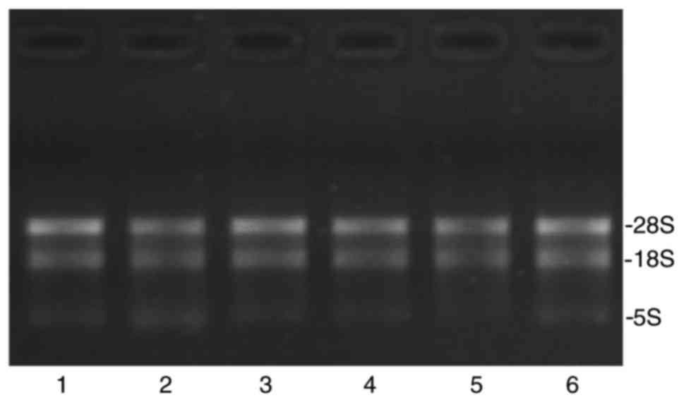
The integrity of RNAs was assessed by
electrophoresis on a denaturing agarose gel. The 28S and 18S
ribosomal RNA bands should appear as relatively sharp, intense
bands. The intensity of the upper band (28S rRNA band) should be
~2x that of the lower band (18S rRNA band). Fig. 1 revealed that the total RNA
obtained was of high purity. Total RNA quantification and quality
were assured using the NanoDrop ND-1000 spectrophotometer. For
spectrophotometry, the optical density A260/A280 ratio should be
close to 2.0 for pure RNA (ratios between 1.8 and 2.1 are
acceptable). The optical density A260/A230 ratio should be >1.8.
In the present study, the total RNA quality of each sample was
demonstrated (Table II),
suggesting that the samples were appropriate for use in further
experiments.
| Table IIRNA quantification and quality
assurance, as assessed using a NanoDrop ND-1000
spectrophotometer. |
Table II
RNA quantification and quality
assurance, as assessed using a NanoDrop ND-1000
spectrophotometer.
| Sample ID | OD260/280 | OD260/230 | Concentration
(ng/µl) | Volume
(µl) | Quantity (ng) | QC result (pass or
fail) |
|---|
| A1 | 2.00 | 2.24 | 1,550.05 | 80 | 124,004.0 | Pass |
| A2 | 2.03 | 2.33 | 1,175.57 | 80 | 94,045.6 | Pass |
| A3 | 1.99 | 2.26 | 888.76 | 100 | 88,876.0 | Pass |
| B1 | 1.97 | 2.27 | 594.67 | 40 | 23,786.8 | Pass |
| B2 | 1.89 | 2.32 | 420.78 | 40 | 16,831.2 | Pass |
| B3 | 1.89 | 2.36 | 357.84 | 40 | 14,313.6 | Pass |
Overview of lncRNA and mRNA profiles
The Arraystar human lncRNA microarray v. 4.0 is
designed for the global expression profiling of human lncRNA and
protein-coding mRNA transcripts. The array is able to detect a
total of 40,173 lncRNAs and 20,730 protein-coding mRNAs. In the
present study, ~4,289 differentially expressed lncRNAs and 4,246
differentially expressed mRNAs were detected between high-grade
ovarian serous cancer tissue samples and healthy fallopian tube
tissue samples, using this fourth-generation lncRNA microarray
(fold change, ≥2.0; P<0.05). Among these lncRNAs, 1,511 were
upregulated and 2,778 were downregulated. The most upregulated
lncRNA was TCONS_l2_00000435 (fold change, 81.26) and the most
downregulated lncRNA was RP11-356K23.1 (fold change, 1221.26). In
addition, 1,834 mRNAs were upregulated while 2,412 mRNAs were
downregulated. The most upregulated mRNA was paired-like
homeodomain 2 (fold change, 333.42) and the most downregulated mRNA
was anterior gradient 3 (fold change, 2696.97). The 10 most
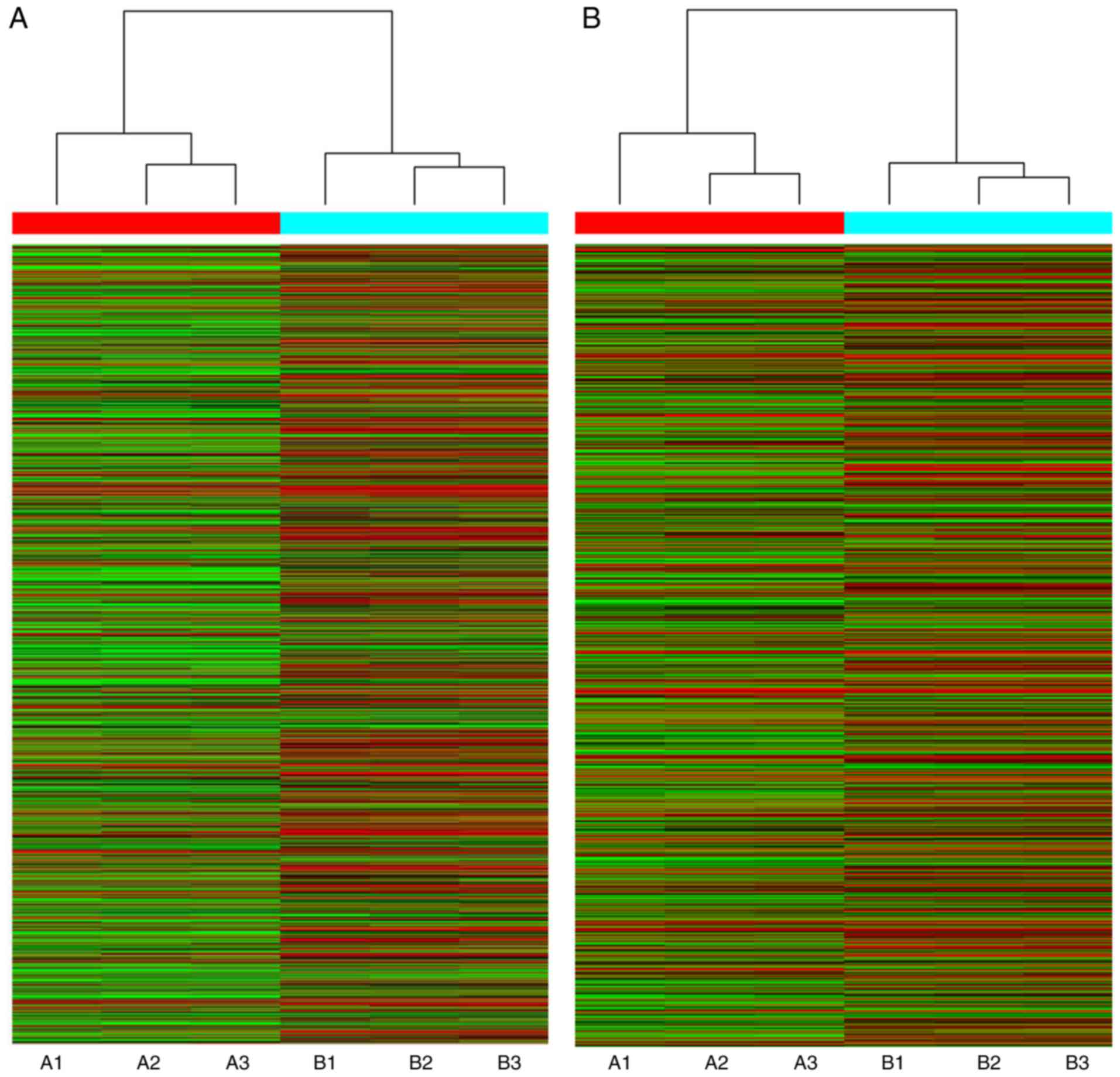
upregulated and downregulated lncRNAs (Table III) and mRNAs (Table IV) were listed. Distinguishable
lncRNA and mRNA expression patterns of the samples are presented
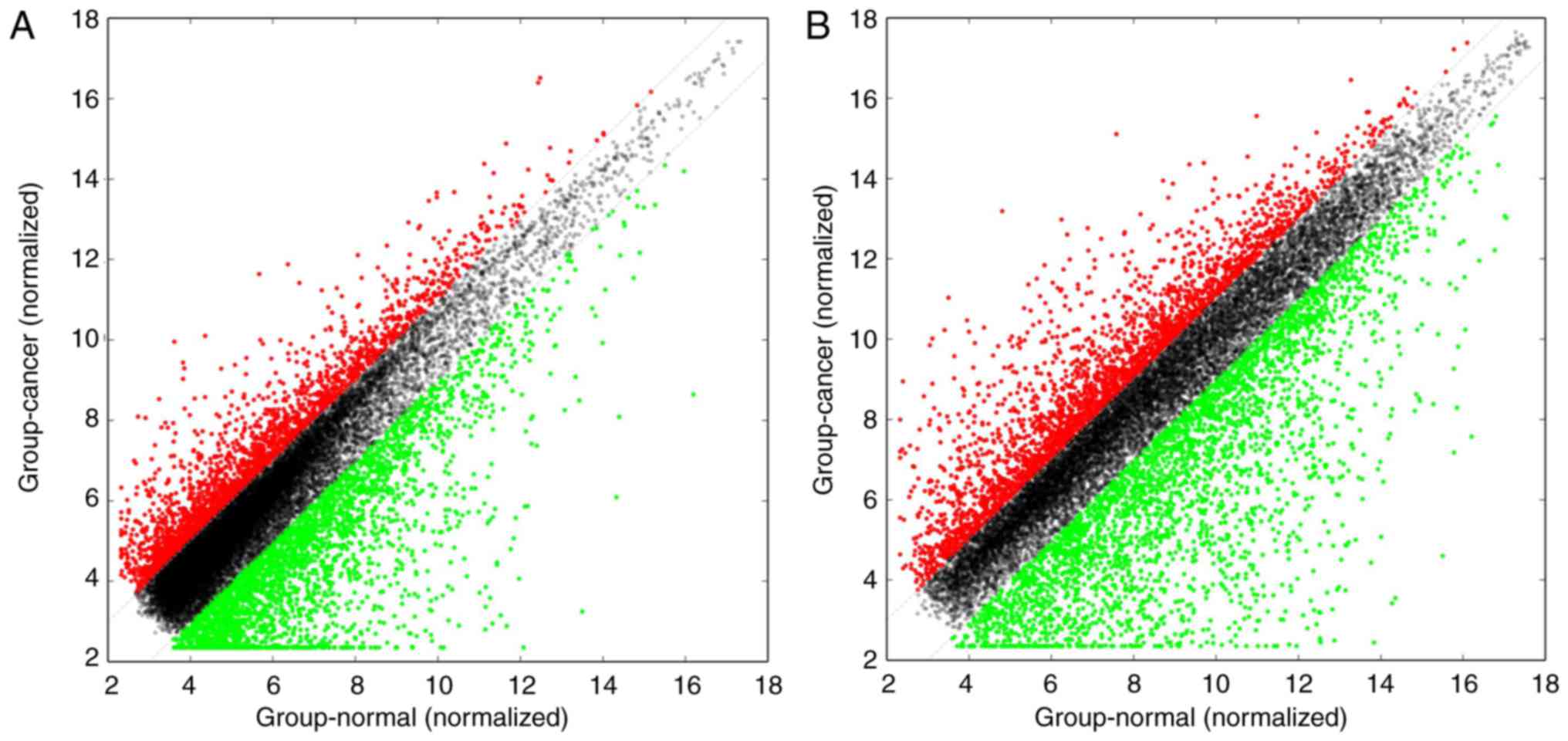
via heat maps of the hierarchical clustering (Fig. 2A and B). Reproducible changes in
gene expression were observed between the two groups through
scatter plots (Fig. 3A and B).
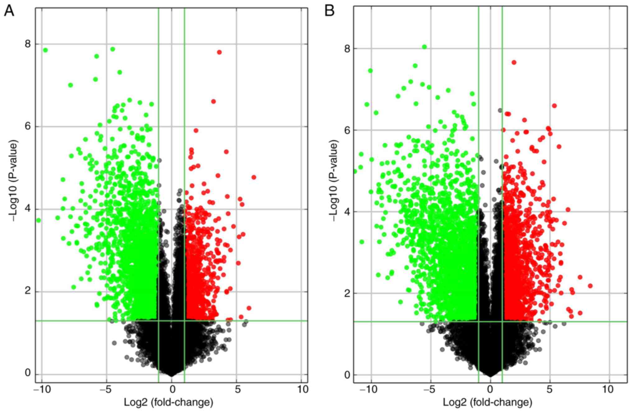
LncRNAs and mRNAs with statistically significant differences in
expression between the two groups (fold change, ≥2.0; P<0.05)
were identified by volcano plot filtering (Fig. 4A and B).
| Table IIIThe 10 most upregulated and
downregulated lncRNAs in high-grade ovarian serous cancer tissues
compared with non-tumor tissues, compared by volcano plot. |
Table III
The 10 most upregulated and
downregulated lncRNAs in high-grade ovarian serous cancer tissues
compared with non-tumor tissues, compared by volcano plot.
A, Upregulated
lncRNAs
|
|---|
| Seq. name | Gene symbol | Fold change
Chromosome | Associated gene
name | | Associated protein
name |
|---|
|
TCONS_12_00000435 |
XLOC_l2_000324 | 81.26 | chr1 | | |
| NR_002947 | TCAM1P | 62.61 | chr17 | | |
| NR_002712 | CXCR2P1 | 45.67 | chr2 | | |
| uc002ywy.3 |
AK027145 | 43.38 | chr21 | KCNJ15 | ATP-sensitive
inward rectifier potassium channel 15 |
|
ENST00000419814 |
RP11-131J3.1 | 41.20 | chr1 | | |
| NR_027072 |
LINC00189 | 38.96 | chr21 | | |
|
ENST00000428667 |
AP000695.4 | 36.94 | chr21 | CLDN14
C | laudin-14 |
|
ENST00000470135 |
RP5-884M6.1 | 35.52 | chr7 | | |
|
ENST00000455309 |
AC017002.1 | 27.37 | chr2 | | |
|
ENST00000606457 |
RP11-1C8.7 | 23.40 | chr8 | | |
|
B, Downregulated
lncRNAs
|
| Seq. name | Gene symbol | Fold change
Chromosome | Associated gene
name | | Associated protein
name |
|
|
ENST00000556942 |
RP11-356K23.1 | 1221.26 | chr14 | FOXN3 | Forkhead box
protein N3 isoform 1 |
|
ENST00000556942 |
RP11-356K23.1 | 1221.26 | chr14 | FOXN3 | Forkhead box
protein N3 isoform 2 |
| uc031qjg.1 |
AK129935 | 845.63 | chr12 | | |
|
ENST00000574178 |
RP11-424M24.5 | 439.06 | chr16 | | |
|
ENST00000584807 |
WI2-1959D15.1 | 331.26 | chr9 |
CR392000.1 | UniProtKB/TrEMBL or
E7EUX6 |
| AL049990 |
AL049990 | 330.93 | chr4 | | |
| NR_110114 |
LOC101927668 | 324.98 | chr7 | | |
| NR_003063 | TUBA4B | 302.06 | chr2 | | |
| uc002xuq.1 |
AK055386 | 239.29 | chr20 | | |
| NR_110916 |
LINC01571 | 221.41 | chr16 | | |
| Table IVThe 10 most upregulated and
downregulated mRNAs in high-grade ovarian serous cancer tissues
compared with non-tumor tissues, compared by volcano plot. |
Table IV
The 10 most upregulated and
downregulated mRNAs in high-grade ovarian serous cancer tissues
compared with non-tumor tissues, compared by volcano plot.
A, Upregulated
mRNAs
|
|---|
| Seq. name | Gene symbol | Fold change | Chromosome | Description |
|---|
| NM_153426 | PITX2 | 333.42 | chr4 | Paired-like
homeodomain 2 |
| NM_001884 | HAPLN1 | 184.94 | chr5 | Hyaluronan and
proteoglycan link protein 1 |
| NM_002125 |
HLA-DRB5 | 184.27 | chr6 | Major
histocompatibility complex, class II, DR beta 5 |
| NM_021192 | HOXD11 | 119.74 | chr2 | Homeobox D11 |
| NM_014391 | ANKRD1 | 112.86 | chr10 | Ankyrin repeat
domain 1 (cardiac muscle) |
| NM_003108 | SOX11 | 111.46 | chr2 | SRY-box 11 |
| NM_001854 | COL11A1 | 106.05 | chr1 | Collagen, type XI,
alpha 1 |
| NM_003469 | SCG2 | 93.91 | chr2 | Secretogranin
II |
| NM_005733 | KIF20A | 91.34 | chr5 | Kinesin family
member 20A |
| NM_022346 | NCAPG | 74.57 | chr4 | Non-SMC condensin I
complex subunit G |
|
B, Downregulated
mRNAs
|
| Seq. name | Gene symbol | Fold change | Chromosome | Description |
|
| NM_176813 | AGR3 | 2696.97 | chr7 | Anterior gradient
3, protein disulphide isomerase family member |
| NM_006408 | AGR2 | 1914.59 | chr7 | Anterior gradient
2, protein disulphide isomerase family member |
| NM_053285 | TEKT1 | 1848.50 | chr17 | Tektin 1 |
| NM_003357 | SCGB1A1 | 1791.03 | chr11 | Secretoglobin
family 1A member 1 |
| NM_031956 | TTC29 | 1334.47 | chr4 | Tetratricopeptide
repeat domain 29 |
| NM_001004303 |
C1orf168 | 1088.35 | chr1 | Chromosome 1 open
reading frame 168 |
| NM_001080537 | SNTN | 1051.19 | chr3 | Sentan, cilia
apical structure protein |
| NM_000777 | CYP3A5 | 1007.52 | chr7 | Cytochrome P450
family 3 subfamily A member 5 |
| NM_024730 | RERGL | 778.24 | chr12 | RERG/RAS-like |
| NM_005634 | SOX3 | 752.28 | chrX | SRY-box 3 |
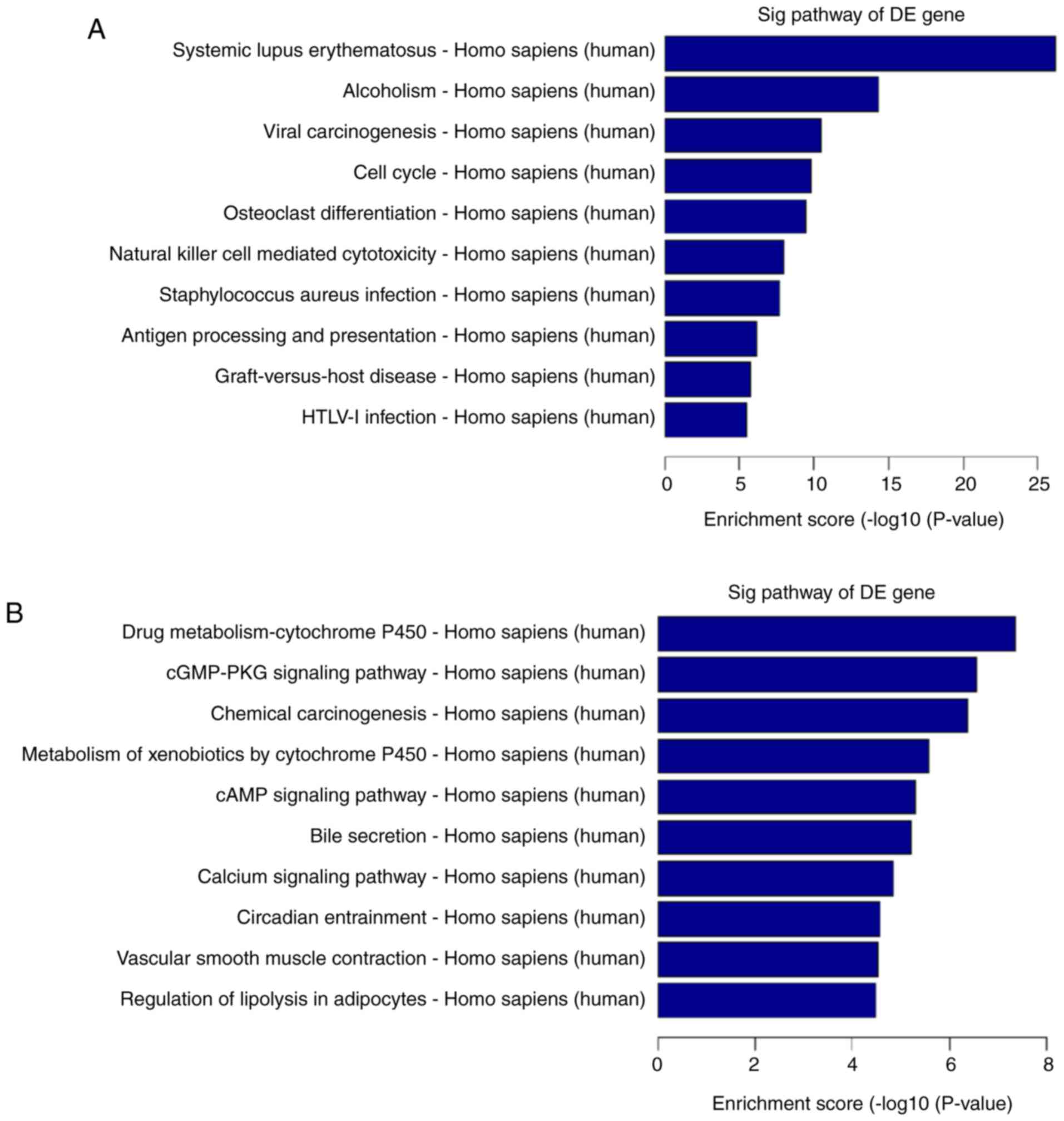
Bioinformatics analysis
Pathway analysis was used to determine biological
pathways where differentially expressed genes were significantly
enriched, according to the Kyoto Encyclopedia of Genes and Genomes
(25), BioCarta (26) and Reactome (27,28) databases (29). Total pathway analysis results
revealed that 121 pathways had significant differences in gene
expression between high-grade ovarian serous cancer tissue samples
and healthy fallopian tube tissue samples. Of these, 61 pathways
were upregulated and 60 were downregulated. The top ten upregulated
and downregulated pathways are presented in Fig. 5A and B. The top three upregulated
pathways included systemic lupus erythematosus, alcoholism and
viral carcinogenesis. The top three downregulated pathways included
drug metabolism-cytochrome P450, cGMP-PKG signaling pathway, and
chemical carcinogenesis.
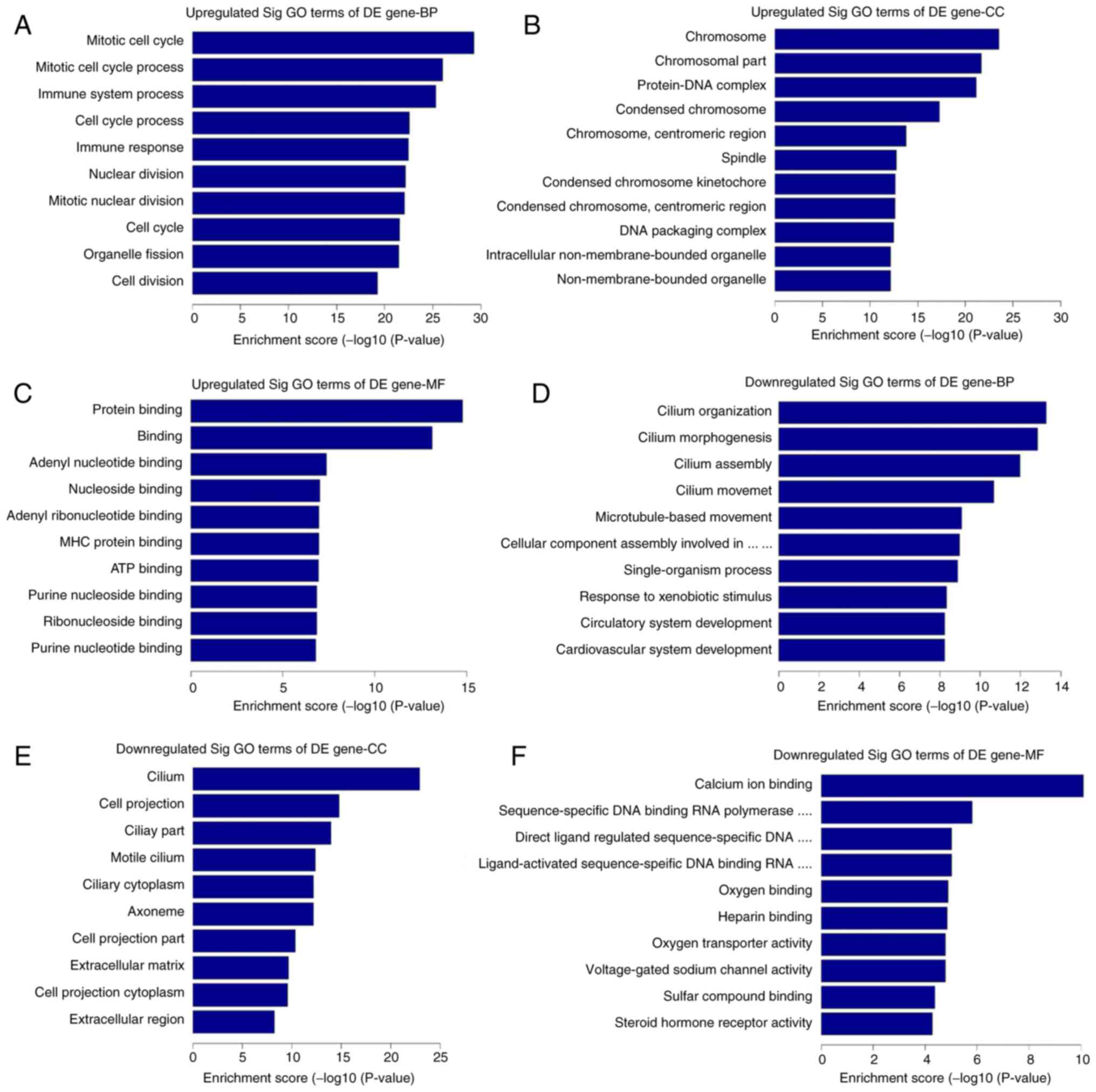
GO analysis was used to analyze the main functions
of the differentially expressed genes, according to the GO
database. The GO database provides the key functional
classifications for the National Center for Biotechnology
Information (22), which
comprises three structured networks: Biological processes, cellular
components and molecular function (30). The most enriched GO terms in each
structured network were mitotic cell cycle, chromosome, protein
binding, cilium organization, cilium, and calcium ion binding. The
first three of these terms were associated with upregulated genes
in high-grade ovarian serous cancer tissue samples compared with
the healthy fallopian tube tissue samples, and the last three terms
were associated with downregulated genes. The top ten upregulated
and downregulated GO functions of score enrichment terms are
presented in Fig. 6.
Subgroup analysis
Subgroup analysis was used to further investigate
the associations between lncRNA expression and high-grade ovarian
serous cancer. The differentially expressed antisense lncRNAs and
their associated mRNAs were integrated to infer the function of the
lncRNAs. The results revealed that 315 antisense lncRNAs and
associated mRNAs were abnormally expressed (fold change, ≥2.0;
P<0.05): 51 of the lncRNA-mRNA pairs were regulated in the up-up
direction, 200 pairs were regulated in the down-down direction, 30
pairs were regulated in the up-down direction, and 34 pairs were
regulated in the down-up direction. A total of 10 differentially
expressed antisense lncRNAs and associated mRNAs are presented in
Table V. In addition, long
intergenic non-coding RNAs (lincRNAs) are of particular interest to
the present study as they may be a novel factor associated with
cancer progression (31).
Statistical analysis of differentially expressed lincRNAs and their
nearby mRNAs (distance <300 kb) was conducted. The results
revealed that 1,807 lincRNAs and nearby mRNAs were abnormally
expressed (fold change, ≥2.0; P<0.05): 475 of the lincRNA-mRNA
pairs were regulated in the up-up direction, 738 pairs were
regulated in the down-down direction, 264 pairs were regulated in
the up-down direction and 330 pairs were regulated in the down-up
direction. A total of 10 differentially expressed lincRNAs and
nearby mRNAs are presented in Table
VI.
| Table VA total of 10 differentially
expressed lncRNAs (antisense) and their associated coding gene
pairs. |
Table V
A total of 10 differentially
expressed lncRNAs (antisense) and their associated coding gene
pairs.
Antisense LncRNAs
| Associated mRNAs
| Nearby protein
name | Direction
(lncRNA-mRNA) |
|---|
| Seq. name | Gene symbol | Nearby gene | Nearby gene
symbol |
|---|
|
ENST00000373226 | RP11-435D7.3 | NM_022111 | CLSPN C | laspin | Up-up |
|
ENST00000433905 | RP4-583P15.10 | NM_025224 | ZBTB46 | Zinc finger and BTB
domain containing 46 | Up-up |
|
ENST00000435915 | RP11-217B7.2 | NM_005502 | ABCA1 | ATP binding
cassette subfamily A member 1 | Up-up |
| ciRS-7 | ciRS-7 | NM_004065 | CDR1 | Cerebellar
degeneration related protein 1 | Down-down |
|
ENST00000411847 | RP11-301L8.2 | NM_021069 | SORBS2 | Sorbin and SH3
domain containing 2 | Down-down |
|
ENST00000412896 | AC107218.3 | NM_024532 | SPAG16 | Sperm associated
antigen 16 | Down-down |
|
ENST00000357045 | AC073834.3 | NM_152275 | TTC30A | Tetratricopeptide
repeat domain 30A | Up-down |
|
ENST00000414633 | SRGAP3-AS1 | NM_014850 | SRGAP3 | SLIT-ROBO Rho
GTPase activating protein 3 | Up-down |
|
ENST00000415004 | RP11-165F24.3 | NM_203447 | DOCK8 | Dedicator of
cytokinesis 8 | Down-up |
|
ENST00000424435 | RP11-350G8.5 | NM_000565 | IL6R | Interleukin 6
receptor | Down-up |
| Table VIA total of 10 cases of differentially
expressed long non-coding RNAs (lincRNAs) and nearby coding gene
pairs (distance <300 kb). |
Table VI
A total of 10 cases of differentially
expressed long non-coding RNAs (lincRNAs) and nearby coding gene
pairs (distance <300 kb).
LincRNAs
| Nearby mRNAs
| Nearby protein
name | Direction
(lncRNA-mRNA) |
|---|
| Seq. name | Gene symbol | Nearby gene | Nearby gene
symbol |
|---|
|
ENST00000412295 |
CTC-338M12.9 | NM_033549 | TRIM41 | Tripartite motif
containing 41 | Up-up |
|
ENST00000412362 |
RP11-236B18.2 | NM_014391 | ANKRD1 | Ankyrin repeat
domain 1 (cardiac muscle) | Up-up |
|
ENST00000412427 |
RP11-380J14.1 | NM_022089 | ATP13A2 | ATPase type
13A2 | Up-up |
| AF070541 |
AF070541 | NM_030632 | ASXL3 | Additional sex
combs like 3, transcriptional regulator | Down-down |
| AF339807 |
AF339807 | NM_015567 | SLITRK5 | SLIT and NTRK like
family member 5 | Down-down |
| AK023372 |
AK023372 | NM_000574 | CD55 | CD55 molecule
(Cromer blood group) | Down-down |
|
ENST00000413304 |
AC098872.3 | NM_004657 | SDPR | Serum deprivation
response | Dp-down |
|
ENST00000417932 |
RP11-10J5.1 | NM_022121 | PERP | PERP, TP53
apoptosis effector | Up-down |
| AK055324 |
AK055324 | NM_005184 | CALM3 | Calmodulin 3
(phosphorylase kinase, delta) | Down-up |
| AL049990 |
AL049990 | NM_024873 | TNIP3 | TNFAIP3 interacting
protein 3 | Down-up |
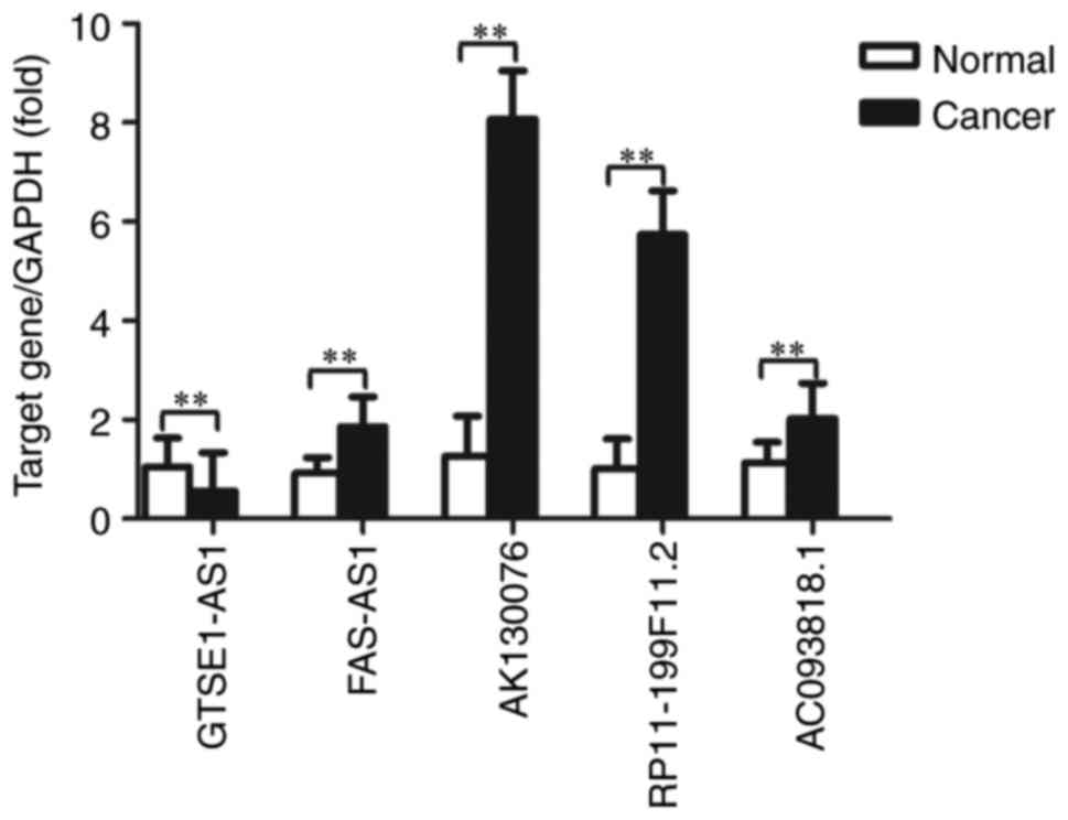
Validation of microarray data with
RT-qPCR
To validate the microarray consistency, RT-qPCR was
performed. A total of 5 abnormally expressed lncRNAs (GTSE1-AS1,
FAS-AS1, AK130076, RP11-199F11.2 and AC093818.1) and
their associated mRNAs [G2 and S-phase expressed 1 (GTSE1),
Fas surface cell death receptor (FAS), phosphatase and
tensin homolog (PTEN), tumor protein p53 (TP53) and
pyruvate dehydrogenase kinase 1 (PDK1)] were selected, and
their expression was analyzed in 20 high-grade ovarian serous
cancer tissue samples and healthy fallopian tube tissue samples,
using GAPDH as an internal control. These mRNAs have been
demonstrated to be associated with cell apoptosis, the p53
signaling pathway, the phosphoinositide 3 kinase (PI3K)/protein
kinase B (Akt) signaling pathway, and other mechanisms that are
associated with ovarian cancer progression and metastasis (32–35); therefore, our group predicted that
these lncRNAs may be involved in the development of ovarian cancer.
The results demonstrated that FAS-AS1, AK130076,
RP11-199F11.2 and AC093818.1 were upregulated and
GTSE1-AS1 was downregulated in high-grade ovarian serous
cancer tissues compared with healthy fallopian tube tissues
(Fig. 7). For mRNA, GTSE1,
TP53 and PDK1 were upregulated and FAS and
PTEN were downregulated in high-grade ovarian serous cancer
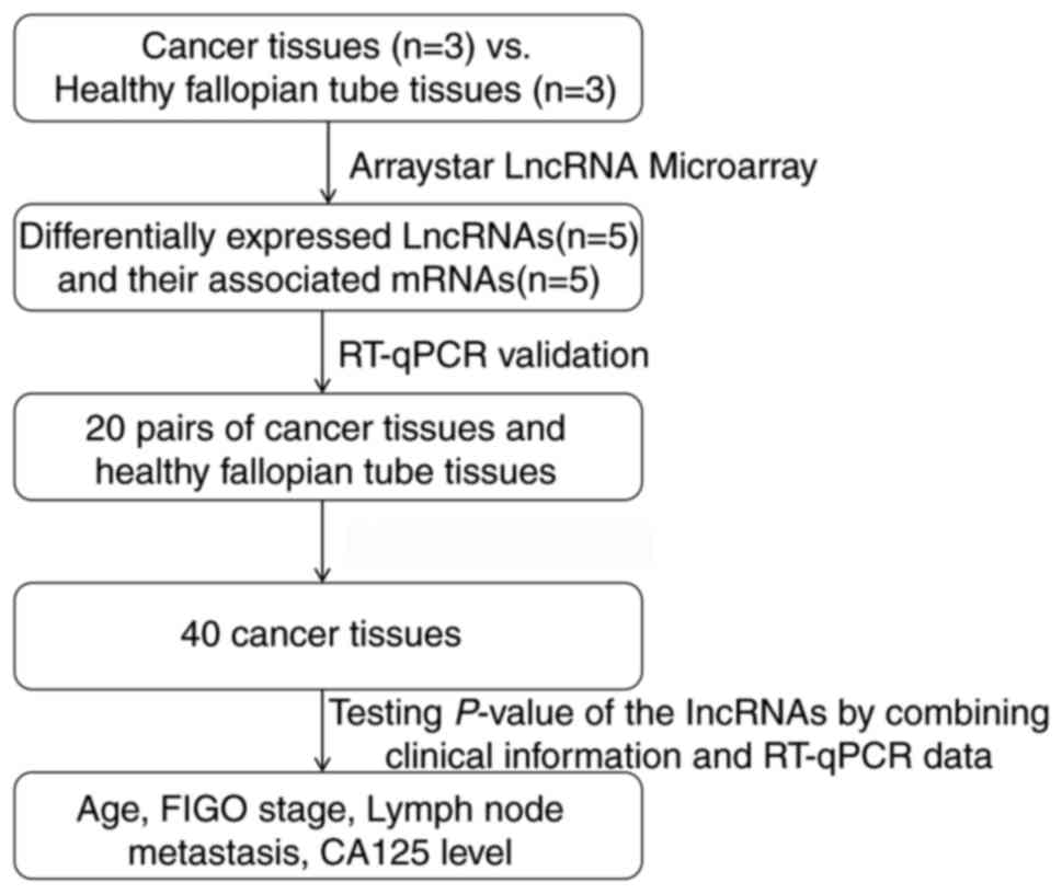
tissues compared with healthy fallopian tube tissues (Fig. 8). The RT-qPCR results matched the
microarray data well. Fig. 9
represents the entire workflow. The associations between the
expression of the 5 lncRNAs and clinicopathological parameters in
patients with high-grade ovarian serous cancer patients are
presented in Table VII. The
microarray results of the selected lncRNAs are presented in
Table VIII. These lncRNAs were
significantly associated with International Federation of
Gynecology and Obstetrics (FIGO) stage (36) and lymph node metastases in ovarian
cancer. These results suggested that these differentially expressed
lncRNAs may be associated with ovarian carcinogenesis and tumor
progression.
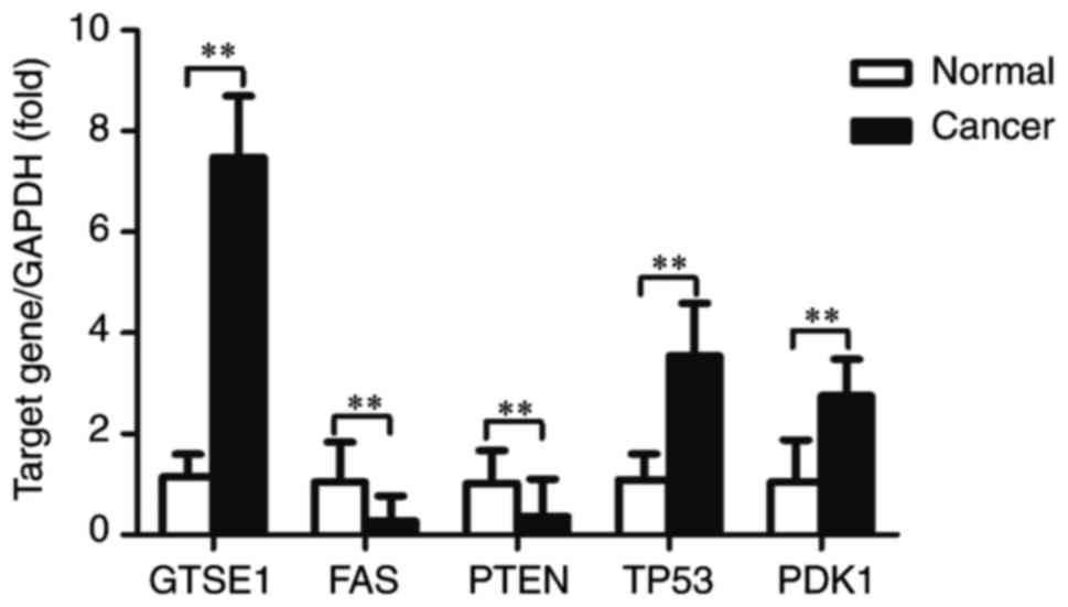 | Figure 8Expression levels of GTSE1, FAS,
PTEN, TP53 and PDK1 in high-grade ovarian serous cancer
tissue samples and healthy fallopian tube tissue samples, measured
by reverse transcription-quantitative polymerase chain reaction.
Expression levels were normalized to GAPDH. Data are expressed as
the mean ± standard deviation. **P<0.01, with
comparisons indicated by lines. GTSE1, G2 and S-phase
expressed 1; FAS, Fas cell surface death receptor;
PTEN, phosphatase and tensin homolog; TP53, tumor
protein p53; PDK1, pyruvate dehydrogenase kinase 1. |
| Table VIIAssociations between long non-coding
RNA expression and clinicopathological parameters in 40 patients
with high-grade ovarian serous cancer. |
Table VII
Associations between long non-coding
RNA expression and clinicopathological parameters in 40 patients
with high-grade ovarian serous cancer.
| Parameter | Age (years)
| FIGO stage
| Lymph node
metastasis
| CA125 level (U/ml)
|
|---|
| <50 | ≥50 | I-II | III-IV | Negative | Positive | <600 | ≥600 |
|---|
|
GTSE1-AS1 | | | | | | | | |
| High no.
cases | 6 | 8 | 5 | 4 | 6 | 4 | 10 | 6 |
| Low no. cases | 10 | 16 | 16 | 15 | 17 | 13 | 15 | 9 |
| P-value | 0.548 | | <0.01 | | <0.01 | | 0.432 | |
| FAS-AS1 |
| High no.
cases | 9 | 15 | 17 | 14 | 16 | 12 | 17 | 8 |
| Low no. cases | 7 | 9 | 4 | 5 | 7 | 5 | 8 | 7 |
| P-value | 0.433 | | <0.01 | | <0.01 | | 0.694 | |
|
AK130076 |
| High no.
cases | 10 | 16 | 18 | 15 | 18 | 12 | 17 | 10 |
| Low no. cases | 6 | 8 | 3 | 4 | 5 | 5 | 8 | 5 |
| P-value | 0.249 | | <0.01 | | <0.01 | | 0.167 | |
|
RP11-199F11.2 |
| High no.
cases | 10 | 15 | 16 | 16 | 20 | 11 | 14 | 9 |
| Low no. cases | 6 | 9 | 5 | 3 | 3 | 6 | 11 | 6 |
| P-value | 0.726 | | <0.01 | | <0.01 | | 0.259 | |
|
AC093818.1 |
| High no.
cases | 11 | 14 | 15 | 14 | 17 | 14 | 16 | 8 |
| Low no. cases | 5 | 10 | 6 | 5 | 6 | 3 | 9 | 7 |
| P-value | 0.673 | | <0.01 | | <0.01 | | 0.464 | |
| Table VIIIMicroarray results of the selected
five long non-coding RNAs. |
Table VIII
Microarray results of the selected
five long non-coding RNAs.
A, Downregulated
|
|---|
| Seq. name | Gene symbol | Fold change | Chromosome | Associated gene
name | Associated protein
name |
|---|
| NR_024009 |
GTSE1-AS1 | 3.11 | chr22 | GTSE1 | G2 and S
phase-expressed protein 1 |
|
B, Upregulated
|
| Seq. name | Gene symbol | Fold change | Chromosome | Associated gene
name | Associated protein
name |
|
| NR_028371 | FAS-AS1 | 2.01 | chr10 | FAS | Tumor necrosis
factor receptor superfamily member 6 isoform 1 precursor |
| uc001kfc.1 |
AK130076 | 6.08 | chr10 | PTEN |
Phosphatidylinositol 3,4,5-trisphosphate
3-phosphatase and dual-specificity protein Phosphatase PTEN |
|
ENST00000571370 |
RP11-199F11.2 | 5.87 | chr17 | TP53 | Cellular tumor
antigen p53 isoform a |
|
ENST00000436922 |
AC093818.1 | 2.54 | chr2 | PDK1 | Pyruvate
dehydrogenase kinase, isozyme 1 |
Discussion
Treatment of high-grade ovarian serous cancer
remains a serious challenge and the disease represents a global
health problem for women, with a 5-year mortality rate of >70%
and a high incidence of metastasis (37). Although the molecular mechanisms
underlying high-grade ovarian serous cancer have been investigated,
the exact pathogenesis of this disease remains unclear. Therefore,
investigation of molecular markers and effective therapeutic
strategies targeting high-grade ovarian serous cancer are of great
value. At present, an increasing number of studies have indicated
that lncRNAs are involved in the regulation of a wide variety of
biological processes (38).
However, the function that lncRNAs serve in cancer is of particular
interest. An increasing number of studies have reported that
several types of tumor are associated with differentially expressed
lncRNAs, which are involved in occurrence, development, invasion,
metastasis, apoptosis and drug resistance, via a series of
complicated mechanisms (39,40). For instance, lncRNA UCA1
was detected in bladder cancer tissues and cell lines, and the
expression levels of UCA1 were significantly higher than
that in a control group. Overexpression of UCA1 increases
the proliferation, invasion, and metastasis of bladder cancer cells
(41). Hou et al (14) discovered that long intergenic non
protein-coding RNA, regulator of reprogramming (LINC-ROR)
was upregulated in breast tumor samples, and ectopic overexpression
of LINC-ROR increased breast cancer cell migration and
invasion. Furthermore, LINC-ROR functioned as a competing
endogenous RNA to microRNA-205 and induced
epithelial-to-mesenchymal transition (14). In addition, dysregulation of
lncRNAs, including HOTAIR, metastasis associated lung
adenocarcinoma transcript 1, antisense non-coding RNA in the INK4
locus, growth arrest-specific 5, cervical carcinoma expressed PCNA
regulatory lncRNA, H19, imprinted maternally expressed transcript
(non-protein coding), and lncRNA-activated by TGFβ, has been
demonstrated to exacerbate several human cancers, including
small-cell lung cancer (12),
prostate cancer (42), gastric
cancer (15), colorectal cancer
(43), cervical cancer (44), liver cancer (45) and pancreatic cancer (46). Therefore, lncRNAs represent
potential indicators of tumor prognosis and diagnosis.
Usually, searching for differentially expressed
lncRNAs with high-throughput microarray technologies in cell lines
and tissues is the first step in the study of genes. Zhou et
al (47) investigated
differences in lncRNA and mRNA expression profiles between
hypopharyngeal squamous cell carcinoma tissues and adjacent
non-tumor tissues by microarray analysis, and identified a series
of significantly dysregulated lncRNAs and mRNAs. These results laid
the foundation for their following research. Similar studies have
been carried out in pancreatic cancer, endometrial cancer and
nasopharyngeal carcinoma (48–50).
In the present study, three pairs of high-grade
ovarian serous cancer tissue samples and healthy fallopian tube
tissue samples were used to investigate the differences in lncRNA
and mRNA expression profiles through microarray analysis. The
results revealed that the expression profiles of lncRNAs and mRNAs
in high-grade ovarian serous cancer tissues were significantly
altered. In addition, compared with the number of upregulated
genes, the number of downregulated genes was larger, indicating
that this may influence the occurrence and development of
high-grade ovarian serous cancer. These differentially expressed
genes were sub sequently organized into hierarchical categories
based on heat maps of hierarchical clustering, and the differences
in lncRNA and mRNA expression between the two groups were analyzed
by scatter plot and volcano plot filtering. Furthermore, GO and
pathway analysis were performed to obtain information on the
biological functions and potential mechanisms underlying the action
of these differentially expressed lncRNAs. However, more detailed
research should be performed to investigate the specific mechanisms
existing between these pathways, networks and genes.
Antisense lncRNA has been the subject of intense
research among lncRNAs (51). In
total, >30% of annotated human transcripts have corresponding
antisense lncRNAs. These antisense lncRNAs regulate the
corresponding sense lncRNAs at transcription or post transcription
level through a variety of mechanisms, and serve an important
biological function. In addition, lincRNAs are a subject of
particular interest to the present study (52). Multiple studies have demonstrated
that a common and important function of lncRNAs is to alter the
expression of nearby coding genes by affecting their transcription
(53–55). Therefore, in the present study,
differentially expressed antisense lncRNAs, lincRNAs, and their
associated mRNAs were integrated in order to infer the function of
lncRNAs in lncRNA-mRNA coexpression, which may predict the target
genes of lncRNAs. Subgroup analysis results identified 315
abnormally expressed antisense lncRNAs and associated mRNAs, as
well as 1807 abnormally expressed lincRNAs and nearby mRNAs.
However, the function of this lncRNA-mRNA coexpression requires
further research.
To confirm the microarray consistency, 5
differentially expressed lncRNAs (GTSE1-AS1, FAS-AS1, AK130076,
RP11-199F11.2 and AC093818.1) and their associated mRNAs
(GTSE1, FAS, PTEN, TP53 and PDK1) were selected to
verify expression consistency by RT-qPCR. FAS has been
reported to be associated with ovarian cancer cell apoptosis
(32). PTEN serves an
important biological function in ovarian cancer cell growth,
proliferation, and migration (34). TP53 is a common tumor
suppressor gene, which influences ovarian cancer cell proliferation
and cell cycle through the p53 signaling pathway (33). PDK1 is involved in the development
and drug resistance mechanisms of ovarian cancer by regulating the
PI3K/Akt signaling pathway (35).
The present study revealed that GTSE1, FAS, PTEN, TP53, and
PDK1 are the associated mRNAs of GTSE1-AS1, FAS-AS1,
AK130076, RP11-199F11.2 and AC093818.1, suggesting that
these lncRNAs may serve a role in the development of ovarian
cancer. In particular, the expression of 4 lncRNAs (FAS-AS1,
AK130076, RP11-199F11.2 and AC093818.1) was
significantly increased in high-grade ovarian serous cancer tissues
compared with healthy fallopian tube tissues, while the expression
of GTSE1-AS1 was significantly lower. The expression of 3
associated mRNAs (GTSE1, TP53 and PDK1) was
significantly increased in high-grade ovarian serous cancer
tissues; that of FAS and PTEN was significantly
decreased compared with normal tissues. These results are
consistent with the microarray data and reflect the variable
expression of lncRNAs and mRNAs in different tissues. Furthermore,
the expression of the selected lncRNAs was significantly associated
with ovarian cancer FIGO stages and lymph node metastases in 40
patients with high-grade ovarian serous cancer. These results
provide more evidence that these differentially expressed lncRNAs
may be associated with ovarian carcinogenesis and tumor
progression. Although the biological functions of a large number of
genes remain unclear, the data from the present study may be useful
for further studies on the pathogenesis and underlying molecular
mechanisms of high-grade ovarian serous cancer. Multiple further
studies are necessary to strengthen the association between lncRNAs
and high-grade ovarian serous cancer. In consequent work, our group
will further explore gene expression and clinical data in
high-grade ovarian serous cancer according to The Cancer Genome
Atlas and Gene Expression Omnibus databases, and analyze the
associations with the prognosis of patients with high-grade ovarian
serous cancer. Further experiments, including immunohistochemistry,
western blot analysis and other in vivo and in vitro
experiments will be performed to identify the specific molecular
mechanisms and biochemical functions between lncRNAs and high-grade
ovarian serous cancer.
In conclusion, to the best of our knowledge, the
present study was the first to examine differences in lncRNA and
mRNA expression profiles between high-grade ovarian serous cancer
tissues and healthy fallopian tube tissues using micro-array
analysis. According to the present study, 4,289 lncRNAs and 4,246
mRNAs were differentially expressed in high-grade ovarian serous
cancer tissues, and may serve a key function in the occurrence and
development of high-grade ovarian serous cancer. Furthermore,
bioinformatics analyses were applied to determine the potential
functions of these abnormally expressed genes. In the near future,
our group will conduct more detailed research on several lncRNAs,
and identify their specific molecular mechanisms and biochemical
functions for the purpose of providing effective methods for the
diagnosis and therapy of high-grade ovarian serous cancer.
Acknowledgments
Not applicable.
References
|
1
|
Bast RC Jr, Hennessy B and Mills GB: The
biology of ovarian cancer: New opportunities for translation. Nat
Rev Cancer. 9:415–428. 2009. View
Article : Google Scholar : PubMed/NCBI
|
|
2
|
Siegel R, Naishadham D and Jemal A: Cancer
statistics, 2013. CA Cancer J Clin. 63:11–30. 2013. View Article : Google Scholar : PubMed/NCBI
|
|
3
|
Yiwei T, Hua H, Hui G, Mao M and Xiang L:
HOTAIR interacting with MAPK1 regulates ovarian cancer skov3 cell
proliferation, migration, and invasion. Med Sci Monit.
21:1856–1863. 2015. View Article : Google Scholar : PubMed/NCBI
|
|
4
|
ENCODE Project Consortium; Birney E,
Stamatoyannopoulos JA, Dutta A, Guigó R, Gingeras TR, Margulies EH,
Weng Z, Snyder M, Dermitzakis ET, et al: Identification and
analysis of functional elements in 1% of the human genome by the
ENCODE pilot project. Nature. 447:799–816. 2007. View Article : Google Scholar : PubMed/NCBI
|
|
5
|
Nagano T and Fraser P: No-nonsense
functions for long noncoding RNAs. Cell. 145:178–181. 2011.
View Article : Google Scholar : PubMed/NCBI
|
|
6
|
Yang Q, Xu E, Dai J, Liu B, Han Z, Wu J,
Zhang S, Peng B, Zhang Y and Jiang Y: A novel long noncoding RNA
AK001796 acts as an oncogene and is involved in cell growth
inhibition by resveratrol in lung cancer. Toxicol Appl Pharmacol.
285:79–88. 2015. View Article : Google Scholar : PubMed/NCBI
|
|
7
|
Gibb EA, Brown CJ and Lam WL: The
functional role of long non-coding RNA in human carcinomas. Mol
Cancer. 10:382011. View Article : Google Scholar : PubMed/NCBI
|
|
8
|
Zhang H, Chen Z, Wang X, Huang Z, He Z and
Chen Y: Long non-coding RNA: A new player in cancer. J Hematol
Oncol. 6:372013. View Article : Google Scholar : PubMed/NCBI
|
|
9
|
Tang SC and Chen YC: Novel therapeutic
targets for pancreatic cancer. World J Gastroenterol.
20:10825–10844. 2014. View Article : Google Scholar : PubMed/NCBI
|
|
10
|
Zeng Z, Bo H, Gong Z, Lian Y, Li X, Li X,
Zhang W, Deng H, Zhou M, Peng S, et al: AFAP1-AS1, a long noncoding
RNA upregulated in lung cancer and promotes invasion and
metastasis. Tumour Biol. 37:729–737. 2016. View Article : Google Scholar
|
|
11
|
Qiu JJ, Wang Y, Ding JX, Jin HY, Yang G
and Hua KQ: The long non-coding RNA HOTAIR promotes the
proliferation of serous ovarian cancer cells through the regulation
of cell cycle arrest and apoptosis. Exp Cell Res. 333:238–248.
2015. View Article : Google Scholar : PubMed/NCBI
|
|
12
|
Ono H, Motoi N, Nagano H, Miyauchi E,
Ushijima M, Matsuura M, Okumura S, Nishio M, Hirose T, Inase N and
Ishikawa Y: Long noncoding RNA HOTAIR is relevant to cellular
proliferation, invasiveness, and clinical relapse in small-cell
lung cancer. Cancer Med. 3:632–642. 2014. View Article : Google Scholar : PubMed/NCBI
|
|
13
|
Han Y, Liu Y, Zhang H, Wang T, Diao R,
Jiang Z, Gui Y and Cai Z: Hsa-miR-125b suppresses bladder cancer
development by down-regulating oncogene SIRT7 and oncogenic long
noncoding RNA MALAT1. FEBS Lett. 587:3875–3882. 2013. View Article : Google Scholar
|
|
14
|
Hou P, Zhao Y, Li Z, Yao R, Ma M, Gao Y,
Zhao L, Zhang Y, Huang B and Lu J: LincRNA-ROR induces
epithelial-to-mesenchymal transition and contributes to breast
cancer tumorigenesis and metastasis. Cell Death Dis. 5:e12872014.
View Article : Google Scholar : PubMed/NCBI
|
|
15
|
Zhang EB, Kong R, Yin DD, You LH, Sun M,
Han L, Xu TP, Xia R, Yang JS, De W and Chen Jf: Long noncoding RNA
ANRIL indicates a poor prognosis of gastric cancer and promotes
tumor growth by epigenetically silencing of miR-99a/miR-449a.
Oncotarget. 5:2276–2292. 2014. View Article : Google Scholar :
|
|
16
|
Xia T, Chen S, Jiang Z, Shao Y, Jiang X,
Li P, Xiao B and Guo J: Long noncoding RNA FER1L4 suppresses cancer
cell growth by acting as a competing endogenous RNA and regulating
PTEN expression. Sci Rep. 5:134452015. View Article : Google Scholar : PubMed/NCBI
|
|
17
|
Qiu JJ, Lin YY, Ye LC, Ding JX, Feng WW,
Jin HY, Zhang Y, Li Q and Hua KQ: Overexpression of long non-coding
RNA HOTAIR predicts poor patient prognosis and promotes tumor
metastasis in epithelial ovarian cancer. Gynecol Oncol.
134:121–128. 2014. View Article : Google Scholar : PubMed/NCBI
|
|
18
|
Richards EJ, Permuth-Wey J, Li Y, Chen YA,
Coppola D, Reid BM, Lin HY, Teer JK, Berchuck A, Birrer MJ, et al:
A functional variant in HOXA11-AS, a novel long non-coding RNA,
inhibits the oncogenic phenotype of epithelial ovarian cancer.
Oncotarget. 6:34745–34757. 2015. View Article : Google Scholar : PubMed/NCBI
|
|
19
|
Gao Y, Meng H, Liu S, Hu J, Zhang Y, Jiao
T, Liu Y, Ou J, Wang D, Yao L, et al: LncRNA-HOST2 regulates cell
biological behaviors in epithelial ovarian cancer through a
mechanism involving microRNA let-7b. Hum Mol Genet. 24:841–852.
2015. View Article : Google Scholar
|
|
20
|
Li J, Zhou D, Wang Z, Tan L, Zhou Y, Li J
and Sheng X: Reversal effect of 5-aza-2-deoxycytidine on the
maternally expressed gene 3 promoter hypermethylation and its
inhibitory effect on the proliferation of epithelial ovarian cancer
cells. Zhonghua Zhong Liu Za Zhi. 37:324–329. 2015.In Chinese.
PubMed/NCBI
|
|
21
|
Yang Y, Jiang Y, Wan Y, Zhang L, Qiu J,
Zhou S and Cheng W: UCA1 functions as a competing endogenous RNA to
suppress epithelial ovarian cancer metastasis. Tumour Biol.
37:10633–10641. 2016. View Article : Google Scholar : PubMed/NCBI
|
|
22
|
Ashburner M, Ball CA, Blake JA, Botstein
D, Butler H, Cherry JM, Davis AP, Dolinski K, Dwight SS, Eppig JT,
et al: Gene ontology: Tool for the unification of biology. The Gene
Ontology Consortium. Nat Genet. 25:25–29. 2000. View Article : Google Scholar : PubMed/NCBI
|
|
23
|
The Gene Ontology Consortium: Expansion of
the Gene Ontology knowledgebase and resources. Nucleic Acids Res.
45(D1): D331–D338. 2017. View Article : Google Scholar :
|
|
24
|
Livak KJ and Schmittgen TD: Analysis of
relative gene expression data using real-time quantitative PCR and
the 2(-Delta Delta C(T)) method. Methods. 25:402–408. 2001.
View Article : Google Scholar
|
|
25
|
Kanehisa M and Goto S: KEGG: Kyoto
encyclopedia of genes and genomes. Nucleic Acids Res. 28:27–30.
2000. View Article : Google Scholar
|
|
26
|
Darryl N: Biotech Software & Internet
Report 2. 117–120. 2004.
|
|
27
|
Fabregat A, Jupe S, Matthews L,
Sidiropoulos K, Gillespie M, Garapati P, Haw R, Jassal B, Korninger
F, May B, et al: The reactome pathway knowledgebase. Nucleic Acids
Res. 46(D1): D649–D655. 2018. View Article : Google Scholar :
|
|
28
|
Milacic M, Haw R, Rothfels K, Wu G, Croft
D, Hermjakob H, D'Eustachio P and Stein L: Annotating cancer
variants and anti-cancer therapeutics in reactome. Cancers (Basel).
4:1180–1211. 2012. View Article : Google Scholar
|
|
29
|
Yang Y, Li H, Hou S, Hu B, Liu J and Wang
J: The noncoding RNA expression profile and the effect of lncRNA
AK126698 on cisplatin resistance in non-small-cell lung cancer
cell. PLoS One. 8:e653092013. View Article : Google Scholar : PubMed/NCBI
|
|
30
|
Cao G, Zhang J, Wang M, Song X, Liu W, Mao
C and Lv C: Differential expression of long non-coding RNAs in
bleomycin-induced lung fibrosis. Int J Mol Med. 32:355-3642013.
View Article : Google Scholar
|
|
31
|
Tsai MC, Spitale RC and Chang HY: Long
intergenic noncoding RNAs: New links in cancer progression. Cancer
Res. 71:3–7. 2011. View Article : Google Scholar : PubMed/NCBI
|
|
32
|
Cohen M, Pierredon S, Wuillemin C, Delie F
and Petignat P: Acellular fraction of ovarian cancer ascites induce
apoptosis by activating JNK and inducing BRCA1, Fas and FasL
expression in ovarian cancer cells. Oncoscience. 1:262–271. 2014.
View Article : Google Scholar
|
|
33
|
Hayano T, Yokota Y, Hosomichi K, Nakaoka
H, Yoshihara K, Adachi S, Kashima K, Tsuda H, Moriya T, Tanaka K,
et al: Molecular characterization of an intact p53 pathway subtype
in high-grade serous ovarian cancer. PLoS One. 9:e1144912014.
View Article : Google Scholar : PubMed/NCBI
|
|
34
|
Li J, Hu K, Gong G, Zhu D, Wang Y, Liu H
and Wu X: Upregulation of MiR-205 transcriptionally suppresses
SMAD4 and PTEN and contributes to human ovarian cancer progression.
Sci Rep. 7:413302017. View Article : Google Scholar : PubMed/NCBI
|
|
35
|
Bugide S, Gonugunta VK, Penugurti V,
Malisetty VL, Vadlamudi RK and Manavathi B: HPIP promotes
epithelial-mesenchymal transition and cisplatin resistance in
ovarian cancer cells through PI3K/AKT pathway activation. Cell
Oncol (Dordr). 40:133–144. 2017. View Article : Google Scholar
|
|
36
|
Zeppernick F and Meinhold-Heerlein I: The
new FIGO staging system for ovarian, fallopian tube, and primary
peritoneal cancer. Arch Gynecol Obstet. 290:839–842. 2014.
View Article : Google Scholar : PubMed/NCBI
|
|
37
|
Jelovac D: Armstrong DK. Recent progress
in the diagnosis and treatment of ovarian cancer. CA Cancer J Clin.
61:183–203. 2011. View Article : Google Scholar : PubMed/NCBI
|
|
38
|
Ji Q, Zhang L, Liu X, Zhou L, Wang W, Han
Z, Sui H, Tang Y, Wang Y, Liu N, et al: Long non-coding RNA MALAT1
promotes tumour growth and metastasis in colorectal cancer through
binding to SFPQ and releasing oncogene PTBP2 from SFPQ/TBP2
complex. Br J Cancer. 111:736–748. 2014. View Article : Google Scholar : PubMed/NCBI
|
|
39
|
Shi Y, Liu Y, Wang J, Jie D, Yun T, Li W,
Yan L, Wang K and Feng J: Downregulated long noncoding RNA BANCR
promotes the proliferation of colorectal cancer cells via
downregualtion of p21 expression. PLoS One. 10:e01226792015.
View Article : Google Scholar : PubMed/NCBI
|
|
40
|
Xue M, Li X, Li Z and Chen W: Urothelial
carcinoma associated 1 is a hypoxia-inducible
factor-1alpha-targeted long noncoding RNA that enhances hypoxic
bladder cancer cell proliferation, migration, and invasion. Tumour
Biol. 35:6901–6912. 2014. View Article : Google Scholar : PubMed/NCBI
|
|
41
|
Srivastava AK, Singh PK, Rath SK, Dalela
D, Goel MM and Bhatt ML: Appraisal of diagnostic ability of UCA1 as
a biomarker of carcinoma of the urinary bladder. Tumour Biol.
35:11435–11442. 2014. View Article : Google Scholar : PubMed/NCBI
|
|
42
|
Wang F, Ren S, Chen R, Lu J, Shi X, Zhu Y,
Zhang W, Jing T, Zhang C, Shen J, et al: Development and
prospective multicenter evaluation of the long noncoding RNA
MALAT-1 as a diagnostic urinary biomarker for prostate cancer.
Oncotarget. 5:11091–11102. 2014.PubMed/NCBI
|
|
43
|
Yin D, He X, Zhang E, Kong R, De W and
Zhang Z: Long noncoding RNA GAS5 affects cell proliferation and
predicts a poor prognosis in patients with colorectal cancer. Med
Oncol. 31:2532014. View Article : Google Scholar : PubMed/NCBI
|
|
44
|
Yang M, Zhai X, Xia B, Wang Y and Lou G:
Long noncoding RNA CCHE1 promotes cervical cancer cell
proliferation via upregulating PCNA. Tumour Biol. 36:7615–7622.
2015. View Article : Google Scholar : PubMed/NCBI
|
|
45
|
Li H, Li J, Jia S, Wu M, An J, Zheng Q,
Zhang W and Lu D: miR675 upregulates long noncoding RNA H19 through
activating EGR1 in human liver cancer. Oncotarget. 6:31958–31984.
2015.PubMed/NCBI
|
|
46
|
Qu S, Yang X, Song W, Sun W, Li X, Wang J,
Zhong Y, Shang R, Ruan B, Zhang Z, et al: Downregulation of
lncRNA-ATB correlates with clinical progression and unfavorable
prognosis in pancreatic cancer. Tumour Biol. 37:3933–3938. 2016.
View Article : Google Scholar
|
|
47
|
Zhou J, Li W, Jin T, Xiang X, Li M, Wang
J, Li G, Pan X and Lei D: Gene microarray analysis of lncRNA and
mRNA expression profiles in patients with hypopharyngeal squamous
cell carcinoma. Int J Clin Exp Med. 8:4862–4882. 2015.PubMed/NCBI
|
|
48
|
Zhou M, Ye Z, Gu Y, Tian B, Wu B and Li J:
Genomic analysis of drug resistant pancreatic cancer cell line by
combining long non-coding RNA and mRNA expression profling. Int J
Clin Exp Pathol. 8:38–52. 2015.PubMed/NCBI
|
|
49
|
Yang L, Zhang J, Jiang A, Liu Q, Li C,
Yang C and Xiu J: Expression profile of long non-coding RNAs is
altered in endometrial cancer. Int J Clin Exp Med. 8:5010–5021.
2015.PubMed/NCBI
|
|
50
|
Yang QQ and Deng YF: Genome-wide analysis
of long non-coding RNA in primary nasopharyngeal carcinoma by
microarray. Histopathology. 66:1022–1030. 2015. View Article : Google Scholar
|
|
51
|
Xie M, Sun M, Zhu YN, Xia R, Liu YW, Ding
J, Ma HW, He XZ, Zhang ZH, Liu ZJ, et al: Long noncoding RNA
HOXA-AS2 promotes gastric cancer proliferation by epigenetically
silencing P21/LK3/DDIT3 expression. Oncotarget. 6:33587–33601.
2015. View Article : Google Scholar : PubMed/NCBI
|
|
52
|
Zhou X, Gao Q, Wang J, Zhang X, Liu K and
Duan Z: Linc-RNA-RoR acts as a 'sponge' against mediation of the
differentiation of endometrial cancer stem cells by microRNA-145.
Gynecol Oncol. 133:333–339. 2014. View Article : Google Scholar : PubMed/NCBI
|
|
53
|
Khalil AM, Guttman M, Huarte M, Garber M,
Raj A, Rivea Morales D, Thomas K, Presser A, Bernstein BE, van
Oudenaarden A, et al: Many human large intergenic noncoding RNAs
associate with chromatin-modifying complexes and affect gene
expression. Proc Natl Acad Sci USA. 106:11667–11672. 2009.
View Article : Google Scholar : PubMed/NCBI
|
|
54
|
Mattick JS and Gagen MJ: The evolution of
controlled multi-tasked gene networks: The role of introns and
other noncoding RNAs in the development of complex organisms. Mol
Biol Evol. 18:1611–1630. 2001. View Article : Google Scholar : PubMed/NCBI
|
|
55
|
Popadin K, Gutierrez-Arcelus M,
Dermitzakis ET and Antonarakis SE: Genetic and epigenetic
regulation of human lincRNA gene expression. Am J Hum Genet.
93:1015–1026. 2013. View Article : Google Scholar : PubMed/NCBI
|