Introduction
Lung cancer is a major cause of mortality from
malignant disease, due to its high incidence, malignant behavior
and lack of major advancements in treatment strategy (1). Lung cancer was the leading indication
for respiratory surgery (47.5%) in 2009 in Japan (2) and more than 30,000 patients underwent
surgery for lung cancer at Japanese institutions in the same year
(2). The clinical behavior of
non-small cell lung cancer (NSCLC) is largely associated with its
stage. NSCLC is only cured by surgery in cases that are in the
early stages of the disease (3).
Recently, the theory that chronic inflammation plays
a significant role in cancer development, including carcinogenesis,
invasion and metastasis, has been proposed (4). It is well known that inflammation
induced by environmental exposure, including tobacco smoking and
inhaled pollutants (silica or asbestos), increase cancer risk
(5–7). It has also been reported that chronic
and subclinical levels of inflammation, for example,
obesity-induced inflammation, may increase cancer risk (8). Angiopoietin-like protein 2 (ANGPTL2)
is a causative mediator of chronic inflammation in obesity and its
related metabolic abnormalities (9). ANGPTL2 is secreted by adipose tissue
and the expression is increased during hypoxia and endoplasmic
reticulum stress (9). The stress is
commonly induced in cancer tissues in progression and metastasis
(10). A previous study suggested
that ANGPTL2 increased inflammatory carcinogenesis in a chemically
induced skin squamous cell carcinoma mouse model (11). ANGPTL2 protein expression has also
been reported in certain tumor cell types, including ovarian cancer
(12), lung cancer (13) and sarcoma (14). Although the role of ANGPTL2 is
controversial, ANGPTL2 may be a critical factor in cancer
development.
The current study investigated ANGPTL2 mRNA
expression in NSCLC and adjacent normal lung tissues using
real-time quantitative polymerase chain reaction (qPCR) using
LightCycler (Roche Molecular Biochemicals, Mannheim, Germany)
(15) in surgically treated cases.
The findings were compared with the clinocopathological features of
the NSCLC and ANGPTL2 gene status.
Patients and methods
Patients
The study group included NSCLC patients who had
undergone surgery at the Department of Surgery, Nagoya City
University Hospital between 2007 and 2009. All tumor samples were
immediately frozen and stored at −80°C until assayed. The clinical
and pathological characteristics of the 110 NSCLC patients for
ANGPTL2 mRNA gene analyses were as follows: 70 (63.6%) were
male, 40 were female; 89 cases (80.9%) were diagnosed as
adenocarcinomas and 18 were diagnosed as squamous cell carcinomas;
69 (62.7%) were smokers, 41 were non-smokers and 73 (66.4%) were
pathological stage I.
PCR assay for ANGPTL2 gene
Total RNA was extracted from NSCLC and adjacent
normal lung tissues using an Isogen kit (Nippon gene, Tokyo, Japan)
according to the manufacturer’s instructions. RNA concentration was
determined by Nano Drop ND-1000 Spectrophotometer (Nano Drop
Technologies Inc., Rockland, DE, USA). Approximately 10 cases were
excluded in each assay as there were too few tumor cells to
sufficiently extract tumor RNA. RNA (1 μg) was reverse
transcribed using a First strand cDNA synthesis kit with 0.5
μg oligo (dT)16 (Roche Diagnostics GmbH, Mannheim, Germany)
according to the manufacturer’s instructions. The reaction mixture
was incubated at 25°C for 15 min, 42°C for 60 min, 99°C for 5 min
and then at 4°C for 5 min. The cDNA concentration was determined by
Nano Drop ND-1000 Spectrophotometer. Approximately 200 ng of each
cDNA was used for PCR analysis. To ensure the fidelity of mRNA
extraction and reverse transcription, all samples were subjected to
qPCR amplification with a β-actin primers kit (Nihon Gene
Laboratory, Miyagi, Japan) using LightCycler-FastStart DNA Master
HybProbe kit (Roche Diagnostics GmbH). The ANGTL2 qPCR assay
reactions were performed using LightCycler FastStart DNA Master
SYBR Green I kit (Roche Diagnostics GmbH) in a 20-μl
reaction volume. The primer sequences for the ANGPTL2 gene
were as follows: forward primer, 5′-GCCACCAAGTGTCAGCCTCA-3′ and
reverse, 5′-TGGACAGTACCAAACATCCAACATC-3′ (134 bp). The cycling
conditions were as follows: initial denaturation at 95°C for 10
min, followed by 40 cycles at 95°C for 10 sec, 57°C for 10 sec and
72°C for 6 sec.
Statistical analysis
Statistical analyses were performed using the
Student’s t-test for unpaired samples and Wilcoxon’s signed rank
test for paired samples. Correlation coefficients were determined
by rank correlation using Spearman’s test. The overall survival
rate of lung cancer patients was examined by the Kaplan-Meier
methods and differences were examined by the Log-rank test. All
analyses were performed using the Stat-View software package
(Abacus Concepts Inc. Berkeley, CA, USA). P<0.05 was considered
to indicate a statistically significant result.
Results
ANGPTL2 mRNA status in Japanese lung
cancer patients
The ANGPTL2 gene status was quantified for
110 NSCLC samples and adjacent normal lung tissues. The
ANGPTL2/β-actin mRNA levels were not significantly
different between lung cancer (1598.481±6465.781) and adjacent
normal lung tissues (2116.639±8337.331, P=0.5453). The tumor/normal
(T/N) ratio of the ANGPTL2/β-actin mRNA level was not
correlated with gender (male vs. female, P= 0.4284), age (age≤65
vs. >65, P= 0.8290) or smoking status (smoker vs. non-smoker,
P=0.8879). The T/N ratio of the ANGPTL2/β-actin mRNA
level was not correlated with pathological stages and pathological
subtypes (adenocarcinoma vs. others, P=0.2652). The T/N ratio of
the ANGPTL2/β-actin mRNA level was significantly
higher in lymph node metastasis cases (2.173±3.151) when compared
with the lymph node metastasis negative cases (1.212±1.778,
P=0.0468) (Table I).
 | Table I.Clinicopathological data of 110 lung
cancer patients. |
Table I.
Clinicopathological data of 110 lung
cancer patients.
| Factors | No. of patients
(%) | T/N ratio of
ANGPTL2/β-actin mRNA levels (mean ± SD) | P-value |
|---|
| Stage | | | |
| I | 73 (66.4) | 1.271±1.841 | NS |
| II | 16 (14.5) | 1.853±3.02 | |
| III–IV | 21 (19.1) | 1.893±2.895 | |
| Tumor status | | | |
| T1 | 50 (45.5) | 1.285±2.070 | NS |
| T2 | 45 (40.9) | 1.603±2.283 | |
| T3 | 3 (2.7) | 3.867±5.077 | |
| T4 | 12 (10.9) | 1.182±2.046 | |
| Lymph node
metastasis | | | |
| Negative | 80 (72.7) | 1.212±1.778 | 0.0468 |
| Positive | 30 (27.3) | 2.173±3.151 | |
| Age (years) | | | |
| ≤65 | 53 (48.2) | 1.425±2.154 | 0.8290 |
| >65 | 57 (51.8) | 1.519±2.376 | |
| EGFR
mutation | | | |
| Positive | 29 (26.4) | 1.168±1.708 | 0.3976 |
| Negative | 81 (73.6) | 1.584±2.429 | |
| Smoking | | | |
| BI=0 | 41 (37.3) | 1.434±1.991 | 0.8879 |
| BI>0 | 69 (62.7) | 1.498±2.422 | |
| Pathological
subtypes | | | |
|
Adenocarcinoma | 89 (80.9) | 1.413±2.037 | 0.5653 |
|
Non-adenocarcinoma | 21 (19.1) | 1.731±3.089 | |
| Gender | | | |
| Male | 70 (63.6) | 1.344±2.176 | 0.4284 |
| Female | 40 (36.4) | 1.701±2.417 | |
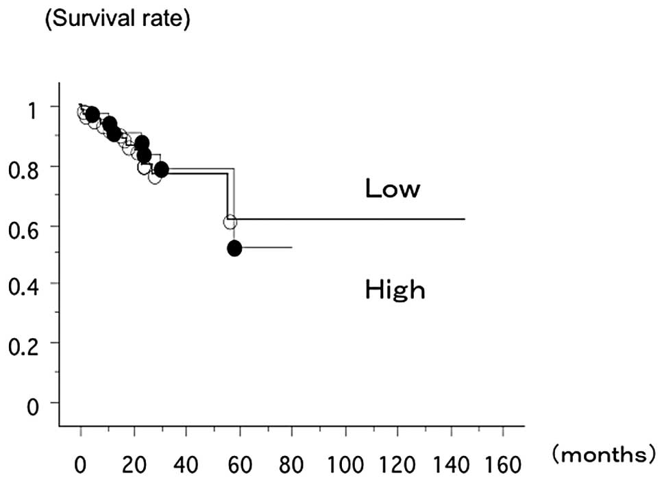
The overall survival rate of 110 lung cancer
patients from Nagoya City University, with a follow up until August
31 2011, was studied in reference to the ANGPTL2 gene
status. The survival rate of the patients with the T/N ratio of
ANGPTL2/β-actin mRNA level>1 (n=39, 7 had
succumbed) and the patient within the T/N ratio of
ANGPTL2/β-actin mRNA level <1 (n=71, 13 had
succumbed) was not significantly different (log-rank test,
P=0.7564; Fig. 1).
Discussion
The current study focused on chronic inflammation
(16) and the angiogenesis-related
gene, ANGPTL2 (17).
ANGPTL2 mRNA expression was correlated with lymph node
metastasis in surgically resected NSCLC using LightCycler.
The increased accumulation of reactive oxygen
species (ROS) and reactive nitrogen intermediates caused by chronic
inflammation may inactivate DNA repair enzymes (18). Previous studies have suggested that
the chronic inflammation status and ROS levels were positively
correlated with ANGPTL2 expression levels (11). ANGPTL2 expression was highly
correlated with the frequency of carcinogenesis in a chemically
induced skin squamous cell carcinoma in mice (11).
The ANGPTL2 gene has been reported to act as
a tumor suppressor in ovarian cancer (4). Decreased ANGPTL2 expression was
associated with a worse prognosis in stage I and II disease,
whereas ANGPLT2 positivity was significantly associated with a
worse survival rate in stage III and IV disease (4). Thus, ANGPTL2 may also act as a
molecule for tumor progression and metastasis in advanced stage
disease. In a xenograft mouse model, tumor cell-derived ANGPTL2
accelerated metastasis and shortened the survival rate, whereas
attenuating ANGPTL2 expression in tumor cells inhibited metastasis
and extended the survival rate (13). ANGPTL2 expression was high and
homogeneous in tumor cells within metastasized tumor sites
(13). In our analysis, ANGPTL2
expression correlated with metastasis but not tumor invasion. The
tumor cells expressing ANGPTL2 may exhibit high metastatic
potential.
A recent study has reported that the ANGPTL2
gene promoted vascular inflammation (19) and enhanced endothelial cell
migration (20). The expression of
cytokines, including tumor necrosis factor-α (21), interlukin-6 and interleukin-1β, were
found to be increased in ANGPTL2 transgenic mice (19). The ANGPTL2-null mice survived and
grew normally. Thus it is predicted that the suppression of ANGPTL2
signaling has few side-effects. Therefore the suppression of
ANGPLT2 signaling as a therapeutic strategy is likely to be
beneficial (20).
However, if the study were expanded, it would not be
possible to perform qPCR in the majority of patients since the
availability of tumor samples in the cohort was limited. The
majority of patients with advanced stage NSCLC had only small
tissue samples and the samples were mostly used for clinical
diagnosis, leaving limited residual samples for molecular
diagnosis. ANGPTLs have a signal sequence in the N-terminals for
protein secretion (20). ANGPTL2 is
predominantly secreted from adipose tissue and the heart (22). Cells transfected with expression
vectors encoding ANGPTLs secreted ANGPTLs proteins into culture
supernatants (23,24). ANGPTLs have been detected in the
systemic circulation (23,24), suggesting that the detection of
ANGPTL2 in blood samples may be useful for cancer patients. Thus,
the development and validation of strategies to improve effective
identification of the patient population with strategies
incorporating immunohistochemistry (IHC) or other techniques are
important and likely to assume a place in clinical practice. A
prospective study is required to compare the usage of RT-PCR, IHC
and detection from blood samples.
In summary, ANGPTL2 may drive metastasis and provide
a candidate for blockade of its function as a strategy to
antagonize the metastatic process. The result of RT-PCR was
validated in a limited number of patients.
Acknowledgements
The authors thank Mrs. Yuka Toda for
her excellent technical assistance. This study was supported by
Grants-in-Aid for Scientific Research, Japan Society for the
Promotion of Science (JSPS, Nos, 24592097, 23659674) and a grant
for cancer research of Program for developing the supporting system
for upgrading the education and research (2009) from the Ministry
of Education, Culture, Sports, Science and Technology of Japan.
References
|
1.
|
RJ GinsbergMK KrisG ArmstrongCancer of the
lung In: Principles and Practice of Oncology4th
editionLippincottPhiladelphia6736821993
|
|
2.
|
R SakataY FujiiH KuwanoThoracic and
cardiovascular surgery in Japan during 2009: annual report by the
Japanese Association for Thoracic SurgeryGen Thorac Cardiovasc
Surg59636667201110.1007/s11748-011-0838-522231795
|
|
3.
|
PE PostmusChemotherapy for non-small cell
lung cancer: the experience of the Lung Cancer Cooperative Group of
the European Organization for Research and Treatment of
CancerChest11328S31S199810.1378/chest.113.1_Supplement.28S9438687
|
|
4.
|
SI GrivennikovFR GretenM KarinImmunity,
inflammation, and
cancerCell140883899201010.1016/j.cell.2010.01.025
|
|
5.
|
C de MartelS FranceschiInfections and
cancer: established associations and new hypothesesCrit Rev Oncol
Hematol70183194200918805702
|
|
6.
|
H TakahashiI TakahashiM HonmaPrevalence of
metabolic syndrome in Japanese psoriasis patientsJ Dermatol
Sci57143144201010.1016/j.jdermsci.2009.11.00220005080
|
|
7.
|
C DestertV PetrilliR Van BruggenInnate
immune activation through Nalp3 inflammasome sensing of asbestos
and silicaScience320674677200810.1126/science.115699518403674
|
|
8.
|
P KantMA HullExcess body weight and
obesity-the link with gastrointestinal and hepatobilliary cancerNat
Rev Gastroenterol
Hepatol8224238201110.1038/nrgastro.2011.2321386810
|
|
9.
|
M TabataT KadomatsuS
FukuharaAngiopoietin-like protein 2 promotes chronic adipose tissue
inflammation and obesity-related systemic insulin resistanceCell
Metab10178188200910.1016/j.cmet.2009.08.00319723494
|
|
10.
|
M BiC NaczkiM KoritzinskyER
stress-regulated translation increases torelance to extreme hypoxia
and promotes tumor growthEMBO
J2434703481200510.1038/sj.emboj.760077716148948
|
|
11.
|
J AoiM EndoT KadomatsuAngiopoietin-like
protein 2 is an important facilitator of inflammatory
carcinogenesis and metastasisCancer
Res7175027512201110.1158/0008-5472.CAN-11-175822042794
|
|
12.
|
R KikuchiH TsudaK KozakiFrequent
inactivation of a putative tumor suppressor, angiopoietin-like
protein 2, in ovarian cancerCancer
Res6850675075200810.1158/0008-5472.CAN-08-006218593905
|
|
13.
|
M EndoM NakanoT KadomatsuTumor
cell-derived angiopoietin-like protein ANGPTL2 is a critical driver
of metastasisCancer
Res7217841794201210.1158/0008-5472.CAN-11-387822345152
|
|
14.
|
BA TeicherSearching for molecular targets
in sarcomaBiochem
Pharmacol84110201210.1016/j.bcp.2012.02.00922387046
|
|
15.
|
CT WittwerKM RirieRV AndrewThe
LightCycler: a microvolume multi sample fluorimeter with rapid
temperature controlBiotechniques2217618119978994665
|
|
16.
|
A OgataM EndoJ AoiThe role of
angiopoietin-like protein 2 in pathogenesis of
dermatomyositisBiochem Biophys Res
Commun418494499201210.1016/j.bbrc.2012.01.05222281496
|
|
17.
|
H TazumeK MiyakeZ TianMacropage-derived
angiopoietin-like protein 2 accelerates development of abdominal
aortic aneurysmArterioscler Thromb Vasc
Biol3214001409201210.1161/ATVBAHA.112.24786622556334
|
|
18.
|
F ColottaP AllavenaA SicaCancer-related
inflammation, the seventh hallmark of cancer: links to genetic
instabilityCarcinogenesis30107381200910.1093/carcin/bgp12719468060
|
|
19.
|
T KadomatsuM TabataY OikeAngiopoietin-like
proteins: emerging targets for treatment of obesity and related
metabolic diseaseFEBS
J278559564201110.1111/j.1742-4658.2010.07979.x21182596
|
|
20.
|
M TabataT KadomatsuS
FukuharaAngiopoietin-like protein 2 promotes chronic adipose tissue
inflammation and obesity-related systemic insulin resistanceCell
Metab10178188200910.1016/j.cmet.2009.08.00319723494
|
|
21.
|
JY ZhengJT ZouWZ WangTumor necrosis
factor-α a increases angiopoietin-like 2 gene expression by
activating Foxo1 in 3T3-L1 adipocytesMol Cell
Endocrinol3391201292011
|
|
22.
|
M KitazawaM NagonoKH
MasumotoAngiopoietin-like 2, a circadian gene, improves type 2
diabetes through potentiation of insulin
sensitivityEndocrinology15225582567201110.1210/en.2010-140721586562
|
|
23.
|
I KimSO MoonKN KohMolecular cloning,
expression, and characterization of angiopoietin-related protein.
Angiopoietin-related protein induces endothelial cell spoutingJ
Biol Chem2742652326528199910.1074/jbc.274.37.2652310473614
|
|
24.
|
Y OikeY ItoH MaekawaAngiopoietin-related
growth factor (AGF) promotes
angiogenesisBlood10337603765200410.1182/blood-2003-04-127214764539
|


