Introduction
Primary choriocarcinoma is an unusual neoplasm of
the posterior mediastinum, which frequently occurs in the gonads
and uterus, and less frequently in the extragonadal sites (1). There are several theories regarding
the development of primary mediastinal choriocarcinoma, including
abnormal germ migration (2) and
tumor cell distortion (3). However,
the precise genetic mechanism remains unknown. The majority of
cases are misdiagnosed by imaging examination and clinical features
prior to surgery (1). Chemotherapy,
radiotherapy and surgery are the main therapeutic strategies,
however, the effect of any method of treatment is poor. The current
study presents a case of primary choriocarcinoma of the posterior
mediastinum in a 40-year-old male and reviews the characteristics
of previously reported cases. The aim of this report is to explore
the diagnosis and treatment of primary choriocarcinoma of the
posterior mediastinum.
Case report
A 40-year-old male presented to the Department of
Thoracic Surgery (Medical College of Yangzhou University, Yangzhou,
China) with left chest pain, which had endured for two months.
Computed tomography (CT) showed a 4.9×5.2-cm mass in the posterior
mediastinum and enlargement of the mediastinal lymph nodes, while
no other lesions were identified in the thoracic cavity (Fig. 1). A CT-guided percutaneous
aspiration of the mediastinal mass led to a diagnosis of
adenocarcinoma. Due to the diagnosis of a malignant tumor, the
patient underwent surgical removal of the entire tumor. After
providing written informed consent, the patient underwent surgical
intervention on March 5, 2012. During surgery, the left upper lung
lobe was found to be invaded and therefore, the tumor and upper
lobe of the left lung was dissected. Simultaneously, lymph node
dissection was performed, which involved radical dissection of the
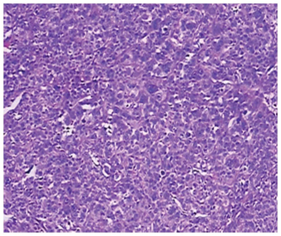
mediastinum. Following surgery, pathological examination revealed a
malignant tumor with a diffuse distribution of abnormal cells, with
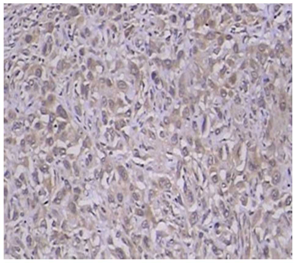
magnocellular, dark-stained and pleomorphic nuclei (Fig. 2). The immunohistochemical reaction
for β-human chorionic gonadotropin (hCG) was positive, and a fine,
granular and brown deposit was observed within the cytoplasm of
multinucleate giant cells in all areas of the sampled tumor
(Fig. 3). Histological examination
and immunohistochemical staining of the surgical specimens
confirmed choriocarcinoma with invasion of the upper lobe of the
left lung. The patient received combination chemotherapy with six
courses of etoposide plus cisplatin. The preoperative serum
concentration of β-hCG was 1.0 mIU/ml (normal range, <100
mIU/ml). The abdominal CT was unremarkable and the testes appeared
to be normal on the ultrasound. Following the six cycles of
chemotherapy, the patient recovered and was discharged. However,
one year post surgery and chemotherapy, the patient was followed up
and evidence of a recurrent tumor in the cerebrum was observed
(Fig. 4).
Discussion
Choriocarcinoma is a highly malignant trophoblastic
tumor, which commonly arises in pregnant females, with a few cases
arising in females and males also being reported. Early metastasis
may occur via blood circulation throughout the body, which results
in bleeding and necrosis of tissues and organs. Primary
choriocarcinoma of the posterior mediastinum in males is
particularly rare and treatment is considered to be ineffective. As
choriocarcinoma grow rapidly, early recurrence and metastasis are
frequently observed. Common symptoms include coughing, fever, chest
pain and breathing difficulties. The histological features of
choriocarcinoma are characterized by a biphasic cellular population
with extensive areas of hemorrhage and necrosis (4). There are two types of cells in this
tumor; cytotrophoblastic and syncytiotrophoblastic. The
cytotrophoblastic type is characterized by round-to-polygonal cells
with clear, polyhedral cytoplasm, round nuclei, sparse chromatin
and prominent nucleoli, while the syncytiotrophoblastic type is
composed of polynuclear giant cells with abundant eosinophilic
cytoplasm (4).
The immunohistochemical reaction for β-hCG is the
predominant method of diagnosis for choriocarcinoma. In addition,
developments in technology and advanced equipment, such as
18F-fluorodeoxyglucose positron emission tomography (FDG-PET), can
be used to assess the diagnosis and prognosis of this type of
tumor. Shukuya et al (5)
reported a case of an elderly male with primary mediastinal
choriocarcinoma, who underwent surgery and four cycles of
chemotherapy. Following treatment, specific sections of the
residual tumors on the CT images showed markedly reduced uptake,
while the other sections showed similar uptake in FDG-PET.
Therefore, FDG-PET may facilitate the evaluation of the viability
of tumors following treatment in cases of mediastinal
choriocarcinoma.
Numerous studies on extragenital choriocarcinomas
occurring in other organs, such as the lung (6), stomach (7) and colon (8), have previously been reported. To the
best of our knowledge, the first description of a posterior
mediastinal choriocarcinoma was by Yamashita et al (9) in 2002 of a 29-year-old male with
posterior mediastinal choriocarcinoma. The patient received two
cycles of combination chemotherapy. However, the patient succumbed
to the disease approximately three months following the diagnosis.
A search was conducted using the electronic database, PubMed, for
all the relevant literature between 2002 and 2013, involving
primary choriocarcinoma of the mediastinum. Since 2002, no similar
neoplasms occurring in the posterior mediastinum in males have been
reported.
The prognosis for patients with mediastinal
choriocarcinoma appears to be poor (9). Lynch and Blewitt (10) reported a case of a 26-year-old male
who succumbed to the disease two weeks following the diagnosis of
choriocarcinoma; the total duration of the illness had been six
weeks. Ramia et al (11)
presented the first case of a patient that presented with
appendicular metastasis from a choriocarcinoma of the mediastinum
who underwent an appendectomy and radical surgery of the
mediastinal tumor. However, during chemotherapy the patient showed
progressive clinical deterioration and succumbed to the disease
five months following the appendectomy. Certain studies have
reported long-term survival. Kathuria et al (12) reported a case of mediastinal
choriocarcinoma where the patient underwent surgery in association
with chemotherapy and at the two-year follow-up, no lymphadenopathy
was observed and the other physical observations were unremarkable.
In addition, Zheng (13) reported a
case of a young male with primary anterior mediastinal
choriocarcinoma that was accompanied by primary cystoid teratoma.
The patient underwent surgery in combination with chemotherapy and
survived for at least one year. According to the study by Moran
et al (4), no patient
survived for more than one year; however, one patient survived for
one year, although, a brain tumor, which was potentially
metastatic, was identified. The level of β-hCG is considered to be
a major factor affecting the prognosis of primary choriocarcinoma
of the posterior mediastinum. The treatment of patients with a
combination of surgery combined with chemotherapy is likely to
improve the long-term outcome (14).
In conclusion, this study presents an extremely rare
case of a patient with primary mediastinal choriocarcinoma, who
underwent successful surgery and chemotherapy. The patient
recovered and was discharged with no issues. Thus, the early
diagnosis and timely treatment of primary mediastinal
choriocarcinoma is key to improving the long-term outcome.
References
|
1
|
Shi H, Cao D, Wei L, Sun L and Guo A:
Primary choriocarcinoma of the liver: a clinicopathological study
of five cases in males. Virchows Arch. 456:65–70. 2010.
|
|
2
|
Sullivan LG: Primary choriocarcinoma of
the lung in a man. Arch Pathol Lab Med. 113:82–83. 1989.
|
|
3
|
Pushchak MJ and Farhi DC: Primary
choriocarcinoma of the lung. Arch Pathol Lab Med. 111:477–497.
1987.
|
|
4
|
Moran CA, Suster S and Koss MN: Primary
germ cell tumors of the mediastinum: III. Yolk sac tumor, embryonal
carcinoma, choriocarcinoma, and combined nonteratomatous germ cell
tumors of the mediastinum - a clinicopathologic and
immunohistochemical study of 64 cases. Cancer. 80:699–707.
1997.
|
|
5
|
Shukuya T, Hirano S, Takeda Y, Ito H,
Hurihata K, Sugiyama H, Kobayashi N and Kudo K: A case of primary
mediastinal choriocarcinoma in which FDG-PET was performed for the
evaluation of the treatment. Nihon Kokyuki Gakkai Zasshi.
45:174–179. 2007.
|
|
6
|
Ibi T, Hirai K, Bessho R, Kawamoto M,
Koizumi K and Shimizu K: Choriocarcinoma of the lung: report of a
case. Gen Thorac Cardiovasc Surg. 60:377–380. 2012.
|
|
7
|
Kobayashi A, Hasebe T, Endo Y, Sasaki S,
Konishi M, Sugito M, Kinoshita T, Saito N and Ochiai A: Primary
gastric choriocarcinoma: two case reports and a pooled analysis of
53 cases. Gastric Cancer. 8:178–185. 2005.
|
|
8
|
Le DT, Austin RC, Payne SN, Dworkin MJ and
Chappell ME: Choriocarcinoma of the colon: report of a case and
review of the literature. Dis Colon Rectum. 46:264–266. 2003.
|
|
9
|
Yamashita S, Ohyama C, Nakagawa H,
Takeuchi A, Watanabe M and Hoshi S: Primary choriocarcinoma in the
posterior mediastinum. J Urol. 167:17892002.
|
|
10
|
Lynch MJ and Blewitt GL: Choriocarcinoma
arising in the male mediastinum. Thorax. 8:157–161. 1953.
|
|
11
|
Ramia JM, Alcalde J, Dhimes P and Cubedo
R: Metastasis from choriocarcinoma of the mediastinum producing
acute appendicitis. Dig Dis Sci. 43:332–334. 1998.
|
|
12
|
Kathuria S and Jablokow VR: Primary
choriocarcinoma of mediastinum with immunohistochemical study and
review of the literature. J Surg Oncol. 34:39–42. 1987.
|
|
13
|
Zheng XM: Primary cystoid teratoma
companied with primary choriocarcinoma in anterior mediastinum: a
case report. J Diagn Pathol (Chin). 8:3712001.
|
|
14
|
Shen HH, Zhang GS and Xu F: Primary
choriocarcinoma in the anterior mediastinum in a man: a case report
and review of the literatures. Chin Med J (Engl). 117:1743–1745.
2004.
|


