Introduction
Central nervous system (CNS) melanoma is a rare type
of neoplasm. It is particularly rare in the brain compared with
other areas of the CNS and accounts for 0.07–0.17% of all
intracranial neoplasms. Depending on the behavior of the tumor,
brain melanomas are classified into the following categories: i)
Well-differentiated melanocytoma; ii) intermediate grade
melanocytoma; and iii) primary malignant melanoma (1). CNS primary malignant melanoma only
accounts for ~1% of all cases of melanoma (2). Primary CNS melanoma is generally
diagnosed following the exclusion of a primary cutaneous or
mucosal/retinal malignant melanoma, as differential histological
diagnosis between primary and metastatic origins is often difficult
(3). Primary leptomeningeal melanoma
is an extremely rare type of primary intracranial melanoma, with a
global incidence of 1 case per 20 million individuals (4). Primary leptomeningeal melanoma may be
classified pathologically into two types based on the behaviour of
the tumor; one type invades the pia mater diffusely and spreads
into the subarachnoid space, while the other causes nodular tumors
(5,6).
Depending on the clinical presentation, treatment options include
surgical resection, whole-brain radiation therapy, stereotactic
radiosurgery and systemic therapy (7). Furthermore, the clinical outcome of
patients with primary CNS melanoma is reported to be better than
that of patients with metastatic disease owing to the possibility
of long-term tumor control (8).
Although the lifespan of patients with solid tumors may be
prolonged with surgery following early diagnosis, generally,
malignant melanomas present poor prognoses, partly due to their
high rate of misdiagnosis. In the current study, 7 cases of
histopathologically diagnosed intracranial malignant melanoma are
presented.
Case report
Patients
The details of 7 patients with intracranial
malignant melanoma were collected from January 1996 to March 2013
in The Chinese PLA General Hospital (Beijing, China). Among these
patients, 3 were hospitalized in the department of neurology, and 4
in the department of neurosurgery. Primary and metastatic melanoma
was the diagnosis for 5 and 2 of the patients, respectively. The
cohort was formed by 1 female, who was affected with primary
melanoma, and 6 males. Among the male patients, 4 were affected
with primary melanoma, 1 with metastasis and 1 with post-surgical
recurrence. The average age of onset of disease was 37.5 years. The
lesions were identified in the following brain regions: The
cerebellum (1 case), left frontal lobe (1 case), foramen magnum (1
case) and cerebral pia mater (3 cases). In 2 cases, the melanoma
was accompanied by subarachnoid hemorrhage. Ethical approval was
obtained from the Medical Ethics Community of the Chinese PLA
General Hospital. In addition, written informed consent was
obtained from all patients.
Clinical features
The majority of the cases presented an insidious
onset, whilst 1 case had a sudden onset. The course of the disease
was 2–3 months. The primary symptoms were headache (2 cases), upper
digestive tract disturbances, including nausea and vomiting (2
cases), dizziness (2 cases) and neck and occipitalia pain (1 case).
During the course of the disease, other symptoms, including
nystagmus, diplopia, seizures and impaired vision and audition
appeared in some but not all cases. In 4 of the 7 patients, >1
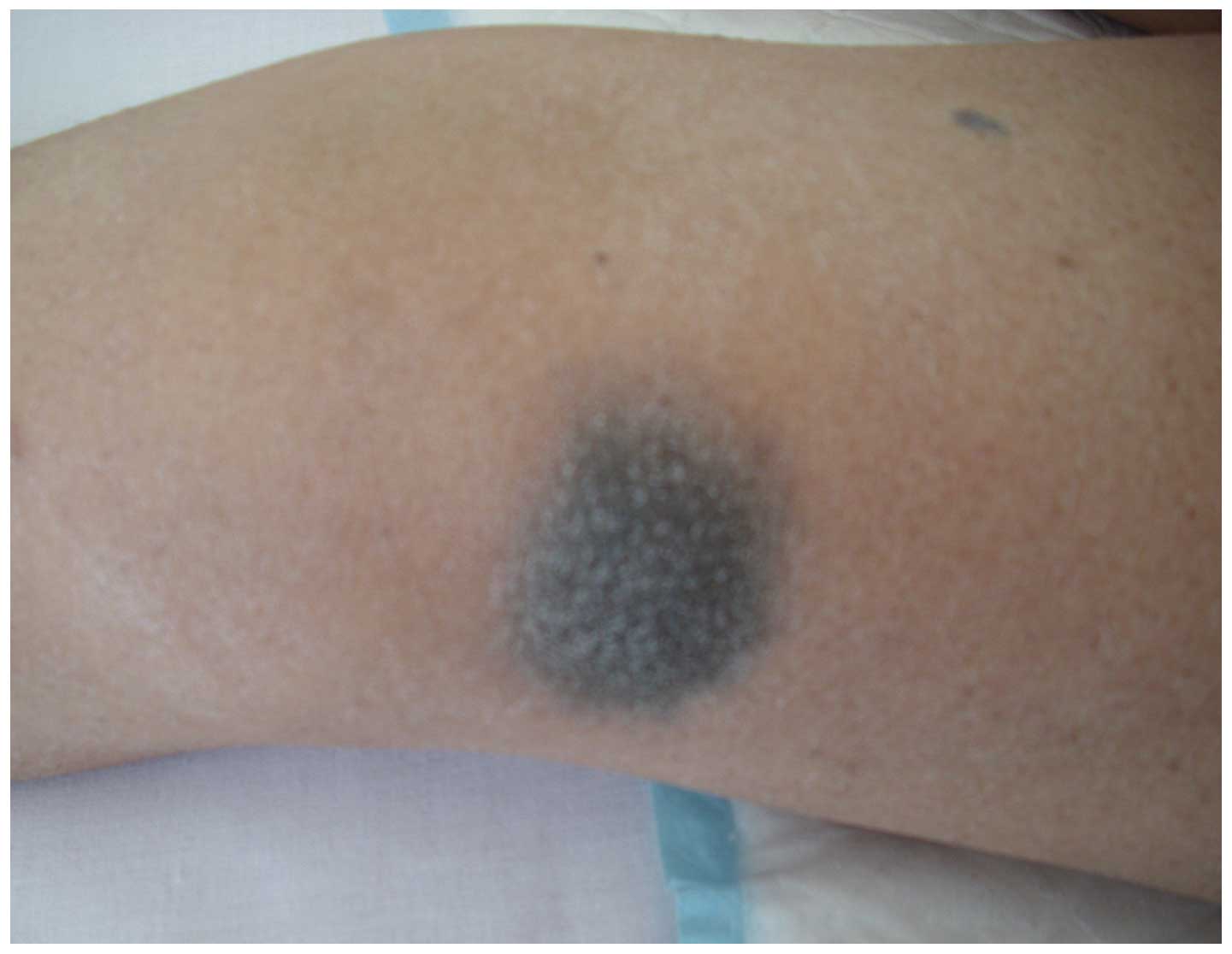
skin melanin pigmented nevus was observed (Fig. 1). The female patient had a history of
epilepsy from childhood.
Cerebrospinal fluid (CSF)
examination
In 3 cases, a solid tumor was not observed following
a lumbar puncture. The intracranial pressure was markedly elevated,
with all cases presenting intracranial pressure >300
mmH2O (normal range in adults, 100–180 mmH2O)
(9), and 1 case >600
mmH2O. In the CSF, the following parameters were
measured: Leukocyte numbers, 2–5×10⁶; glucose
concentration, 0.17–2.44 mmol/l; levels of chlorides were normal;
and protein concentration, 0.26–4.45 g/l. Cytological analysis of
the CSF identified heterocysts with a large cellular volume and
occasional double nuclei. Immunohistochemical analysis demonstrated
positive staining for the melanoma marker human melanoma black 45
(HMB-45) and the leukocyte antigen cluster of differentiation 20
(CD20).
Imageology examination
Table I summarizes the
results of the imageology performed on the 7 cases presented in the
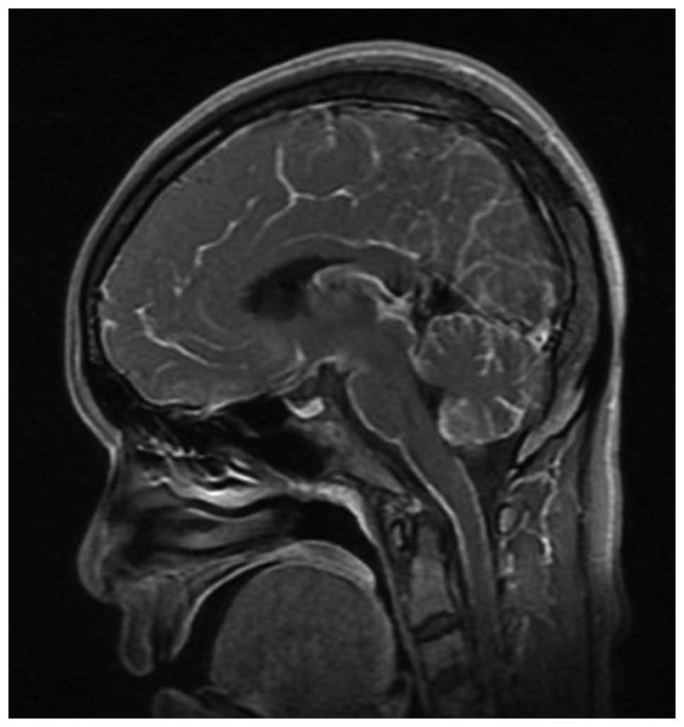
current report. Fig. 2 illustrates
the enhanced diffused meninges of case 6, observed by magnetic
resonance imaging (MRI) examination.
 | Table I.Imageology summary of 7 cases of
intracranial malignant melanoma. |
Table I.
Imageology summary of 7 cases of
intracranial malignant melanoma.
| Case | CT | Enhanced CT | MRI | Enhanced MRI | MRA | DSA | PET-CT |
|---|
| 1 | Left cerebellum
lateral high density | Uniformly
enhanced | NA | NA | NA | NA | NA |
| 2 | Multiple metastatic
tumor | NA | NA | NA | NA | NA | NA |
| 3 | Forehead lesion (with
hemorrhage) | NA | Equal T1 short T2
uneven sign surrounded by forehead | NA | - | NA | NA |
| 4 | NA | - | Foramen magnum and
right CPA lesion partially equal T1, short T2 | NA | NA | - | NA |
| 5 | Left temporal
hemorrhage with SAH | NA | Left temporal
multiple short T1, short T2 | NA | - | NA | NA |
| 6 | Left forehead sulcus
high density | NA | - | Leptomeninges
enhanced | - | - | - |
| 7 | SAH | Uniformly
enhanced | - | Leptomeninges
enhanced | NA | - | NA |
Pathology
During the surgery, the cerebral dura mater of the
patients appeared to be black in 4 cases. In addition, the
arachnoid appeared foliated and black in color in 4 cases. The
tumors were observed to be tenacious and enveloped, rich in blood
supply, and occasionally accompanied by obsolete hemorrhage. A
diagnosis of melanoma was confirmed by postoperative pathology in 4
of the 7 patients, with 2 primary and 2 metastasized. CSF cytology
confirmed the final diagnosis in the other 3 cases.
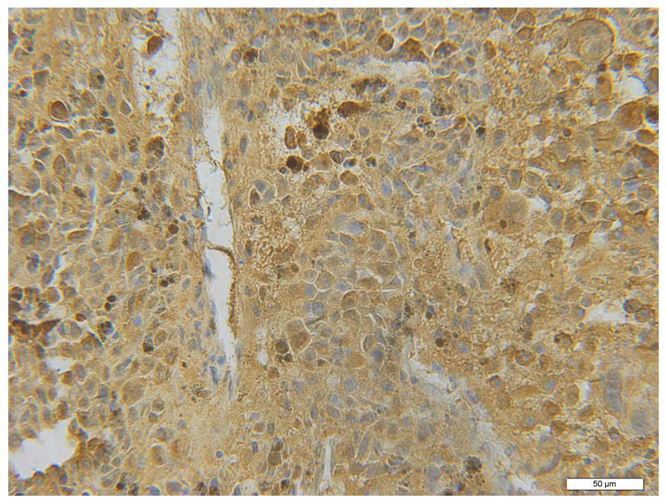
Immunohistochemical analysis demonstrated positive staining for
melan-A/S100 (Fig. 3); HMB-45
(Fig. 4); Ki-67 (50–75% positive
staining) (Fig. 5); and vimentin
(VIM) (Fig. 6). The staining for
epithelial membrane antigen (EMA) was negative in 2 cases.
Treatment outcome
The symptoms of headache, dizziness, radicular pain
and unsteady walking reduced in 4 cases following resection of the
tumor mass. Since the treatment for leptomeninges melanoma was not
effective, 1 patient discontinued the treatment, whereas 2 others
refused chemoradiation, and were discharged following dehydration
using mannitol.
Follow-up
Following surgery, 2 of the patients with metastatic
malignant melanoma deceased after 2 months, while 2 survived for
9–15 months. Among the patients with leptomeninges melanoma, 1
patient succumbed several days following discharge, 1 survived for
4 months subsequent to chemotherapy and cytokine-induced killer
cell therapy (four injections, every 2 days for 12 days), and 1 was
unable to be contacted.
Discussion
Melanoma originates from melanocytes, which are
present in the human skin, mucous membranes, leptomeninges,
cerebral parenchyma and uvea (2).
Intracranial melanoma is classified as primary or metastatic, and
primary CNS melanoma accounts for 1% of all melanomas (10). Primary melanoma derives from the
leptomeningeal melanocytes, which are abundant in the skull base of
the cervical cord (11,12). Primary meningeal melanoma are
classified into 2 distinct groups, including diffuse tumors of the
meninges, particularly around the base of the brain or spinal cord,
which classically presents as communicating hydrocephalus; and
discrete foci of melanoma, which are associated with diffuse
meningeal involvement (13). Arantes
et al (14) reported 13 cases
of primary malignant melanoma derived from the pineal body.
Pedersen et al (15)
established the first model of melanoma driven by the oncogenic
NRAS gene, which was expressed endogenously and did not require any
additional genetic engineering of the mice, or exposure to
carcinogens or tumor promoters. The authors reported 2 cases of
children with melanoma of the CNS that presented mutations in the
NRAS gene (15), and highlighted that
melanoma of the CNS is associated with mutations in the GNAQ and
GNA11 genes, whereas mutations in the NRAS gene are rare in adults
(16,17).
Lang (18) reported
that 90% of the cases of intracranial metastatic melanoma
investigated were induced by skin melanoma metastases, whereas 2%
arose from melanoma in the ocular region, and in 8%, the original
lesion was not identified. In the current report, 4 patients
presented with skin melanin pigmented nevus, 3 of which were
affected by primary leptomeninges melanoma, and in 1 case the
intracranial melanoma was secondary to the skin melanoma. Thus, in
patients with skin melanin pigmented nevus concomitant with
subarachnoid space hemorrhage, headache or intracranial
hypertension, a diagnosis of intracranial malignant melanoma should
be considered.
In the present study, the brain computed tomography
(CT) plain scans of intracranial melanomas commonly displayed
circular lesions with high densities, which were strengthened to
varying degrees or presented a ring-shaped enhancement. In
addition, the affected meninges displayed similar characteristics
to those observed in subarachnoid space hemorrhage. The assessment
of melanoma by MRI was complicated, since the melanin pigment is
paramagnetic, and the results of the MRI mainly depend on the
content of melanin pigment and the volume of blood in the tumor.
Previous MRI results of metastatic melanomas were reviewed by
Isiklar et al (19), who
classified them into 4 groups as follows, based on the putative
pattern: i) Melanotic pattern. Hyper, hypo and iso or hyperintense
compared with cortex T1-, T2- and proton density-weighted images,
respectively; ii) amelanotic pattern. Hypo or isointense compared
with cortex on T1-weighted images, and hyper or isointense compared
with cortex on T2-and proton density-weighted images; iii)
indeterminate or mixed pattern. MRI characteristics do not conform
to those of categories i) or ii); and iv) hematoma pattern. MRI
features exhibit hematoma characteristics. The results of a brain
MRI plain scan may be normal if solely the cerebral pia mater is
involved (19). In these cases,
enhancement MRI must be performed, since the meninges may be
enhanced (20). In the present study,
whole-body F-18 fluorodeoxyglucose (FDG) position emission
tomography (PET)/CT eliminated the possibility of an extracranial
origin for the melanoma lesions, and aided in the assessment of the
therapeutic response in the early phase of the disease. Post-hoc
PET/MR fusion images facilitated the correlation between the PET
and MR images, and revealed the hypermetabolic lesions more
accurately than the unenhanced PET/CT images did. Whole-body F-18
FDG PET/CT and post-hoc PET/MR images may aid clinicians in
determining the best therapeutic strategy for patients with primary
meningeal melanomatosis (21).
In the current report, 3 cases presented with
primary leptomeninges melanoma, which is rare; its clinical
manifestations, which are diverse and atypical, include the
following: Subacute onset, behavior of chronic meningeal
carcinomatosis, tendency to hemorrhage and repeated spontaneous
subarachnoid space hemorrhage. Examination of the CSF demonstrated
the following: Its color was pale yellow, due to repeated
hemorrhage; sugar content was low; and protein concentration was
>4.45 g/l (normal range, 0.15–0.45 g/l) (22). Further cytological examination of the
CSF indicated the presence of heterocysts, and immunohistochemical
analysis demonstrated positive staining for HMB-45, S100 and VIM.
The latter results are important in the differential diagnosis of
melanoma, particularly the positive staining for HMB-45, since this
is a specific biomarker for melanoma (23).
The pathogenesis of leptomeninges melanoma is
malignant, and its prognosis is poor (24). This is partly due to the lack of
expertise in radio and chemotherapy in a large number of hospitals.
However, it has not been reported that these treatments are capable
of prolonging the lifespan of patients with leptomeninges melanoma.
This disease should be differentiated from primary meninges
melanocytoma, which is benign, and from neurocutaneous melanosis,
which may cancerate to neurocutaneous melanoma. The patient of case
7 in the present study, who had a history of melanin pigmented
nevus, presented with sheet melanin pigmented nevus on her face and
body. The patient also suffered from epilepsy from a young age,
which was revealed by right-sided rotation of the head, concomitant
with lateral spasm. The brain CT scan of the patient demonstrated a
high-density shadow in the left frontal lobe and sulcus. Although
the presence of 5×10⁹ red cells/l in the CSF suggested a
subarachnoid space hemorrhage, no alterations were observed in the
6 repeated brain CT scans. In addition, brain enhanced MRI
suggested a focal enhanced lesion. Therefore, it was hypothesized
that the patient suffered from meninges melanocytosis that
cancerated to malignant, and developed a subarachnoid space
hemorrhage. This hypothesis was supported by the fact that the high
density of the brain CT scan corresponded to the presence of
melanocytic neoplastic cells in the subarachnoid space.
Furthermore, the cytological analysis of the CSF demonstrated the
presence of heterocysts, which also suggested canceration.
Meningeal melanocytomas and primary CNS melanomas
share a similar origin, and represent the benign and malignant ends
of the spectrum, respectively (25).
Malignant transformation of melanocytoma to malignant melanoma has
been previously reported (26–29).
Intracranial malignant melanoma often progresses, as it is not
eradicated easily and tends to recur. Primary intracranial melanoma
should be treated by thorough resection, followed by post-surgical
radio and chemotherapy (30).
Currently, the most common and effective chemotherapeutic agent is
dacarbazine, which presents an effectiveness rate of 16–20%, and is
administered intravenously following surgery or radiotherapy
(31). Previous studies have
demonstrated that stereotaxic radiosurgery (SRS) is able to treat
1–3 lesions, and is more effective at treating metastatic
intracranial melanoma than traditional whole-brain radiotherapy,
since a single exposure to a large dose of SRS may overcome
resistance to radiation, limit the damage to peripheral brain
tissue, and enable the control of focal transfer. SRS, on its own
or with the addition of whole-brain radiotherapy, may prolong the
lifespan of patients with metastatic intracranial melanoma, and
improve the control rate of CNS lesions (31,32).
Salpietro et al (33) reported that specific immunotherapy was
an important adjuvant method in the treatment of small residual
malignant melanoma lesions, and presented low toxicity. High doses
of IFNβ or IFNα-2b may improve disease control and prolong survival
time, but the dosage required is disputed and difficult to
tolerate. In 2008, Hunder et al (34) reported the bioremediation experienced
by a patient with multiple metastasis melanoma, who was in
remission for 2 years, but did not present with any metastatic
lesion in the brain.
In conclusion, the prognosis of intracranial
malignant melanoma is determined by the following factors: i) The
type of lesion; ii) the involvement of the leptomeninges; iii) the
extent of tumor excised; and iv) the molecular immunology borstel
number 1 (MIB 1) antibody index, which is the most relevant factor
for prognosis in this type of cancer (1).
References
|
1
|
Bhandari L, Alapatt J, Govindan A and
Sreekumar T: Primary cerebellopontine angle melanoma: A case report
and review. Turk Neurosurg. 22:469–474. 2012.PubMed/NCBI
|
|
2
|
GrecoCrasto S, Soffietti R, Bradac GB and
Boldorini R: Primitive cerebral melanoma: Case report and review of
the literature. Surg Neurol. 55:163–168. 2001. View Article : Google Scholar : PubMed/NCBI
|
|
3
|
Ferraresi V, Ciccarese M, Zeuli M and
Cognetti F: Central nervous system as exclusive site of disease in
patients with melanoma: Treatment and clinical outcome of two
cases. Melanoma Res. 15:467–469. 2005. View Article : Google Scholar : PubMed/NCBI
|
|
4
|
Helseth A, Helseth E and Unsgaard G:
Primary meningeal melanoma. Acta Oncol. 28:103–104. 1989.
View Article : Google Scholar : PubMed/NCBI
|
|
5
|
Allcutt D, Michowiz S, Weitzman S, et al:
Primary leptomeningeal melanoma: An unusually aggressive tumor in
childhood. Neurosurgery. 32:721–729. 1993. View Article : Google Scholar : PubMed/NCBI
|
|
6
|
Fish LA, Friedman DI and Sadun AA:
Progressive cranial polineuropathy caused by primary central
nervous system melanoma. J Clin Neuroophthalmol. 10:41–44.
1990.PubMed/NCBI
|
|
7
|
Tarhini AA and Agarwala SS: Management of
brain metastases in patients with melanoma. Curr Opin Oncol.
16:161–166. 2004. View Article : Google Scholar : PubMed/NCBI
|
|
8
|
Freudenstein D, Wagner A, Bornemann A,
Ernemann U, Bauer T and Duffner F: Primary melanocytic lesions of
the CNS: Report of five cases. Zentralbl Neurochir. 65:146–153.
2004. View Article : Google Scholar : PubMed/NCBI
|
|
9
|
Victor M and Ropper AH: Principles of
Neurology. 7th. McGraw-Hill; New York: pp. 142001
|
|
10
|
Shah I, Imran M, Akram R, Rafat S, Zia K
and Emaduddin M: Primary intracranial malignant melanoma. J Coll
Physicians Surg Pak. 23:157–159. 2013.PubMed/NCBI
|
|
11
|
Bydon A, Guttierez JA and Mahmood A:
Meningeal melanocytoma: An aggressive course for a benign tumor. J
Neurooncol. 64:259–263. 2003. View Article : Google Scholar : PubMed/NCBI
|
|
12
|
Rades D and Schild SE: Dose-response
relationship for fractionated irradiation in the treatment of
spinal meningeal melanocytomas: A review of the literature. J
Neurooncol. 77:311–314. 2006. View Article : Google Scholar : PubMed/NCBI
|
|
13
|
Gibson JB, Burrows D and Weir WP: Primary
melanoma of the meninges. J Pathol Bacteriol. 74:419–438. 1957.
View Article : Google Scholar
|
|
14
|
Arantes M, Castro AF, Romão H, Meireles P,
Garcia R, Honavar M, Vaz AR and Resende M: Primary pineal malignant
melanoma: Case report and literature review. Clin Neurol Neurosurg.
113:59–64. 2011. View Article : Google Scholar : PubMed/NCBI
|
|
15
|
Pedersen M, Küsters-Vandevelde HV, Viros
A, Groenen PJ, Sanchez-Laorden B, Gilhuis JH, van Engen-van
Grunsven IA, Renier W, Schieving J, Niculescu-Duvaz I, et al:
Primary melanoma of the CNS in children is driven by congenital
expression of oncogenic NRAS in melanocytes. Cancer Discov.
3:458–469. 2013. View Article : Google Scholar : PubMed/NCBI
|
|
16
|
Küsters-Vandevelde HV, Klaasen A, Küsters
B, Groenen PJ, van Engen-van Grunsven IA, van Dijk MR, Reifenberger
G, Wesseling P and Blokx WA: Activating mutations of the GNAQ gene:
A frequent event in primary melanocytic neoplasms of the central
nervous system. Acta Neuropathol. 119:317–323. 2010. View Article : Google Scholar : PubMed/NCBI
|
|
17
|
Murali R, Wiesner T, Rosenblum MK and
Bastian BC: GNAQ and GNA11 mutations in melanocytomas of the
central nervous system. Acta Neuropathol. 123:457–459. 2012.
View Article : Google Scholar : PubMed/NCBI
|
|
18
|
Lang PG: Current concepts in the
management of patients with melanoma. Am J Clin Dermatol.
3:401–426. 2002. View Article : Google Scholar : PubMed/NCBI
|
|
19
|
Isiklar I, Leeds NE, Fuller GN and Kumar
AJ: Intracranial metastatic melanoma: Correlation between MR
imaging characteristics and melanin content. AJR Am J Roentgenol.
165:1503–1512. 1995. View Article : Google Scholar : PubMed/NCBI
|
|
20
|
Chen CJ, Hsu YI, Ho YS, et al:
Intracranial meningeal melanocytoma: CT and MRI. Neuroradiology.
39:811–814. 1997. View Article : Google Scholar : PubMed/NCBI
|
|
21
|
Lee HJ, Ahn BC, Hwang SW, Cho SK, Kim HW,
Lee SW, Hwang JH and Lee J: F-18 fluorodeoxyglucose PET/CT and post
hoc PET/MRI in a case of primary meningeal melanomatosis. Korean J
Radiol. 14:343–349. 2013. View Article : Google Scholar : PubMed/NCBI
|
|
22
|
Williams HR, Oliver NS, Murphy F, Howell
M, Badman MK, Hillson RM and Thomas DJ: The role of the
biochemistry department in the diagnosis of pituitary apoplexy. Ann
Clin Biochem Mar. 41:162–165. 2004. View Article : Google Scholar
|
|
23
|
Liubinas SV, Maartens N and Drummond KJ:
Primary melanocytic neoplasms of the central nervous system. J Clin
Neurosci. 17:1227–1232. 2010. View Article : Google Scholar : PubMed/NCBI
|
|
24
|
Chu WC, Lee V, Chan YL, Shing MM, Chik KW,
Li CK and Ma KC: Neurocutaneous melanomatosis with a rapidly
deteriorating course. AJNR Am J Neuroradiol. 24:287–290.
2003.PubMed/NCBI
|
|
25
|
Faillace WJ, Okawara SH and McDonald JV:
Neurocutaneous melanosis with extensive intracerebral and spinal
cord involvement. Report of two cases. J Neurosurg. 61:782–785.
1984. View Article : Google Scholar : PubMed/NCBI
|
|
26
|
Rades D, Schild SE, Tatagiba M, Molina HA
and Alberti W: Therapy of meningeal melanocytomas. Cancer.
100:2442–2447. 2004. View Article : Google Scholar : PubMed/NCBI
|
|
27
|
Roser F, Nakamura M, Brandis A, Hans V,
Vorkapic P and Samii M: Transition from meningeal melanocytoma to
primary cerebral melanoma. Case report. J Neurosurg. 101:528–531.
2004. View Article : Google Scholar : PubMed/NCBI
|
|
28
|
Uozumi Y, Kawano T, Kawaguchi T, Kaneko Y,
Ooasa T, Ogasawara S, Yoshida H and Yoshida T: Malignant
transformation of meningeal melanocytoma: A case report. Brain
Tumor Pathol. 20:21–25. 2003. View Article : Google Scholar : PubMed/NCBI
|
|
29
|
Wang F, Li X, Chen L and Pu X: Malignant
transformation of spinal meningeal melanocytoma. Case report and
review of the literature. J Neurosurg Spine. 6:451–454. 2007.
View Article : Google Scholar : PubMed/NCBI
|
|
30
|
Kurita H, Segawa H, Shin M, Ueki K, Ichi
S, Sasaki T, Tago M and Kirino T: Radiosurgery of meningeal
melanocytoma. J Neurooncol. 46:57–61. 2000. View Article : Google Scholar : PubMed/NCBI
|
|
31
|
Douglas JG and Margolin K: The treatment
of brain metastases from malignant melanoma. Semin Oncol.
29:518–24. 2002. View Article : Google Scholar : PubMed/NCBI
|
|
32
|
Mingione V, Oliveira M, Prasad D, Steiner
M and Steiner L: Gamma surgery for melanoma metastases in the
brain. J Neurosurg. 96:544–551. 2002. View Article : Google Scholar : PubMed/NCBI
|
|
33
|
Salpietro FM, Alafaci C, Gervasio O, La
Rosa G, Baio A, Francolini DC, Batolo D and Tomasello F: Primary
cervical melanoma with brain metastases. Case report and review of
the literature. J Neurosurg. 89:659–666. 1998. View Article : Google Scholar : PubMed/NCBI
|
|
34
|
Hunder NN, Wallen H, Cao J, Hendricks DW,
Reilly JZ, Rodmyre R, Jungbluth A, Gnjatic S, Thompson JA and Yee
C: Treatment of metastatic melanoma with autologous CD4+ T cells
against NY-ESO-1. N Engl J Med. 358:2698–2703. 2008. View Article : Google Scholar : PubMed/NCBI
|


