Introduction
Urothelial carcinoma (UC) is the most common type of
cancer of the bladder and upper urinary tract (1,2). In the
United States, there are 76,960 incident cases and 31,540
cancer-associated mortalities caused by bladder cancer each year
(3). Furthermore, UC accounts for
~80,500 incident cases and 32,900 mortalities in China annually
(4). With an annual incidence of
0.69–0.73/100,000 per year, upper tract urothelial carcinoma (UTUC)
is less common compared with bladder urothelial carcinoma (BUC)
(5), but UTUC is often diagnosed
concurrently with BUC. Although the majority of tumors are
non-muscle-invasive when initially diagnosed, UC is characterized
by a high risk of recurrence and progression. The ‘Tumor, Node,
Metastases’ (TNM) classification system is widely used for staging
UC (6). Of patients with the stage
Ta-T2, ~60% relapse within three years of initial treatment
(7,8),
and multiple or recurrent lesions may indicate a lifelong risk of
progression (9). UC is associated
with a number of risk factors, with tobacco smoking considered to
be the most important (10,11). A previous study confirms that smoking
exposure confers risk to the recurrence rate for smokers and
non-smokers (12).
Diagnosis and surveillance of UC generally involve
invasive procedures. Physical examination typically finds a limited
number of clues, whilst in rare and advanced cases masses may be
palpable. Cystoscopy, ureteroscopy and biopsy remain the standard
diagnostic tools for UC. However, endoscopy is usually associated
with complications including hemorrhage, infection and perforation,
and has serious limitations in certain patients with anatomical
abnormalities or history of urinary tract reconstruction, for
example urinary diversion or ureteroileal anastomosis (13).
Urine-based diagnostic tests are non-invasive and
convenient, but inaccurate. Urine cytology for exfoliated cells
exhibits excellent specificity, but the overall sensitivity is low,
particularly for low-grade tumors, ranging between 4–31% (13,14).
Numerous biomarkers for tumor-associated proteins including human
complement factor H-related protein and nuclear matrix protein 22,
and the diagnostic test ImmunoCyt have been introduced to achieve a
higher sensitivity. However, as these tests are immunology-based
approaches, these markers are susceptible to urinary tract
infections and intravesical therapies, resulting in a trade-off
between sensitivity and specificity (15).
Fluorescence in situ hybridization (FISH) is
a technique that allows the visualization of genetic aberrations in
many types of tumor. In UC, the most prominent types of genetic
changes are the loss of the p16 gene at 9p21 and aneuploidies of
chromosomes 3, 7 and 17. Therefore, multitargeted multicolor FISH
probes that are specific for these aberrations have been developed
and tested for the diagnosis of UC (16).
Over the previous decade, several studies have
evaluated the diagnostic value of FISH in UC, but the clinical
value and cost-efficiency remains incompletely characterized
(17,18). As important data has arisen from
case-control designs (19–21) or focused on BUC or UTUC exclusively
(22–25), the present study was designed to
assess the diagnostic value of FISH for detecting UC prospectively,
and to evaluate the utility of FISH in association with cytology as
a non-invasive diagnostic technique.
Patients and methods
Between November 2010 and June 2012, patients with
suspected UC in West China Hospital, Sichuan University (Chengdu,
China) were enrolled consecutively in a prospective,
cross-sectional study. The majority of patients presented with
gross hematuria, unexplained hydroureterosis or hydronephrosis or
abnormal imaging findings in the urinary tract, for example; masses
or filling defects.
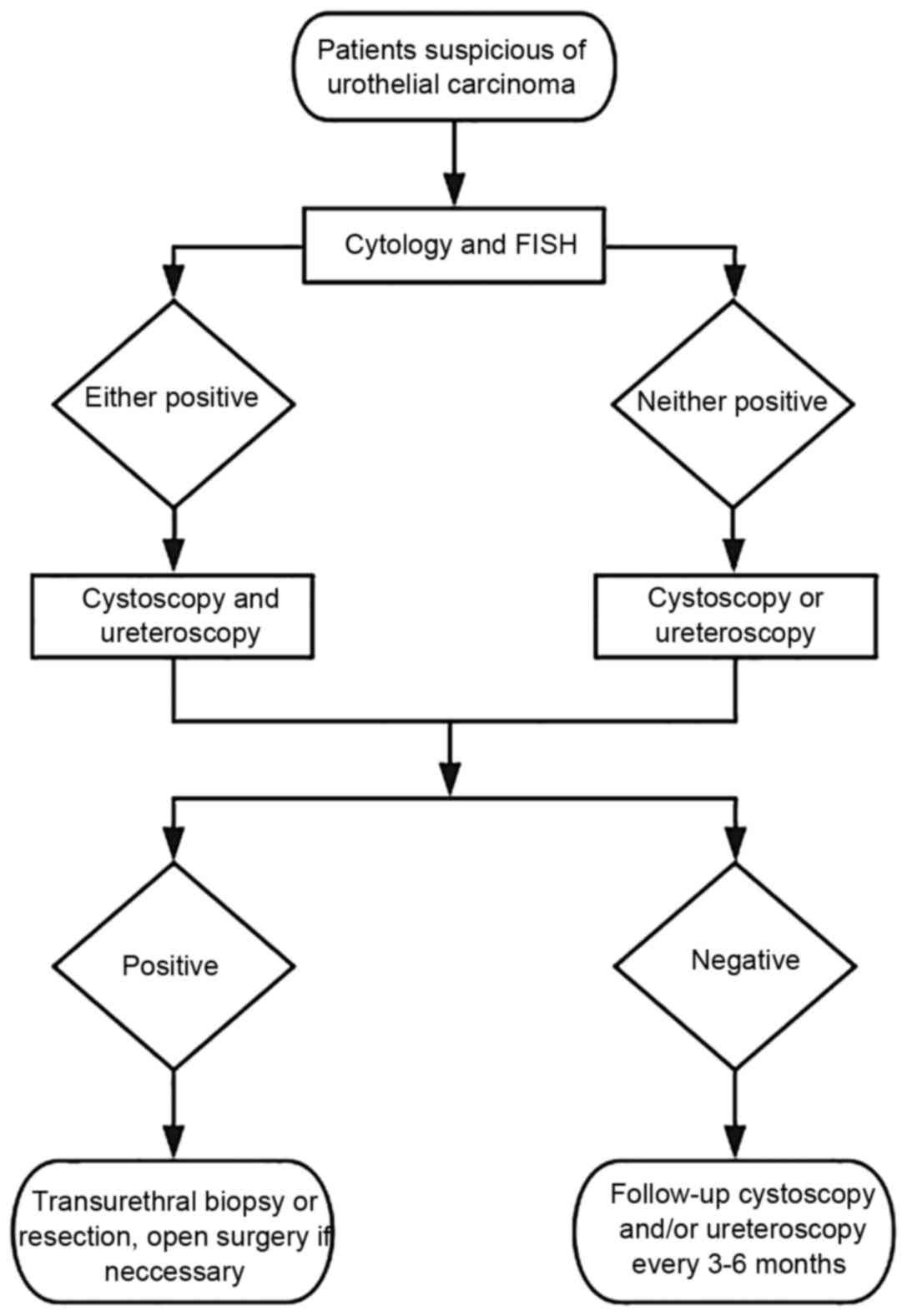
Once recruited, patient samples underwent urine
cytology and FISH assays, followed by cystoscopy and/or
ureteroscopy within one week of urine collection. In patients with
negative cystoscopy but either positive cytology or FISH,
ureteroscopy was mandatory. If indicated, a biopsy or resection was
performed. Patients negative for endoscopy in the initial
assessment underwent a follow-up endoscopy every 3–6 months. The
diagnostic procedures are illustrated in Fig. 1. The present study was approved by the
West China Hospital (Chengdu, China) Ethics Committee and written
informed consent was obtained from each patient.
Prior to urine collection, each patient was
instructed to empty the bladder completely, then drink 500 ml
water. After 1–2 h, two urine specimens of voided midstream urine
were collected, ≥100 ml per sample, and analyzed within one
hour.
For cytology, the urine specimens were centrifuged
at 600 × g for 10 min at room temperature, and the sediment
fractions were sectioned for Papanicolaou staining and cytological
diagnosis. Atypical or equivocate interpretations were reported as
non-diagnostic.
The urine FISH assays were conducted with
multitargeted, multicolor and commercially available kits (Bladder
Cancer kits, Beijing GP Medical Technologies, Ltd., Beijing, China)
according to the protocol of the manufacturer. The slides were
prepared using the same procedure as the cytological analysis, and
were denaturized and hybridized with two pairs of probes:
Chromosome enumeration probe (CEP) 3/7 for chromosomes 3 and 7, and
glucagon-like receptor cyclin-dependent kinase inhibitor 2A
(p16)/CEP17 for the locus 9p21 and chromosome 17, respectively.
Subsequent to washing of the slides, the nuclei were counterstained
with 4,6-diamidino-2-phenylindole and were analyzed using a
fluorescence microscope. A total of 100 unoverlapped cells were
evaluated consecutively. An analyzable reading required a minimum
80% hybridization rate.
One key step was to determine the cutoff for a
positive reading. The fluorescent signals in fresh voided urine
specimens collected from 20 healthy control individuals in West
China Hospital were analyzed, and the cutoff was determined as the
mean percentage in healthy controls + 3x standard deviation. For
polysomy of CEP3, CEP7 and CEP17, the cutoff was 7, 8 and 8%
respectively. For p16, >10% of the counted nuclei with either
homozygous or hemizygous loss were considered abnormal. A positive
FISH result was given if any single cutoff was exceeded.
To determine the sensitivity and specificity
profiles, endoscopy and subsequent histological diagnosis were used
as the reference standard. The biopsy was performed by
transurethral resection or open surgery, and the tissues were
sectioned by routine procedures. The tumor was staged and graded in
accordance with the TNM classification (6) and the World Health Organization 2004
classification (26).
All tests were examined by two pathologists
independently. The consensus was made and reported, and a third
senior pathologist was consulted if required. All reviewers were
blinded to the results of other tests and the clinical records
except for suspicion of UC.
In order to define the difference in the diagnostic
values between FISH and cytology in a sufficient number of
patients, a binomial McNemar test for paired data and χ2
test for unpaired data were used. The correct sample size was
estimated following a method for paired diagnostic tests (27,28). The
probable difference in the sensitivity and the specificity between
the two assays was assumed based on a previous meta-analysis
(14), and an 80% power was chosen to
identify the disagreement. P<0.05 was considered to indicate a
statistically significant difference. All statistical analyses were
performed using IBM SPSS Statistics version 23.0.
Results
Demographic and clinical
characteristics of the patients
To compare the diagnostic value between FISH and
cytology, a minimum of 65 patients were required and the present
study recruited 123. A total of 119 patients qualified for the
final analysis, and 4 were excluded due to loss to follow-up.
The demographic and clinical characteristics of the
qualified patients are listed in Table
I. A total of 16 patients exhibited histologically confirmed
BUC with at least one transurethral resection prior to enrollment.
A total of 13 had been diagnosed with other urinary disorders,
including benign prostate hyperplasia and renal cysts. Only 1
patient exhibited a previous lateral nephrectomy due to renal
tuberculosis. Gross hematuria was exhibited in 103 patients, and
the most common findings on imaging were masses, followed by
filling defects in the upper urinary tracts. As 33.6% of the
patients in the present study were associated with a current or
prior smoking history, the FISH results, listed by smoking status
in Table II, demonstrate that
tobacco smoking contributed significantly to UC prevalence
(P<0.05). However, the correlation between smoking status and
FISH sensitivity was not established in the present study
(P>0.05).
 | Table I.Demographic and clinical
characteristics of 119 patients with suspected urothelial
carcinoma. |
Table I.
Demographic and clinical
characteristics of 119 patients with suspected urothelial
carcinoma.
| Characteristic | Value |
|---|
| Age, years |
|
| Mean ±
standard deviation | 60.4±17 |
|
Range | 25–89 |
| Female sex, n
(%) | 31 (26.1) |
| Current or prior
tobacco smoking, n (%) | 40 (33.6) |
| History of urinary
disorders, n (%) |
|
|
Urothelial carcinoma | 16 (13.4) |
| Benign
disorders | 13 (10.9) |
| Gross hematuria, n
(%) | 103 (86.6) |
| Positive findings
in ultrasound, | 76 (63.9) |
| IVU, CTU or MRU
imaging, n (%) |
|
 | Table II.Prevalence of urothelial carcinoma
and the sensitivity of FISH by smoking status. |
Table II.
Prevalence of urothelial carcinoma
and the sensitivity of FISH by smoking status.
|
|
|
|
|
|
Urothelial
carcinoma-positive cases detected with FISH |
|---|
|
|
|
|
|
|
|
|---|
| Characteristic | Urothelial
carcinoma confirmed by histology |
| Overall Low-grade
carcinoma | High-grade
carcinoma |
|
|
|
|
|---|
|
|
|
|
|
|
|---|
| Smoking status | Total, n | +, n | -, n | Prevalence, % | +, n | -, n | Sen, % | Total, n | +, n | -, n | Sen, % | Total, n | +, n | -, n | Sen, % |
|---|
| Current or previous
smoker | 40 | 31 | 9 | 77.5 | 24 | 7 | 77.4 | 17 | 14 | 3 | 82.4 | 14 | 10 | 4 | 71.4 |
| ≥30 pack-years | 20 | 18 | 2 | 90.0 | 14 | 4 | 77.8 | 13 | 10 | 3 | 76.9 | 5 | 4 | 1 | 80.0 |
| Non-smoker | 79 | 42 | 37 | 53.2 | 34 | 8 | 82.9 | 16 | 11 | 5 | 68.9 | 26 | 23 | 3 | 88.5 |
The final histological diagnoses of the 73 confirmed
UC are summarized in Table III. A
total of 5 cases of renal cell carcinoma were identified, and the
remaining 41 patients were tumor-free throughout the present study.
The patients were followed up for an average period of 18.7 months,
ranging from 12–30.
 | Table III.Tumor characteristics of 73 patients
diagnosed with urothelial carcinoma by histological diagnosis. |
Table III.
Tumor characteristics of 73 patients
diagnosed with urothelial carcinoma by histological diagnosis.
| Characteristic | Value, n (%) |
|---|
| Location of
lesion |
|
| Urinary
bladder | 52 (71.2) |
| Upper
urinary tract | 19 (26) |
|
Both | 2 (2.7) |
| Tumor grade |
|
|
Low | 33 (45.2) |
|
High | 40 (54.8) |
| Tumor stage |
|
|
pTa | 30 (41.1) |
|
pTis | 1 (1.4) |
|
pT1 | 22 (30.1) |
|
pT2 | 9 (12.3) |
|
pT3 | 6 (8.2) |
|
pT4 | 5 (6.8) |
Diagnostic values of cytology and
FISH
To compare the performance of cytology, FISH and the
two as simultaneous tests for diagnosing UC, the sensitivities,
specificities and predictive values were calculated and are listed
in Table IV. Urine cytology was
positive for malignancy in 24 of 73 patients and negative in 49. A
total of 9 samples were non-diagnostic, due to atypical or
equivocal interpretations, and were considered negative in the
analysis. FISH was positive in 59 and negative in 14 UC cases. A
total of 4 samples that lacked sufficient cells were considered
negative, and comprised 2 patients with BUC, 1 patient with kidney
cancer, and 1 true negative. Additional analysis of the benefit of
combining cytology and FISH as a simultaneous test revealed that
the increase in sensitivity and negative predictive value (NPV) was
1.4%. In contrast, combining the tests decreased the specificity
and positive predictive value (PPV) by 10.9 and 7.7%,
respectively.
 | Table IV.Sensitivity, specificity and
predictive value of cytology, FISH and both as a simultaneous test
for diagnosis of UC in 119 patients. |
Table IV.
Sensitivity, specificity and
predictive value of cytology, FISH and both as a simultaneous test
for diagnosis of UC in 119 patients.
|
| +, n | -, n | Total, n | Sen % | Spe % | PPV % | NPV % |
|---|
| Cytology |
|
|
| 32.9 | 100 | 100 | 48.4 |
| + | 24 | 0 | 24 |
| − | 49 | 46 | 95 |
| FISH |
|
|
| 80.8 | 89.1 | 92.2 | 74.5 |
| + | 59 | 5 | 64 |
| − | 14 | 41 | 55 |
| Simultaneous |
|
|
| 82.2 | 89.1 | 92.3 | 75.9 |
| + | 60 | 5 | 65 |
| − | 13 | 41 | 54 |
| Total | 73 | 46 | 119 |
|
|
|
|
As the major criticism on urine FISH and cytology is
the lack of sensitivity for low-grade and early stage UC, the data
were examined by tumor grade, stage and location. As demonstrated
in Table V, cytology was considerably
less effective for highly differentiated or non-muscle-invasive
tumors. Out of 33 low-grade UC, only 4 were identified by cytology.
Conversely, the sensitivity of FISH was stable for tumor grade and
stage.
 | Table V.Sensitivity of FISH and cytology for
73 urothelial carcinoma by tumor grade, stage and location. |
Table V.
Sensitivity of FISH and cytology for
73 urothelial carcinoma by tumor grade, stage and location.
| Characteristic | n | Cytology
sensitivity, % | FISH sensitivity,
% |
|---|
| Grade |
|
|
|
|
Low | 33 | 12.1 | 75.8 |
|
High | 40 | 50 | 85 |
| Stage |
|
|
|
| Ta,
Tis, T1 | 53 | 28.3 | 81.1 |
| T2, T3,
T4 | 20 | 45 | 80 |
| Location |
|
|
|
|
Bladder | 52 | 32.7 | 84.6 |
| Upper
urinary tract | 19 | 31.6 | 73.7 |
|
Both | 2 | 50 | 100 |
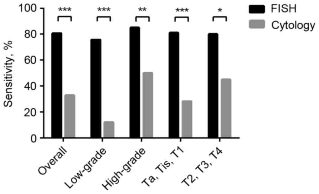
Compared with cytology, FISH exhibited higher
sensitivity for detecting UC, as illustrated in Fig. 2. The discrepancy was particularly
conspicuous between low-grade and early stage UC, as the
sensitivity for low grade FISH was 75.8% vs. 12.1% for low grade
cytology, and the sensitivity for early stage FISH was 81.1% vs.
28.3% for early stage cytology, respectively (FISH vs. cytology,
P<0.001). In 19 patients with UTUC without concomitant BUC, FISH
exhibited significantly higher sensitivity compared with cytology
(P<0.01). When determining specificity, 5 patients with negative
histology were diagnosed with UC by FISH, whereas cytology
exhibited no false positives. However, the difference was not
significant (P=0.063).
Additionally, of the 9 cytologically non-diagnostic
patients, 8 were positive using FISH, including all 6 patients with
UC in this group. There was 1 false positive patient who exhibited
a history of UC prior to the present study, and the other was
diagnosed with kidney cancer. The sensitivity and specificity of
FISH when used to diagnose cytologically equivocal samples were 100
and 33.3% respectively.
Discussion
The purpose of the present study was to evaluate the
diagnostic value of FISH for diagnosing UC, and to assess the
clinical consequences of FISH-based diagnostics in association with
cytology. In a cohort of patients with suspected UC, it was
revealed that FISH exhibited a higher sensitivity, PPV and NPV
compared with cytology across tumor grade, stage and location.
Cytology is associated with an excellent specificity, but the
sensitivity is greatly reduced in low-grade UC. The utility of the
two methods as a simultaneous test was investigated, but the data
suggest that the combination did not improve the accuracy of
FISH.
FISH is effective, but not always efficient, in the
diagnosis of UC. The advantage of FISH in sensitivity over cytology
is reduced subsequent to the exclusion of superficial cancer from
the analysis, indicating that tumor grade and stage may affect the
accuracy (29). Although tobacco
smoking is suggested to affect the wider prevalence of UC, and
there is a higher detection rate in heavy smokers (30), the present study was only able to
validate the former.
The data of the present study suggest that the high
sensitivity of FISH across UC stage and grade is not coincidental
(31). Potential explanations for the
discrepancy between the results of the present study and other
observations may be attributed to selection and observer bias.
However, the approaches to the urine samples of the present study
should also be considered. Firstly, only fresh voided urine
specimens were examined. In addition, the pathology laboratory
performing the tests was in the vicinity of the outpatient building
and urology unit of the West China Hospital (Chengdu, China),
allowing the specimens to be processed and prepared within short
time periods, to reduce degeneration of the samples as much as
possible. All FISH assays were performed on independent samples
rather than the remains or the same slides from cytology to
minimize the interference from previous manipulations. Lastly, the
hybridization rate was considered a key indicator of the quality
control, for example; samples that failed to reach an 80%
hybridization rate were retested until the criteria were met. As
only 4/119, 3.4%, FISH results were deemed ‘uninformative’, careful
handling and strict standard of quality control may improve the
sensitivity of FISH assay for obscure abnormalities in urine
specimens. Another explanation for the observed discrepancy between
the present study and previous studies may be the heterogeneity in
the diagnostic criteria across different studies.
As the first commercially available FISH kit for
diagnosing bladder cancer, UroVysion Bladder Cancer Kit (Abbott
Molecular, Inc., USA), was approved by the United States Food and
Drug Administration in 2001, the UroVysion criteria has been widely
used in clinical practice and studies, with continuous adaptions.
Bubendorf et al (32)
suggested that the detection of ≤4 tetrasomic cells in each sample
should be considered normal in order to improve diagnostic value.
Concurrently, Reynolds et al (33) demonstrated that FISH had improved
sensitivity vs. cytology, but decreased specificity due to a
greater number of false-positive results. Considering the lack of
validation of the UroVysion criteria in the Chinese population, the
threshold was determined by performing FISH assays on samples from
healthy donors. Additionally, Huang et al (22) reported similar results in BUC with
this approach, and a direct comparison with the UroVysion criteria
established by Cui et al (34)
demonstrated that the donor-derived criteria is more effective.
From the literature, FISH possesses a substantially
higher sensitivity for UTUC (25,35–37),
whereas cytology is only positive in 20% patients without
concurrent bladder UC, including invasive or high-grade UTUC
(38,39). As UTUC shares identical karyotypic
profiles with BUC (40), it is
hypothesized that FISH serves an effective role for detecting UTUC.
However, the generally small sample size in previous studies, owing
to the low incidence of UTUC, undermines their strength of
evidence.
Another application of FISH is to supplement
cytological analysis. Previous studies have demonstrated that FISH
possesses a remarkably high accuracy in patients with suspicious,
atypical or negative cytology, and the potential to predict UC
development-patients with atypical cytology and false positive FISH
at the initial assessment commonly develop UC within 15–22 months
(41,42), and a preceding positive FISH result is
associated with tumor relapse in 86% of UC surveillance cases,
including all high-grade recurrences (43). These data suggest that ‘false
positive’ FISH reports should not be ignored, as they may indicate
underlying tumorigenesis.
However, the FISH assay possesses limitations. This
approach is more complicated and expensive compared with cytology,
requiring a fluorescence microscope with commercial kits and
specially trained laboratory staffs to visualize and interpret the
results. Considering these resources, performing FISH assays
subsequent to positive cytology results is not realistic, as
positive cytology results are reliable due to the high specificity
of cytology for UC. FISH is also not cost-effective when low
incidences of UC are expected, due to the reduced chance of
diagnosing tumors. In particular, the performance of FISH is
suboptimal when used as a reflex test concomitantly with cytology
and UC screening in high risk populations (24,44).
Given these arguments, refinements are required to
maximize the potential of urine FISH assays. Bladder and upper
urinary tract washing may increase the probability of the detection
of UC, as low-grade tumors are prone to shed fewer neoplastic cells
into the urinary tract compared with advanced tumors, thus
remaining undetected by urine-based tests (45). Adequate training and calibrated
protocol for the performance of urine FISH assays may reduce the
misinterpretation caused by sample handling and improper
criteria.
Notably, the present study is based on a cohort from
a single institution, which may potentially suffer from limited
patient numbers and selection bias. Notwithstanding the
limitations, these data support the importance of urine FISH assays
for diagnosing bladder and upper urinary tract UC. They also
underscore the value of these assays in patients with low-grade and
early stage tumors, in which cytology-based methods are
unreliable.
Acknowledgements
The present study was supported/partially supported
by Clinical Application Fund for Molecular Biology of the Chinese
Medical Association (grant no. CAMB032010) and China Scholarship
Council. The authors thank our colleagues from Department of
Pathology who provided insight and expertise that greatly assisted
the study.
References
|
1
|
Babjuk M, Oosterlinck W, Sylvester R,
Kaasinen E, Böhle A, Palou-Redorta J and Rouprêt M: European
Association of Urology (EAU): EAU guidelines on non-muscle-invasive
urothelial carcinoma of the bladder, the 2011 update. Eur Urol.
59:997–1008. 2011. View Article : Google Scholar : PubMed/NCBI
|
|
2
|
Ries LAG, Young JL, Keel GE, Eisner MP,
Lin YD and Horner MJ: SEER survival monograph: Cancer survival
among adults: U.S. seer program, 1988–2001, patient and tumor
characteristicsNational Cancer Institute, SEER Program. NIH Pub.
No. 07-6215. Bethesda, MD: 2007
|
|
3
|
Siegel RL, Miller KD and Jemal A: Cancer
statistics, 2016. CA Cancer J Clin. 66:7–30. 2016. View Article : Google Scholar : PubMed/NCBI
|
|
4
|
Chen W, Zheng R, Baade PD, Zhang S, Zeng
H, Bray F, Jemal A, Yu QX and Jie H: Cancer statistics in China,
2015. CA Cancer J Clin. 66:115–132. 2016. View Article : Google Scholar : PubMed/NCBI
|
|
5
|
Munoz JJ and Ellison LM: Upper tract
urothelial neoplasms: Incidence and survival during the last 2
decades. J Urol. 164:1523–1525. 2000. View Article : Google Scholar : PubMed/NCBI
|
|
6
|
Sobin LH, Gospodariwicz MK and Wittekind
C: TNM classification of malignant tumours. UICC International
Union Against Cancer (7th). 262–265. 2009.
|
|
7
|
Lutzeyer W, Rübben H and Dahm H:
Prognostic parameters in superficial bladder cancer: An analysis of
315 cases. J Urol. 127:250–252. 1982.PubMed/NCBI
|
|
8
|
Heney NM, Ahmed S, Flanagan MJ, Frable W,
Corder MP, Hafermann MD and Hawkins IR: Superficial bladder cancer:
Progression and recurrence. J Urol. 130:1083–1086. 1983.PubMed/NCBI
|
|
9
|
Herr HW: Natural history of superficial
bladder tumors: 10- to 20-year follow-up of treated patients. World
J Urol. 15:84–88. 1997. View Article : Google Scholar : PubMed/NCBI
|
|
10
|
Freedman ND, Silverman DT, Hollenbeck AR,
Schatzkin A and Abnet CC: Association between smoking and risk of
bladder cancer among men and women. JAMA. 306:737–745. 2011.
View Article : Google Scholar : PubMed/NCBI
|
|
11
|
Zeegers MP, Tan FE, Dorant E and van Den
Brandt PA: The impact of characteristics of cigarette smoking on
urinary tract cancer risk: A meta-analysis of epidemiologic
studies. Cancer. 89:630–639. 2000. View Article : Google Scholar : PubMed/NCBI
|
|
12
|
Crivelli JJ, Xylinas E, Kluth LA, Rieken
M, Rink M and Shariat SF: Effect of smoking on outcomes of
urothelial carcinoma: A systematic review of the literature. Eur
Urol. 65:742–754. 2014. View Article : Google Scholar : PubMed/NCBI
|
|
13
|
Sanderson KM and Rouprêt M: Upper urinary
tract tumour after radical cystectomy for transitional cell
carcinoma of the bladder: An update on the risk factors,
surveillance regimens and treatments. BJU Int. 100:11–16. 2007.
View Article : Google Scholar : PubMed/NCBI
|
|
14
|
Hajdinjak T: Urovysion FISH test for
detecting urothelial cancers: Meta-analysis of diagnostic accuracy
and comparison with urinary cytology testing. Urol Oncol.
26:646–651. 2008. View Article : Google Scholar : PubMed/NCBI
|
|
15
|
Lotan Y and Roehrborn CG: Sensitivity and
specificity of commonly available bladder tumor markers versus
cytology: Results of a comprehensive literature review and
meta-analyses. Urology. 61:109–118. 2003. View Article : Google Scholar : PubMed/NCBI
|
|
16
|
Sokolova IA, Halling KC, Jenkins RB,
Burkhardt HM, Meyer RG, Seelig SA and King W: The development of a
multitarget, multicolor fluorescence in situ hybridization assay
for the detection of urothelial carcinoma in urine. J Mol Diagn.
2:116–123. 2000. View Article : Google Scholar : PubMed/NCBI
|
|
17
|
Bubendorf L: Multiprobe fluorescence in
situ hybridization (UroVysion) for the detection of urothelial
carcinoma-FISHing for the right catch. Acta Cytol. 55:113–119.
2011. View Article : Google Scholar : PubMed/NCBI
|
|
18
|
Mowatt G, Zhu S, Kilonzo M, Boachie C,
Fraser C, Griffiths TR, N'Dow J, Nabi G, Cook J and Vale L:
Systematic review of the clinical effectiveness and
cost-effectiveness of photodynamic diagnosis and urine biomarkers
(FISH, ImmunoCyt, NMP22) and cytology for the detection and
follow-up of bladder cancer. Health Technol Assess. 14:1–331,
iii-iv. 2010. View
Article : Google Scholar
|
|
19
|
Kang JU, Koo SH, Jeong TE, Kwon KC, Park
JW and Jeon CH: Multitarget fluorescence in situ hybridization and
melanoma antigen genes analysis in primary bladder carcinoma.
Cancer Genet Cytogenet. 164:32–38. 2006. View Article : Google Scholar : PubMed/NCBI
|
|
20
|
Marín-Aguilera M, Mengual L, Ribal MJ,
Burset M, Arce Y, Ars E, Oliver A, Villavicencio H, Algaba F and
Alcaraz A: Utility of a multiprobe fluorescence in situ
hybridization assay in the detection of superficial urothelial
bladder cancer. Cancer Genet Cytogenet. 173:131–135. 2007.
View Article : Google Scholar : PubMed/NCBI
|
|
21
|
Dimashkieh H, Wolff DJ, Smith TM, Houser
PM, Nietert PJ and Yang J: Evaluation of urovysion and cytology for
bladder cancer detection: A study of 1835 paired urine samples with
clinical and histologic correlation. Cancer Cytopathol.
121:591–597. 2013. View Article : Google Scholar : PubMed/NCBI
|
|
22
|
Huang JW, Mu JG, Li YW, Gan XG, Song LJ,
Gu BJ, Fu Q, Xu YM and An RH: The utility of fluorescence in situ
hybridization for diagnosis and surveillance of bladder urothelial
carcinoma. Urol J. 11:1974–1979. 2014.PubMed/NCBI
|
|
23
|
Li HX, Wang MR, Zhao H, Cao J, Li CL and
Pan QJ: Comparison of fluorescence in situ hybridization, NMP22
bladderchek and urinary liquid-based cytology in the detection of
bladder urothelial carcinoma. Diagn Cytopathol. 41:852–857.
2013.PubMed/NCBI
|
|
24
|
Banek S, Schwentner C, Täger D, Pesch B,
Nasterlack M, Leng G, Gawrych K, Bonberg N, Johnen G, Kluckert M,
et al: Prospective evaluation of fluorescence-in situ-hybridization
to detect bladder cancer: Results from the uroscreen-study. Urol
Oncol. 31:1656–1662. 2013. View Article : Google Scholar : PubMed/NCBI
|
|
25
|
Huang WT, Li LY, Pang J, Ruan XX, Sun QP,
Yang WJ and Gao X: Fluorescence in situ hybridization assay detects
upper urinary tract transitional cell carcinoma in patients with
asymptomatic hematuria and negative urine cytology. Neoplasma.
59:355–360. 2012. View Article : Google Scholar : PubMed/NCBI
|
|
26
|
Montironi R and Lopez-Beltran A: The 2004
WHO classification of bladder tumors: A summary and commentary. Int
J Surg Pathol. 13:143–153. 2005. View Article : Google Scholar : PubMed/NCBI
|
|
27
|
Connor RJ: Sample size for testing
differences in proportions for the paired-sample design.
Biometrics. 43:207–211. 1987. View
Article : Google Scholar : PubMed/NCBI
|
|
28
|
Beam CA: Strategies for improving power in
diagnostic radiology research. AJR Am J Roentgenol. 159:631–637.
1992. View Article : Google Scholar : PubMed/NCBI
|
|
29
|
Zhou AG, Hutchinson LM and Cosar EF: Urine
cytopathology and ancillary methods. Surg Pathol Clin. 7:77–88.
2014. View Article : Google Scholar : PubMed/NCBI
|
|
30
|
Sarosdy MF, Kahn PR, Ziffer MD, Love WR,
Barkin J, Abara EO, Jansz K, Bridge JA, Johansson SL, Persons DL
and Gibson JS: Use of a multitarget fluorescence in situ
hybridization assay to diagnose bladder cancer in patients with
hematuria. J Urol. 176:44–47. 2006. View Article : Google Scholar : PubMed/NCBI
|
|
31
|
Chen N, Gong J, Zeng H, Wei Q, Zhu YC,
Chen M and Zhou Q: Value of fluorescence in situ hybridization of
urine exfoliative cells in diagnosis of urinary bladder neoplasms.
Sichuan Da Xue Xue Bao Yi Xue Ban. 42:109–113. 2011.(In Chinese).
PubMed/NCBI
|
|
32
|
Bubendorf L, Grilli B, Sauter G, Mihatsch
MJ, Gasser TC and Dalquen P: Multiprobe FISH for enhanced detection
of bladder cancer in voided urine specimens and bladder washings.
Am J Clin Pathol. 116:79–86. 2001. View Article : Google Scholar : PubMed/NCBI
|
|
33
|
Reynolds JP, Voss JS, Kipp BR, Karnes RJ,
Nassar A, Clayton AC, Henry MR, Sebo TJ, Zhang J and Halling KC:
Comparison of urine cytology and fluorescence in situ hybridization
in upper urothelial tract samples. Cancer Cytopathol. 122:459–467.
2014. View Article : Google Scholar : PubMed/NCBI
|
|
34
|
Cui X, Jiang Y, Luo Y, Zhao J and Zhao L:
The impact of various standards in the diagnosis of bladder cancer
by fluorescence in situ hybridization. J Clin Urology. 24:889–891,
894. 2009.
|
|
35
|
Marin-Aguilera M, Mengual L, Ribal MJ,
Musquera M, Ars E, Villavicencio H, Algaba F and Alcaraz A: Utility
of fluorescence in situ hybridization as a non-invasive technique
in the diagnosis of upper urinary tract urothelial carcinoma. Eur
Urol. 51:409–415. 2007. View Article : Google Scholar : PubMed/NCBI
|
|
36
|
Chen AA and Grasso M: Is there a role for
FISH in the management and surveillance of patients with upper
tract transitional-cell carcinoma? J Endourol. 22:1371–1374. 2008.
View Article : Google Scholar : PubMed/NCBI
|
|
37
|
Mian C, Mazzoleni G, Vikoler S, Martini T,
Knüchel-Clark R, Zaak D, Lazica A, Roth S, Mian M and Pycha A:
Fluorescence in situ hybridisation in the diagnosis of upper
urinary tract tumours. Eur Urol. 58:288–292. 2010. View Article : Google Scholar : PubMed/NCBI
|
|
38
|
Smith AK, Larson BT, Berger A and Jones
SJ: Is there a role for cytology in the diagnosis of upper tract
urothelial cancer? J Urology. 181:1322009. View Article : Google Scholar
|
|
39
|
Messer J, Shariat SF, Brien JC, Herman MP,
Ng CK, Scherr DS, Scoll B, Uzzo RG, Wille M, Eggener SE, et al:
Urinary cytology has a poor performance for predicting invasive or
high-grade upper-tract urothelial carcinoma. BJU Int. 108:701–705.
2011.PubMed/NCBI
|
|
40
|
Fadl-Elmula I: Chromosomal changes in
uroepithelial carcinomas. Cell Chromosome. 4:12005. View Article : Google Scholar : PubMed/NCBI
|
|
41
|
Skacel M, Fahmy M, Brainard JA, Pettay JD,
Biscotti CV, Liou LS, Procop GW, Jones JS, Ulchaker J, Zippe CD and
Tubbs RR: Multitarget fluorescence in situ hybridization assay
detects transitional cell carcinoma in the majority of patients
with bladder cancer and atypical or negative urine cytology. J
Urol. 169:2101–2105. 2003. View Article : Google Scholar : PubMed/NCBI
|
|
42
|
Daniely M, Rona R, Kaplan T, Olsfanger S,
Elboim L, Freiberger A, Lew S and Leibovitch I: Combined
morphologic and fluorescence in situ hybridization analysis of
voided urine samples for the detection and follow-up of bladder
cancer in patients with benign urine cytology. Cancer. 111:517–524.
2007. View Article : Google Scholar : PubMed/NCBI
|
|
43
|
Gofrit ON, Zorn KC, Silvestre J, Shalhav
AL, Zagaja GP, Msezane LP and Steinberg GD: The predictive value of
multi-targeted fluorescent in-situ hybridization in patients with
history of bladder cancer. Urol Oncol. 26:246–249. 2008. View Article : Google Scholar : PubMed/NCBI
|
|
44
|
Ferra S, Denley R, Herr H, Dalbagni G,
Jhanwar S and Lin O: Reflex urovysion testing in suspicious urine
cytology cases. Cancer. 117:7–14. 2009.PubMed/NCBI
|
|
45
|
Yoder BJ, Skacel M, Hedgepeth R, Babineau
D, Ulchaker JC, Liou LS, Brainard JA, Biscotti CV, Jones JS and
Tubbs RR: Reflex urovysion testing of bladder cancer surveillance
patients with equivocal or negative urine cytology: A prospective
study with focus on the natural history of anticipatory positive
findings. Am J Clin Pathol. 127:295–301. 2007. View Article : Google Scholar : PubMed/NCBI
|


