Introduction
Osteosarcoma (OS) is the third most frequent type of
cancer in adolescents and represents >56% of all bone tumors
(1). The median age of patients with
OS is 16 years old, with a male predominance (2,3). The high
incidence of OS during the adolescent growth spurt indicates there
is an association between this disease and bone development
(4,5).
The introduction of preoperative high-dose combined chemotherapy in
the last three decades has significantly improved the disease-free
5-year survival rate of young patients (<40 years old) to ~50%
(6). However, OS is highly aggressive
and numerous patients with OS develop metastases, primarily in the
lung, even following resection of the primary tumor (2,7).
Furthermore, metastatic OS frequently exhibits resistance to
conventional chemotherapies that were effective for treatment of
the primary tumor and >30% of metastatic OS cases do not respond
to chemotherapy (4,8,9). The
chemoresistance of malignant OS limits the effectiveness of current
cytotoxic drugs (10). Therefore,
elucidation of the mechanisms underlying the metastasis of OS and
the development of novel drugs to overcome chemoresistance in this
disease are warranted to improve the survival rate of patients with
OS (1).
The development of novel drugs is a time-consuming
and labor-intensive process. Drug repositioning, which explores
potential novel uses for known drugs, has become an effective and
innovative approach to the drug development process, particularly
with the development of system biology and availability of
biochemical information in public databases (10). For example, the Gene Expression
Omnibus (GEO) database and the Connectivity Map (CMap) database
have provided numerous microarray datasets under disease or
drug-induced conditions (11).
In the present study, a bioinformatics method based
on metabolic subpathway analysis was used to identify potential
drugs for the treatment of metastatic OS. Differentially expressed
genes (DEGs) between patients with OS who relapsed and those who
did not were identified. In addition, existing small molecule drugs
capable of targeting metabolic subpathways associated with OS
metastasis were considered as potential novel agents for the
treatment of metastatic OS. Furthermore, it was experimentally
verified that lansoprazole could inhibit the invasion of U2OS
cells. The candidate drugs identified by the approach used in the
current study may improve the survival of patients with metastatic
OS in the future.
Materials and methods
Microarray data and DEG analysis
The microarray dataset GSE14827 was downloaded from
the GEO database (National Center of Biotechnology Information,
Bethesda, MD, USA; www.ncbi.nlm.nih.gov/geo/query/acc.cgi?acc=GSE14827).
The dataset included biopsy samples from 9 patients with OS who
developed pulmonary metastases ≤4 years following neoadjuvant
chemotherapy and curative resection, and 18 patients who did not
relapse in this time frame (7). All
tumor samples in the dataset were obtained through diagnostic
incisional biopsies from primary sites of OS prior to neoadjuvant
chemotherapy at the National Cancer Center Hospital (Tokyo, Japan)
between March 1996 and September 2007 (7).
Raw microarray data and probe annotation files from
the dataset were downloaded for analysis. The data downloaded
included the expression levels of 54,613 probe sets across 27
samples from patients with OS (age, 8–38 years; 13 males and 14
females) that were analyzed using the GeneChip® Human
Genome U133 Plus 2.0 array (Affymetrix, Inc., Santa Clara, CA,
USA). In the present study, the fold change values for each probe
were determined. The fold change value for each probe is the
average expression value of OS samples divided by that of normal
samples. Subsequently, each probe was converted into an Entrez Gene
ID (www.ncbi.nlm.nih.gov/gene). If a gene
mapped onto >1 probe, the mean expression value of all
corresponding probes was considered the expression value of the
gene. Genes with a fold change value of >1.5 or <0.667 were
identified as DEGs.
Identification of subpathways
associated with OS metastasis
The Subpathway Miner R package (version 1.0;
cran.r-project.org/src/contrib/Archive/SubpathwayMiner),
which is a flexible subpathway identification software, was used to
obtain subpathways associated with OS metastasis (12). DEGs between patients with OS who did
or did not develop metastases were imported into Subpathway Miner,
which identified significantly enriched subpathways using
hypergeometric tests (12). This
software converts pathway structure data from the Kyoto
Encyclopedia of Genes and Genomes (KEGG) database (www.genome.jp/kegg) into undirected R graph objects.
In the pathways, proteins are considered nodes and any two nodes
belonging to the same reaction are connected by an edge. Finally,
Subpathway Miner divides the entire pathway into subpathways using
the ‘k-clique’ method, which is defined as a sub-graph in which the
distance between any two nodes is <k (12). In the present study, k was set at 3,
thus the distance between proteins in a single subpathway was
<3. P<0.05 was considered to indicate a statistically
significant subpathway.
Analysis of the association between
the drugs and metabolic subpathways identified
The global associations between drugs and metabolic
subpathways were obtained from a study performed by Li et al
(13), in order to identify drugs
that serve a role in OS metastasis-associated subpathways. In the
above study (12), microarray data
from cancer cells treated with or without small molecule drugs were
obtained from the CMap database (Broad Institute, Cambridge, MA,
USA) (10) and DEGs were identified
using a fold change threshold of >2 or <0.05. Subsequently,
drug-affected subpathways were identified for each drug if the
corresponding drug-affected genes could be significantly
(P<0.01) enriched in the subpathways using Subpathway Miner
(13). A total of 3,925 associations
were identified between 488 drugs and 403 subpathways in this study
(12). These associations were
downloaded and the intersecting subpathways between OS metastasis
and the drugs were obtained for use in the present study. Among the
403 subpathways, 9 subpathways corresponding to 98 drugs were
associated with OS metastasis. Detailed information and Anatomical
Therapeutic Chemical classification for these drugs were obtained
from the Drug Bank database (www.drugbank.ca).
Transwell invasion assay
U2OS cells (obtained from the American Type Culture
Collection, Manassas, VA, USA) were starved through culturing in
serum-free medium [RPMI-1640 (MGC-803); Gibco; Fisher Scientific,
Inc., Waltham, MA, USA] for 12 h under standard conditions, as
described by the American Type Culture Collection. The cells were
subsequently seeded into 6-well plates at a density of
2.5×10⁵ cells/well. Following treatment with 100 µmol
lansoprazole for 24 h at 37°C, cells were fixed and stained and
their invasiveness was investigated using a Transwell chamber
(Corning Incorporated, Corning, NY, USA). BD Matrigel™ Basement
Membrane Matrix (50 mg/l; BD Biosciences, San Jose, CA, USA) was
diluted with serum-free medium at a ratio of 1:8, and each
Transwell chamber was coated with 60 µl of this solution. Prior to
use, the polycarbonate membrane (pore size, 8 µm) was hydrated with
50 µl of serum-free medium containing 10 g/l bovine serum albumin
(Beyotime Institute of Biotechnology, Haimen, China) at 37°C for 30
min. Treated cells (8×10⁵cells/well) were subsequently
seeded into the upper chamber in 200 µl of serum-free medium, and
600 µl of medium containing 10% fetal bovine serum (Stemcell
Technologies, Inc., Vancouver, BC, Canada) was added to the lower
chamber as an attractant. Following incubation for 24 h at 37°C in
5% CO2, the cells that had invaded the lower surface of
the filter were fixed with 75% ethanol and stained with 0.1%
crystal violet. Cells were counted by eye in three random areas in
each chamber (magnification, ×200) using a light microscope.
Wound healing assay
The migration ability of U2OS cells was determined
using a wound healing assay. U2OS cells were seeded into 6-well
plates (2×10⁵). When the cells reached 70% confluence
they were treated with lansoprazole (200 µM). The control group was
treated with DMSO.A sterile 10 µl pipette tip was used to draw
straight lines on the confluent monolayer. Photographs of the
wounds were taken at 0 and 24 h post-treatment. The width of the
wound at these time points was measured to measure cell
migration.
Statistical analysis
All data were from ≥3 independent experiments and
are presented as the mean ± standard deviation. The differences
between groups were analyzed using the Student's t-test or one way
analysis of variance with SPSS 19.0 software (IBM SPSS, Armonk, NY,
USA) and Graph-Pad Prism 5.0 (GraphPad Software, Inc., La Jolla,
CA, USA). P<0.05 was considered to indicate a statistically
significance difference.
Results
DEG analysis between metastatic and
non-metastatic OS samples
The microarray dataset GSE14827 from the GEO
database was downloaded and used to identify DEGs between
metastatic and non-metastatic OS samples. A total of 546 genes were
identified as DEGs (data not shown).
Investigating the underlying mechanism
of OS metastasis based on subpathway enrichment analysis
To investigate the underlying mechanism of OS
metastasis, dysregulated metabolic subpathways were identified
through the use of Subpathway Miner (12). Following integration of the DEGs with
metabolic subpathways (k=3), 9 enriched metabolic subpathways
corresponding to 6 entire metabolic pathways were identified
(Table I). The 9 metabolic
subpathways were considered significantly associated with the
development of OS metastasis (all P<0.05; Table I).
 | Table I.Enriched subpathway analysis. |
Table I.
Enriched subpathway analysis.
| Entire pathway | Subpathway Miner
ID | P-value |
|---|
| Steroid hormone
biosynthesisa |
path:00140_17 | 0.00006 |
| Tyrosine
metabolisma |
path:00350_12 | 0.01581 |
| Tyrosine
metabolisma | path:00350_4 | 0.01120 |
| Tyrosine
metabolisma | path:00350_5 | 0.02960 |
| Tyrosine
metabolisma | path:00350_6 | 0.04660 |
| Ether lipid
metabolisma | path:00565_4 | 0.01900 |
| Arachidonic acid
metabolisma | path:00590_6 | 0.02960 |
| Metabolism of
xenobiotics by cytochrome P450a | path:00980_3 | 0.00221 |
| Drug
metabolism-cytochrome P450a |
path:00982_12 | 0.02960 |
Drug repositioning for the treatment
of metastatic OS
To determine if any existing drugs could be
repositioned to treat metastatic OS, candidate small molecule drugs
that were capable of targeting OS metastasis-associated metabolic
subpathways were identified. The global associations between drugs
and metabolic subpathways were downloaded from a study performed by
Li et al (12), in which 403
metabolic subpathways affected by 488 known drugs were identified
through the use of Subpathway Miner (k=3).
A total of 9 overlapping subpathways associated with
OS metastasis and affected by small molecule drugs were identified.
In addition, 98 small molecule drugs were identified in these
subpathways (Table II). Detailed
information and Anatomical Therapeutic Chemical classification for
these drugs were obtained from the Drug Bank database (www.drugbank.ca).
| Table II.Candidate small molecule drugs for the
treatment of metastatic osteosarcoma. |
Table II.
Candidate small molecule drugs for the
treatment of metastatic osteosarcoma.
| Drug name | No. of overlapping
subpathways involved in | ATC
classification | Drug information |
|---|
| Lansoprazole | 8 | A02BC03 | Proton pump
inhibitor |
| Omeprazole | 7 | A02BC01 | Proton pump
inhibitor |
| Phentolamine | 6 | C04AB01 | Imidazoline
derivative |
| Amodiaquine | 2 | P01BA06 |
Aminoquinoquinoline |
| Apomorphine | 2 | G04BE07 | Drug used in erectile
dysfunction |
| Butamben | 2 |
|
|
| Carbamazepine | 2 | N03AF01 | Carboxamide
derivative |
| Cefalonium | 2 |
|
|
| Clioquinol | 2 |
|
|
| Colforsin | 2 |
|
|
| Etanidazole | 2 |
|
|
| Isotretinoin | 2 | D10AD04 | Retinoid for topical
use in acne |
| Leflunomide | 2 | L04AA13 | Selective
immunosuppressant |
| Lisuride | 2 | G02CB02 | Prolactine
inhibitor |
| Mefloquine | 2 | P01BC02 |
Methanolquinoline |
| Megestrol | 2 |
|
|
| Methoxsalen | 2 | D05AD02 | Psoralen for topical
use |
| Nabumetone | 2 | M01AX01 | Other
anti-inflammatory and anti-rheumatic agents, non-steroidal |
| Naringenin | 2 |
|
|
| Phenazopyridine | 2 | G04BX06 | Other urological
agent |
| Phenelzine | 2 | N06AF03 | Monoamine oxidase
inhibitor, non-selective |
| Pipenzolate
bromide | 2 |
|
|
| Progesterone | 2 | G03DA04 | Pregnen
derivative |
| Raubasine | 2 |
|
|
| Riboflavin | 2 |
|
|
| Risperidone | 2 | N05AX08 | Other
antipsychotic |
| Sulindac | 2 | M01AB02 | Acetic acid
derivative and related substances |
| Tetracycline | 2 | A01AB13 | Anti-infective and
antiseptic for local oral treatment |
| Tiapride | 2 |
|
|
| Trioxysalen | 2 |
|
|
| Yohimbine | 2 | G04BE04 | Drug used in
erectile dysfunction |
| Astemizole | 1 | R06AX11 | Other antihistamine
for systemic use |
| Atracurium
besilate | 1 |
|
|
| Azapropazone | 1 | M01AX04 | Other
anti-inflammatory and anti-rheumatic agent, non-steroidal |
| Aztreonam | 1 | J01DF01 | Monobactam |
| Carmustine | 1 | L01AD01 | Nitrosourea |
| Chenodeoxycholic
acid | 1 | A05AA01 | Bile acid
preparation |
| Dinoprostone | 1 | G02AD02 | Prostaglandin |
|
Diphenhydramine | 1 | D04AA32 | Antihistamine for
topical use |
| Disulfiram | 1 | N07BB01 | Sulfur containing
product |
| Dosulepin | 1 |
|
|
| Dyclonine | 1 | N01BX02 | Other local
anesthetic |
| Econazole | 1 | D01AC03 | Imidazole and
triazole derivative |
| Etacrynic acid | 1 | C03CC01 | Aryloxyacetic acid
derivative |
| Ethosuximide | 1 | N03AD01 | Succinimide
derivative |
| Etilefrine | 1 |
|
|
| Fenspiride | 1 | R03BX01 | Other drug for
obstructive airway diseases, inhalant |
| Gliquidone | 1 | A10BB08 | Sulfonylurea |
| Heptaminol | 1 |
|
|
|
Homochlorcyclizine | 1 |
|
|
| Hydralazine | 1 | C02DB02 |
Hydrazinophthalazine derivative |
| Imipenem | 1 |
|
|
| Ketorolac | 1 | M01AB15 | Acetic acid
derivative and related substances |
| Lymecycline | 1 | J01AA04 | Tetracycline |
| Meteneprost | 1 |
|
|
| Meticrane | 1 |
|
|
| Metrifonate | 1 |
|
|
| Monobenzone | 1 | D11AX13 | Other
dermatological |
| Moracizine | 1 | C01BG01 | Other
antiarrhythmic, class I and III |
| Myricetin | 1 |
|
|
| Nadide | 1 |
|
|
| Netilmicin | 1 | J01GB07 | Other
aminoglycoside |
| Nifedipine | 1 | C08CA05 | Dihydropyridine
derivative |
| Nifenazone | 1 |
|
|
| Nifuroxazide | 1 |
|
|
| Norfloxacin | 1 | J01MA06 |
Fluoroquinolone |
| Nortriptyline | 1 | N06AA10 | Non-selective
monoamine reuptake inhibitor |
| Oxybenzone | 1 |
|
|
|
Oxyphenbutazone | 1 |
|
|
| Ozagrel | 1 |
|
|
| Pentoxifylline | 1 | C04AD03 | Purine
derivative |
|
Phenoxybenzamine | 1 | C04AX02 | Other peripheral
vasodilator |
| Pioglitazone | 1 | A10BG03 |
Thiazolidinedione |
| Pivmecillinam | 1 | J01CA08 | Penicillin with
extended spectrum |
| Pizotifen | 1 |
|
|
| Praziquantel | 1 | P02BA01 | Quinoline
derivative and related substances |
|
Prochlorperazine | 1 | N05AB04 | Phenothiazine with
piperazine structure |
| Profenamine | 1 |
|
|
| Proxyphylline | 1 |
|
|
| Quercetin | 1 |
|
|
| Ribavirin | 1 | J05AB04 | Nucleosides and
nucleotides |
| Rilmenidine | 1 |
|
|
| Rimexolone | 1 | H02AB12 | Glucocorticoid |
| Ritodrine | 1 | G02CA01 | Sympathomimetic,
labor repressant |
| Salbutamol | 1 | R03AC02 | Selective
β2-adrenoreceptor agonist |
| Semustine | 1 |
|
|
| Sertaconazole | 1 | D01AC14 | Imidazole and
triazole derivative |
| Simvastatin | 1 | C10AA01 | HMG-CoA reductase
inhibitor |
| Tanespimycin | 1 |
|
|
| Terbutaline | 1 | R03AC03 | Selective
β2-adrenoreceptor agonist |
| Thalidomide | 1 | L04AX02 | Other
immunosuppressant |
| Tiabendazole | 1 | D01AC06 | Imidazole and
triazole derivative |
| Tiletamine | 1 |
|
|
| Tracazolate | 1 |
|
|
|
Tranylcypromine | 1 | N06AF04 | Monoamine oxidase
inhibitor, non-selective |
| Tretinoin | 1 | D10AD01 | Retinoid for
topical use in acne |
| Triamterene | 1 | C03DB02 | Other
potassium-sparing agent |
| Troglitazone | 1 |
|
|
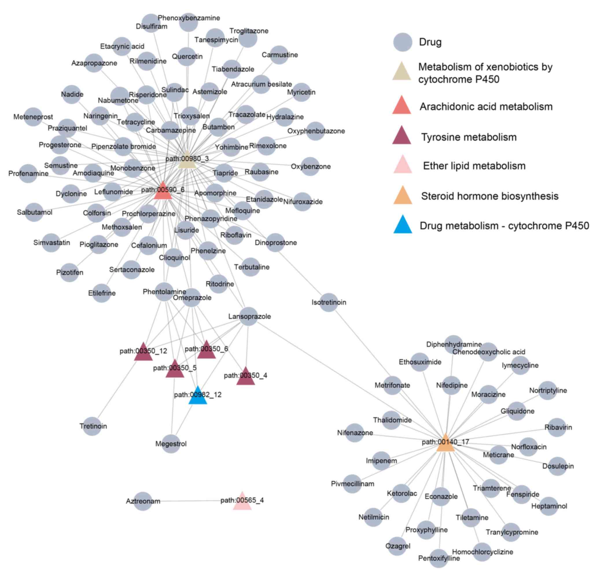
A bipartite network of the drugs identified and the
overlapping metabolic subpathways was built (Fig. 1). In this network, certain drugs could
affect several metabolic subpathways. For example, lansoprazole and
omeprazole perturbed 8 and 7 subpathways, respectively, while
others affected fewer subpathways.
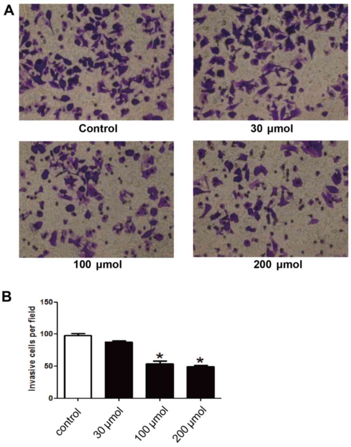
Lansoprazole inhibits the invasiveness
of U2OS cells
In order to investigate the effect of the drugs
identified on the invasiveness of OS cells, a Transwell assay was
performed on U2OS cells following treatment with lansoprazole, due
to it affecting the most subpathways. As illustrated in Fig. 2, lansoprazole significantly inhibited
the invasion of U2OS cells compared with the control group (P=0.02)
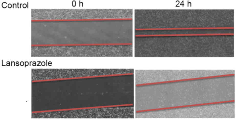
and did so in a dose-dependent manner. Furthermore, cell migration
was assessed using a wound healing assay. The results of this assay
demonstrated a marked reduction in cell migration following
lansoprazole treatment (Fig. 3).
Discussion
Microarray analysis using high-throughput screening
technology is an important tool for studying gene expression
patterns. In addition, this tool can be used to identify potential
therapeutic targets for improving therapeutic interventions. In the
present study, the gene expression profiles of biopsy specimens
from patients who did or did not develop metastatic OS were used to
investigate the mechanisms underlying the development of metastatic
OS. A total of 546 DEGs were identified between the two groups.
Results from Subpathway Miner analysis demonstrated that 9
metabolic subpathways corresponding to 6 entire pathways were
associated with OS metastasis. A total of 98 candidate small
molecule drugs involved in the regulation of OS metastasis were
subsequently identified through integrating OS
metastasis-associated and drug-affected subpathways.
Subpathway analysis focuses on a specific area of a
pathway, rather than the entire pathway. Thus, it can identify more
subtle subpathways that may be neglected through the analysis of
entire pathways, and may be more suitable and flexible compared
with entire pathway analysis for the identification of disease
mechanisms and drug responses (12,13). In
the present study, 9 subpathways corresponding to 6 entire KEGG
metabolic pathways were identified. A number of these subpathways
have been revealed to serve important roles in OS metastasis, such
as steroid hormone biosynthesis (path:00140_17;
P=6.10×10⁻⁵). A report by Fang et al (14) indicated that various concentrations of
estrogen lead to changes in several physiological processes in OS,
such as cell proliferation, migration, invasion and
epithelial-mesenchymal transition. In addition, the enzymes
(Cyclooxygenase 1 and 2) that are essential for arachidonic acid
metabolism (path:00590_6; P=0.029560322) have been identified to be
involved in the apoptosis of OS cells (15,16).
Furthermore, 4/9 subpathways originated from the tyrosine
metabolism pathway (path:00350), which has been demonstrated to
influence the growth of OS cells (17).
In the present study, a group of known drugs with
potential therapeutic efficacy for OS metastasis were identified. A
total of 98 small molecule drugs that have common subpathways with
OS metastasis were identified, including a number of anti-cancer
drugs. For example, semustine, a chloroethyl nitrosourea, is known
to be therapeutically effective against murine models of several
tumor types (18–20). In addition, a number of the drugs
identified have been proven to possess potential anti-OS effects.
For example, progesterone may inhibit the proliferation of OS cells
and increase the expression of c-Fos and c-Jun (21). Notably, the proton pump inhibitor
lansoprazole, which could influence 8/9 OS metastasis-associated
subpathways in the current study, has not, to the best of our
knowledge, been demonstrated to modulate the metastasis of OS.
However, lansoprazole has been revealed to contribute to
proliferation and invasion of breast cancer cells during
tumorigenesis and metastasis (22).
To further investigate the effects of lansoprazole
on OS metastasis, OS U2OS cells were treated with different
concentrations of lansoprazole. Lansoprazole was demonstrated to
inhibit invasion of U2OS cells in a dose-dependent manner. In
addition, a wound healing assay demonstrated that lansoprazole
markedly decreased U2OS cell migration. In conclusion, the present
study presents a bioinformatics approach based on subpathway
analysis to identify potential agents that regulate OS metastasis,
such as lansoprazole. However, further experiments are warranted to
investigate the safety of lansoprazole and explore the underlying
molecular mechanisms of its effects on OS metastasis.
References
|
1
|
He H, Ni J and Huang J: Molecular
mechanisms of chemoresistance in osteosarcoma (Review). Oncol Lett.
7:1352–1362. 2014.PubMed/NCBI
|
|
2
|
Chou AJ and Gorlick R: Chemotherapy
resistance in osteosarcoma: Current challenges and future
directions. Expert Rev Anticancer Ther. 6:1075–1085. 2006.
View Article : Google Scholar : PubMed/NCBI
|
|
3
|
Longhi A, Errani C, de Paolis M, Mercuri M
and Bacci G: Primary bone osteosarcoma in the pediatric age: State
of the art. Cancer Treat Rev. 32:423–436. 2006. View Article : Google Scholar : PubMed/NCBI
|
|
4
|
He JP, Hao Y, Wang XL, Yang XJ, Shao JF,
Guo FJ and Feng JX: Review of the molecular pathogenesis of
osteosarcoma. Asian Pac J Cancer Prev. 15:5967–5976. 2014.
View Article : Google Scholar : PubMed/NCBI
|
|
5
|
Broadhead ML, Clark JC, Myers DE, Dass CR
and Choong PF: The molecular pathogenesis of osteosarcoma: A
review. Sarcoma. 2011:9592482011. View Article : Google Scholar : PubMed/NCBI
|
|
6
|
Bielack SS, Kempf-Bielack B, Delling G,
Exner GU, Flege S, Helmke K, Kotz R, Salzer-Kuntschik M, Werner M,
Winkelmann W, et al: Prognostic factors in high-grade osteosarcoma
of the extremities or trunk: An analysis of 1,702 patients treated
on neoadjuvant cooperative osteosarcoma study group protocols. J
Clin Oncol. 20:776–790. 2002. View Article : Google Scholar : PubMed/NCBI
|
|
7
|
Kobayashi E, Masuda M, Nakayama R,
Ichikawa H, Satow R, Shitashige M, Honda K, Yamaguchi U, Shoji A,
Tochigi N, et al: Reduced argininosuccinate synthetase is a
predictive biomarker for the development of pulmonary metastasis in
patients with osteosarcoma. Mol Cancer Ther. 9:535–544. 2010.
View Article : Google Scholar : PubMed/NCBI
|
|
8
|
Hughes DP: Strategies for the targeted
delivery of therapeutics for osteosarcoma. Expert Opin Drug Deliv.
6:1311–1321. 2009. View Article : Google Scholar : PubMed/NCBI
|
|
9
|
Bielack SS, Carrle D, Hardes J, Schuck A
and Paulussen M: Bone tumors in adolescents and young adults. Curr
Treat Options Oncol. 9:67–80. 2008. View Article : Google Scholar : PubMed/NCBI
|
|
10
|
Osaki S, Tazawa H, Hasei J, Yamakawa Y,
Omori T, Sugiu K, Komatsubara T, Fujiwara T, Sasaki T, Kunisada T,
et al: Ablation of MCL1 expression by virally induced microRNA-29
reverses chemoresistance in human osteosarcomas. Sci Rep.
6:289532016. View Article : Google Scholar : PubMed/NCBI
|
|
11
|
Lamb J, Crawford ED, Peck D, Modell JW,
Blat IC, Wrobel MJ, Lerner J, Brunet JP, Subramanian A, Ross KN, et
al: The connectivity map: Using gene-expression signatures to
connect small molecules, genes, and disease. Science.
313:1929–1935. 2006. View Article : Google Scholar : PubMed/NCBI
|
|
12
|
Li C, Li X, Miao Y, Wang Q, Jiang W, Xu C,
Li J, Han J, Zhang F, Gong B and Xu L: SubpathwayMiner: A software
package for flexible identification of pathways. Nucleic Acids Res.
37:e1312009. View Article : Google Scholar : PubMed/NCBI
|
|
13
|
Li C, Shang D, Wang Y, Li J, Han J, Wang
S, Yao Q, Wang Y, Zhang Y, Zhang C, et al: Characterizing the
network of drugs and their affected metabolic subpathways. PLoS
One. 7:e473262012. View Article : Google Scholar : PubMed/NCBI
|
|
14
|
Fang D, Yang H, Lin J, Teng Y, Jiang Y,
Chen J and Li Y: 17β-estradiol regulates cell proliferation, colony
formation, migration, invasion and promotes apoptosis by
upregulating miR-9 and thus degrades MALAT-1 in osteosarcoma cell
MG-63 in an estrogen receptor-independent manner. Biochem Biophys
Res Commun. 457:500–506. 2015. View Article : Google Scholar : PubMed/NCBI
|
|
15
|
Moalic S, Liagre B, Corbière C, Bianchi A,
Dauça M, Bordji K and Beneytout JL: A plant steroid, diosgenin,
induces apoptosis, cell cycle arrest and COX activity in
osteosarcoma cells. FEBS Lett. 506:225–230. 2001. View Article : Google Scholar : PubMed/NCBI
|
|
16
|
Moalic S, Liagre B, Le Bail JC and
Beneytout JL: Dose-dependent modulation of apoptosis and
cyclooxygenase-2 expression in human 1547 osteosarcoma cells by
NS-398, a selective cyclooxygenase-2 inhibitor. Int J Oncol.
18:533–540. 2001.PubMed/NCBI
|
|
17
|
Tinti L, Taylor AM, Santucci A, Wlodarski
B, Wilson PJ, Jarvis JC, Fraser WD, Davidson JS, Ranganath LR and
Gallagher JA: Development of an in vitro model to investigate joint
ochronosis in alkaptonuria. Rheumatology. 50:271–277. 2011.
View Article : Google Scholar : PubMed/NCBI
|
|
18
|
Wang X, Chen JX, Liu JP, You C, Liu YH and
Mao Q: Gain of function of mutant TP53 in glioblastoma: Prognosis
and response to temozolomide. Ann Surg Oncol. 21:1337–1344. 2014.
View Article : Google Scholar : PubMed/NCBI
|
|
19
|
Saha P, Debnath C and Bérubé G:
Steroid-linked nitrogen mustards as potential anticancer
therapeutics: A review. J Steroid Biochem Mol Biol. 137:271–300.
2013. View Article : Google Scholar : PubMed/NCBI
|
|
20
|
Bartzatt R: Anticancer agents for
treatment of tumors in the central nervous system by correspondent
substituent substitution and elucidation by pattern recognition
methods. Med Chem. 8:138–144. 2012. View Article : Google Scholar : PubMed/NCBI
|
|
21
|
Liang M, Liao EY, Xu X, Luo XH and Xiao
XH: Effects of progesterone and 18-methyl levonorgestrel on
osteoblastic cells. Endocr Res. 29:483–501. 2003. View Article : Google Scholar : PubMed/NCBI
|
|
22
|
Zhang S, Wang Y and Li SJ: Lansoprazole
induces apoptosis of breast cancer cells through inhibition of
intracellular proton extrusion. Biochem Biophys Res Commun.
448:424–429. 2014. View Article : Google Scholar : PubMed/NCBI
|