Introduction
Cancer is one of the leading causes of mortality in
humans worldwide. In 2016, there will be an estimated 1,685,210 new
cancer cases diagnosed and 595,690 cancer-associated mortalities in
the USA (1). Tumor metastasis, which
is defined as the ability of cancer cells to spread to distant
organs or tissues in a patient, is responsible for ~90% of
cancer-related mortalities (2).
Therefore, in addition to limiting the growth of existing tumors,
blocking their metastasis is critical to improve the survival of
cancer patients. Targeting cancer cell motility and invasion are
the main strategies in the attempt to prevent metastasis (3).
Paeonia suffruticosa from the section
Moutan of the genus Paeonia is an important Chinese
medicinal herb. Previous investigations of this medicinal plant
focused on paeonol, paeoniflorin and their analogs as the major
bioactive constituents in the root bark (also called Cortex Moutan)
(4,5).
Nevertheless, researchers have also been interested in other parts
of this medicinal plant, such as the flowers, fruits and seeds
(6–8).
In particular, recent studies demonstrated that the seeds of this
herb are rich and unique sources of oligostilbenes (9,10).
Oligostilbenes are oligomers of the natural molecule
resveratrol and have been reported to exhibit a broad variety of
biological activities, including antioxidant, antitumor,
anti-inflammatory and antimalarial activities (11–13). As
one of the most promising naturally derived cancer chemopreventive
agents, the in vitro and in vivo antitumor activity
of resveratrol has been extensively characterized (13,14). It is
of particular interest to determine whether naturally occurring
oligostilbenes have comparable antitumor activities to resveratrol.
Satyajit et al (15) first
identified three oligostilbenes, suffruticosol A-C, from the seeds
of P. suffruticosa in 1999. In 2002, Kim et al
(6) isolated six oligostilbenes,
trans-resveratrol, trans-ε-viniferin,
cis-ε-viniferin, trans-gnetin H, suffruticosol A and
suffruticosol B, from Paeonia lactiflora and evaluated their
cytotoxicity against five cancer cell lines, HepG2 (human liver
cancer cell line), MCF-7 (human breast cancer cell line), HeLa
(human cervix cancer cell line), C6 (rat brain cancer cell line)
and HT-29 (human colon cancer cell line). In 2003, Kang et
al (7) isolated viniferin,
trans-gnetin H and suffruticosol B from P. lactiflora
and evaluated their effects on the proliferation and apoptosis of
HL-60 cells. In our previous studies, we conducted several
phytochemical analyses of the seedcases of P. suffruticosa
and found that oligostilbenes were the major active ingredients,
accounting for ~20% of the content (up to 200 mg/g) (9,10,16,17). In
addition to their high abundance, some oligostilbenes identified in
the seeds of Paeonia, such as suffruticosol A-C, have never
been reported in other plants. Therefore, a better understanding of
the biological and pharmacological activities of these
oligostilbenes is of great interest in the field of naturally
derived cancer chemopreventive agents.
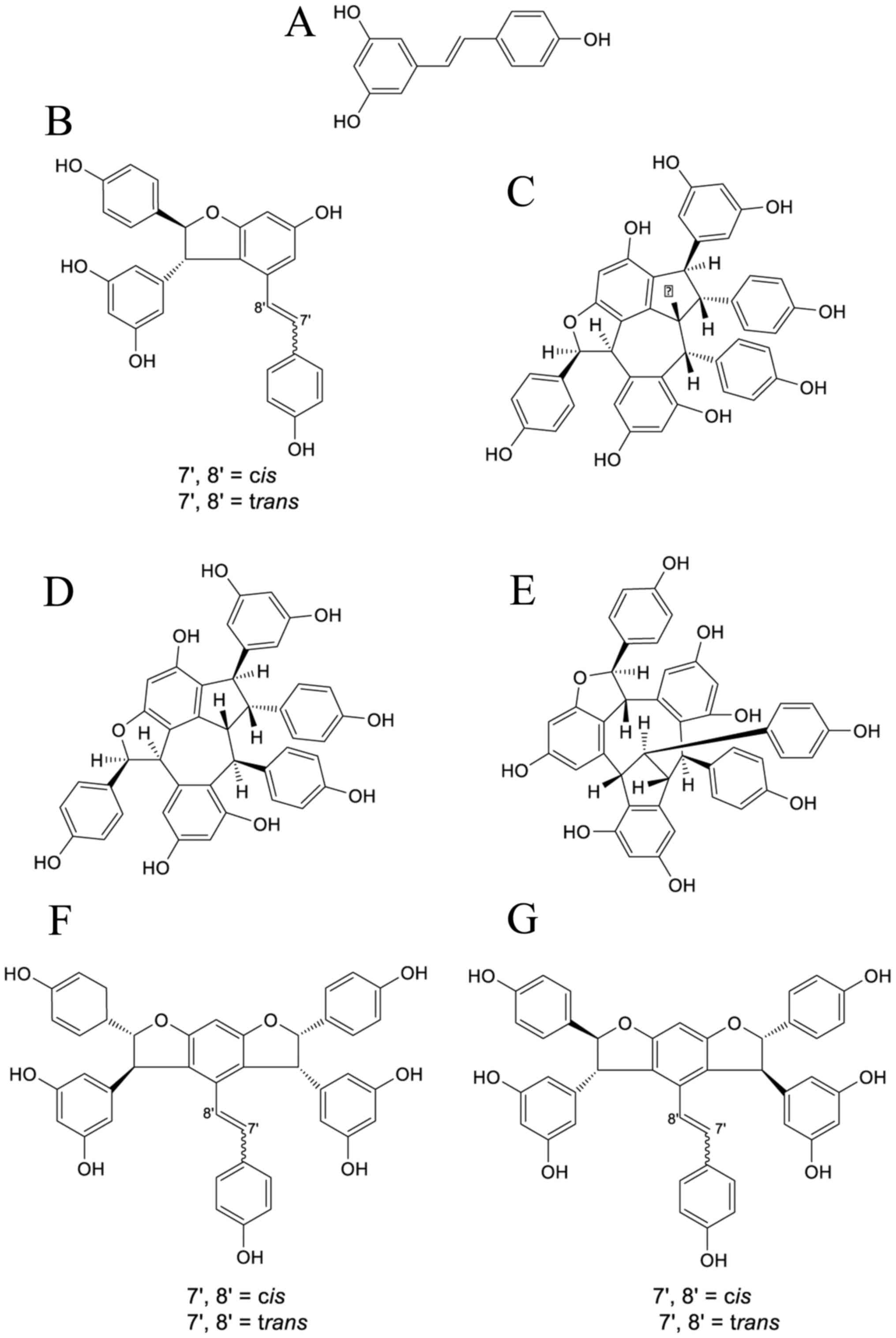
Previously, we identified ten oligostilbenes,
trans-resveratrol, cis-ε-viniferin,
trans-ε-viniferin, suffruticosol A, suffruticosol B,
suffruticosol C, cis-suffruticosol D,
trans-suffruticosol D, cis-gnetin H and
trans-gnetin H, from the seedcases of P. suffruticosa
(Fig. 1) (16). Recently, we investigated the
biological effects of cis- and trans-gnetin H and
cis- and trans-suffruticosol D, and showed that they
suppressed the proliferation of cancer cells (18,19). In
the present study, the antitumor activity of this unique and
comprehensive collection of oligostilbenes was systematically
evaluated, and their structure-activity relationships were
determined based on their anti-proliferative and anti-metastasis
effects.
Materials and methods
Plant material
The seeds of P. suffruticosa (1.2 kg) were
collected in Tongling, Anhui province, China, and identified in
2012. The sample was authenticated by Professor Peigen Xiao and Dr
Chunnian He from the Institute of Medicinal Plant Development,
Chinese Academy of Medical Sciences. A voucher specimen (2012001)
was deposited in the Seed Resource Bank of the Institute of
Medicinal Plant Development and Peking Union Medical College,
Beijing, China.
Simultaneous purification of ten
oligostilbenes
The ten oligostilbenes, trans-resveratrol,
cis-ε-viniferin, trans-ε-viniferin, suffruticosol A,
suffruticosol B, suffruticosol C, cis-suffruticosol D,
trans-suffruticosol D, cis-gnetin H and
trans-gnetin H, were simultaneously purified from the dried
seeds of P. suffruticosa as described previously (16). Their structures were characterized by
ultraviolet (UV), infrared (IR), mass and nuclear magnetic
resonance (NMR) spectroscopy, and the purities of all compounds
were determined to be >95% (16).
The compounds were resuspended in dimethyl sulfoxide (DMSO)
(Sigma-Aldrich; Merck Millipore, Darmstadt, Germany) to yield a
concentration of 10 mM, and stored at 4°C.
Cell culture
Six human cancer cell lines were used in this study,
including lung carcinoma (A549), breast carcinoma (BT20, MCF-7 and
MDA-MB-231), osteosarcoma (U2OS) and cervix adenocarcinoma (HeLa).
All cell lines were purchased from American Type Culture Collection
(Manassas, VA, USA), except for the HPL1A cell line, which was
obtained from Nagoya University, Japan. A549, BT20 and HeLa cells
were grown in RPMI-1640 medium (Sigma-Aldrich; Merck Millipore).
MCF-7 and MDA-MB-231 cells were grown in Dulbecco's Modified
Eagle's Medium (DMEM) (Sigma-Aldrich; Merck Millipore) supplemented
with 2 mM L-glutamine (Sigma-Aldrich; Merck Millipore). U2OS cells
were grown in McCoy's 5A medium (ATCC). HPL1A cells were grown in
DMEM/F-12 medium (Sigma-Aldrich; Merck Millipore). All mediums were
supplemented with 10% fetal bovine serum (FBS) (Thermo Fisher
Scientific, Inc., Waltham, MA, USA), 1% penicillin and
streptomycin, and incubated in a humidified atmosphere containing
5% CO2 at 37°C.
Anti-proliferation assay and half
maximal inhibitory concentration (IC50) determination
Anti-proliferation activity was determined using a
fluorescent staining assay. Cells were seeded into a 96-well tissue
culture plate at a density of 4,000 cells/well and treated with the
one of the ten compounds at final concentrations of 100, 50, 25,
12.5, 6.25 or 3.13 µM for 48 h. Cells treated with vehicle (1%
DMSO) only was also included as an experimental control.
Subsequently, 10% AlamarBlue dye (Invitrogen; Thermo Fisher
Scientific, Inc.) was added to the medium and the cells were
incubated at 37°C in the CO2 incubator for 1 h. The
fluorescence intensity was read in a SpectraMax M5 microplate
reader (Molecular Devices, LLC, Sunnyvale, CA, USA) at
excitation/emission (Ex/Em) wavelengths of 550/590 nm. The results
are expressed as a percentage relative to the untreated control,
and the IC50 values were calculated using non-linear
regression analysis with GraphPad Prism software 6.0 (GraphPad
Software, Inc., La Jolla, CA, USA).
Multiplex apoptosis assay by
high-content screening
MDB-MA-231 cells were seeded into a 96-well plate at
a density of 8,000 cells/well, and treated with the test compound
at 50 µM for 24 h. Apoptosis was assessed using a live-cell assay
by fluorescein isothiocyanate (FITC)-Annexin V and propidium iodide
(PI) staining, as described previously (20,21).
Briefly, 10 µl of 10X binding buffer containing 1 µl Hoechst 33342,
5 µl FITC-Annexin V and 5 µl mg/ml PI (BD Biosciences, Franklin
Lakes, NJ, USA) was added to cell culture medium following
treatment with test compounds. Staurosporine (Sigma-Aldrich; Merck
Millipore) at a concentration of 1 µM served as a positive control
and cells treated with vehicle only served as a negative control.
Removal of cell culture medium from wells was avoided because
necrotic and poorly attached cells would be detached and removed
during this process. Immediately following incubation at room
temperature in the dark for 10 min, cells were imaged using an
ArrayScan VTI High-Content Screening (HCS) reader (Thermo Fisher
Scientific, Inc.) and the fluorescence intensity of each channel
(Hoechst 33342, FITC-Annexin V and PI) was analyzed using HCS
Studio software 6.5.0.
Caspase-3/7 assay
Caspase-3/7 activity was determined using the
SensoLyte Homogeneous AMC Caspase-3/7 Assay kit (AnaSpec, Fremont,
CA, USA), according to the manufacturer's instructions. Briefly,
MDA-MB-231 cells (8,000 cells/well) were seeded into a 96-well
plate and incubated at 37°C overnight. The cells were treated with
test compounds at 50 µM at 37°C for 24 h. Staurosporine at a
concentration of 1 µM served as a positive control and cells
treated with vehicle only served as a negative control. After 24 h,
50 µl caspase-3/7 substrate solution was added to each well, and
the plate was incubated in the dark at room temperature for 1 h.
Fluorescence intensity was measured using a SpectraMax M5
microplate reader at Ex/Em wavelengths of 350/440 nm. Caspase-3/7
activities were calculated by subtracting the fluorescence levels
of wells containing medium only.
In vitro cell migration assay
Prior to performing the in vitro migration
and invasion assay, the cytotoxicity of the test compound was
determined after a 16-h treatment. Only the concentration that had
<20% toxicity on MDA-MB-231 cells was used for the migration and
invasion assay. The in vitro migration assay was performed
as described previously (22), with
slight modifications. Millicell® Cell Culture Inserts
(8-µm pore size; EMD Millipore, Billerica, MA, USA) were placed
into a 24-well cell culture plate. MDA-MB-231 cells
(2×10⁵ cells/ml) in 500 µl serum-free medium were seeded
into the upper chamber. The lower chamber was filled with 500 µl
culture medium supplemented with 10% FBS. Each compound was added
to the medium in both the upper and lower chambers to the desired
concentration (10 µM). Doxycycline (Sigma-Aldrich; Merck Millipore)
at a concentration of 100 µM served as a positive control and cells
treated with vehicle only served as a negative control. The cells
were incubated for 16 h to allow the cells to migrate through the
filter pores. Subsequently, cells on the upper surface of the
filter membrane were removed using a sterile cotton swab. Cells
that had migrated to the lower surface were stained with 10%
AlamarBlue dye at 37°C for 1 h and read in a SpectraMax M5
microplate reader at Ex/Em wavelengths of 550/590 nm.
In vitro Matrigel invasion assay
The in vitro invasion assay followed the same
procedure as the in vitro migration assay except that the
filter inserts were pre-coated with Matrigel matrix. Briefly, 100
µl Matrigel matrix coating (Corning Incorporated, Corning, NY, USA)
at 5 mg/ml was added to each insert and incubated at 37°C for 2 h
to form a continuous thin layer.
Statistical analysis
All experiments were performed in triplicate, and
each experiment was repeated 3 times. Data were presented as the
mean ± standard deviation. Statistical analysis was performed using
GraphPad Prism 6.0 (GraphPad Software, Inc.). Data were analyzed
using one-way analysis of variance tests, where P<0.05 was
considered to indicate a statistically significant difference.
Results
Ten oligostilbenes exhibit
anti-proliferation activity in human breast, lung and bone cancer
cells
The anti-proliferative activity of ten
oligostilbenes was initially evaluated in six different human
cancer cell lines (A549, MCF-7, BT20, MDA-MB-231, U2OS and HeLa).
All oligostilbenes showed mild-to-potent in vitro
cytotoxicity against these human cancer cells, and the oligomers of
resveratrol showed superior antitumor activities compared with the
resveratrol monomer (Table I).
Concentration-dependent anti-proliferation effects were observed
for all oligostilbenes in most of the tested cancer cell lines
after 48 h of treatment. Cis- and trans-gnetin H
showed the most potent anti-proliferation activities, with
IC50 values ranging from 0.9 to 10.0 µM against the six
cancer cell lines, representing a >20-fold increase in potency
compared with resveratrol (data not shown). Generally,
trans-ε-viniferin, trans-suffruticosol D and
trans-gnetin H were more potent than their respective
cis-forms, cis-ε-viniferin, cis-suffruticosol
D and cis-gnetin H. Additionally, in most of the cancer cell
lines, with the exception of A549, the trimers of resveratrol,
suffruticosol A-C, cis- and trans-suffruticosol D and
cis- and trans-gnetin H were more potent than the
dimers of resveratrol cis- and trans-ε-viniferin.
 | Table I.IC50 values (µM) of ten
oligostilbenes against six different types of human cancer cell
lines (A549, BT20, MCF-7, MDA-MB-231, U2OS and HeLa). |
Table I.
IC50 values (µM) of ten
oligostilbenes against six different types of human cancer cell
lines (A549, BT20, MCF-7, MDA-MB-231, U2OS and HeLa).
|
|
| IC50
(µM) |
|---|
|
|
|
|
|---|
| Name | DP | A549 | BT20 | MCF7 | MDA-MB-231 | U2OS | HeLa |
|---|
| Resveratrol
(E)-form | 1 | 53.6a | 91.3a |
>100a | >100 |
>100a | 17.1 |
|
cis-ε-viniferin | 2 | 15.5 | 19.2 | 68.0 | 72.6 | 75.5 | 10.5 |
|
trans-ε-viniferin | 2 | 6.0 | 13.1 | 53.5 | 59.0 | 59.7 |
8.7 |
| Suffruticosol
A | 3 | 3.4 | 5.1 | 27.7 | 34.6 | 31.3 |
4.3 |
| Suffruticosol
B | 3 | 4.8 | 10.9 | 17.8 | 16.7 | 12.2 |
3.3 |
| Suffruticosol
C | 3 | 9.2 | 18.4 | 8.7 | 9.9 | 10.7 |
2.4 |
|
cis-suffruticosol D | 3 | 17.1b | 13.4b | 46.8b | >100 | 24.6b |
5.5 |
|
trans-suffruticosol D | 3 | 11.9b | 9.9b | 15.8b | 38.9 | 11.3b |
2.3 |
| cis-gnetin
H | 3 | 2.8a | 2.4a | 10.1a | 9.7 | 15.9a |
1.3 |
| trans-gnetin
H | 3 | 2.6a | 2.4a | 7.7a | 7.1 | 4.5a |
0.9 |
It is notable that all the tested oligostilbenes
inhibited the proliferation of three representative subtypes of
breast carcinoma cells, including Basal A phenotype BT20 cells
[estrogen receptor (ER)− progesterone (PR)−
human epidermal growth factor receptor 2 (HER2)−],
Luminal A phenotype MCF-7 cells
(ER+PR+HER2−) and Basal B
phenotype MDA-MB-231 cells
(ER−PR−HER2−). Generally they
showed higher potency against BT20 cells than MCF-7 and MDA-MB-231
cells. Since Basal B cells are much more invasive than Basal A and
luminal cells (23), Basal B
phenotype MDA-MB-231 cells were selected for use in the following
experiments to evaluate the anti-metastasis activity of the
compounds.
Ten oligostilbenes induce apoptosis in
human breast cancer cells
In an examination of the cancerous cells using an
inverted microscope, it was observed that A549, BT20, MCF-7,
MDA-MB-231, U2OS and HeLa cells treated with oligostilbenes at
various concentrations (3.13–100 µM) for 24 and 48 h showed the
characteristic morphological changes of apoptosis, including cell
shrinkage and apoptotic bodies (data not shown). Therefore, a
multiplex apoptosis assay of MDA-MB-231 cells was conducted using
HCS to confirm the occurrence of apoptosis. Hoechst 33342 was used
to stain the nucleus, and FITC-Annexin V and PI were used as
indicators of apoptosis. To compare the apoptotic effect, the same
dose of 50 µM was used for all oligostilbenes and the cells were
treated for 24 h. As shown in Fig.
2A, cells treated with vehicle only exhibited blue fluorescence
due to Hoechst staining, cells undergoing early apoptosis exhibited
green fluorescence due to Hoechst and FITC-Annexin V staining, and
cells undergoing late apoptosis exhibited green and red
fluorescence due to FITC-Annexin V and PI staining, respectively.
In cells treated with 50 µM oligostilbenes, both FITC-Annexin V and
PI staining exhibited a significant increase compared with the
negative control (P<0.01 or P<0.001; Fig. 2B). These results suggest that the
oligostilbenes inhibit cancer cell growth by inducing apoptosis in
the cancer cells.
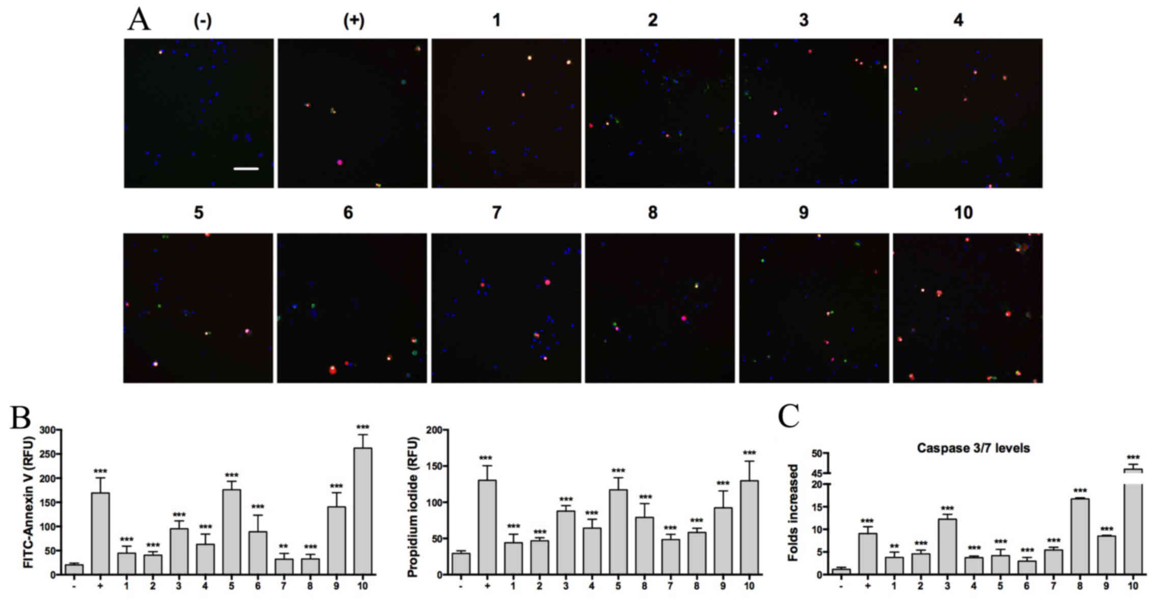 | Figure 2.Effects of ten oligostilbenes on
MDA-MB-231 cell apoptosis and caspase-3/7 activity. MDA-MB-231
cells were treated with 50 µM of the test compounds for 24 h.
Staurosporine (1 µM) served as a positive control and cells treated
with vehicle only served as a negative control. Cells were assessed
for apoptosis using multiplex fluorescence staining and a HCS
reader. (A) Representative HCS images indicative of Hoechst 33342,
FITC-Annexin V and PI staining (scale bar=100 µm). (B) Quantitative
fluorescent intensity of FITC-Annexin V and PI staining. (C)
Caspase-3/7 activity measurements. Cells were assessed for
caspase-3/7 activity using the SensoLyte® Homogeneous
AMC Caspase-3/7 assay kit. Data are presented as the mean ±
standard deviation (n=6). **P<0.01 and ***P<0.001 vs. the
negative control. HCS, high-content screening; 1, resveratrol
(E)-form; 2, cis-ε-viniferin; 3,
trans-ε-viniferin; 4, suffruticosol A; 5, suffruticosol B;
6, suffruticosol C; 7, cis-suffruticosol D; 8,
trans-suffruticosol D; 9, cis-gnetin H; 10,
trans-gnetin H; FITC, fluorescein isothiocyanate; PI,
propidium iodide; RFU, relative fluorescence units. |
To further examine the activities of caspase-3/7,
which are the major effectors activated by the formation of the
apoptosome in apoptotic cells (24),
a fluorescent assay of MDA-MB-231 cells was performed. Subsequent
to a 24-h treatment with oligostilbenes, caspase-3/7 levels were
significantly increased in cancer cells. All oligostilbenes at 50
µM concentration had induced caspase-3/7 activity levels by at
least 3-folds (P<0.001; Fig. 2C).
Among the oligostilbenes, trans-gnetin H showed the most
significant effect. The caspase-3/7 levels in trans-gnetin
H-treated cells were 46-fold higher compared with the negative
control. This result was consistent with the multiplex apoptosis
assay where trans-gnetin H also showed the highest Annexin V
and PI staining.
Ten oligostilbenes exhibit
anti-migration and anti-invasion activities in human breast cancer
cells at a low-toxicity dosage
Subsequently, the effects of oligostilbenes at a
low-toxicity dosage on the migration and invasion of MDA-MB-231
breast cancer cells were investigated. Since all oligostilbenes
displayed <20% cytotoxicity on the MDA-MB-231 cells at the dose
of 10 µM following 18 h of treatment (Fig. 3A), we selected this concentration for
the subsequent migration and invasion assays. All oligostilbenes
inhibited the migration and invasion of MDA-MB-231 cells in
vitro. In the migration assay, all oligostilbenes significantly
affected the number of cells that migrated through the pores of the
filter insert; the inhibition rate ranged from 24.4 to 88.9%
(P<0.001; Fig. 3B). In the
invasion assay, only those cells that passed through the Corning
Matrigel Matrix layer and the filter membrane were detected, and
the inhibition rate by oligostilbenes ranged from 22.0 to 54.6%
(P<0.001; Fig. 3C).
 | Figure 3.Effects of ten oligostilbenes on
MDA-MB-231 cell migration and invasion. MDA-MB-231 cells were
treated with 10 µM of the test compounds for 16 h. Doxycycline (100
µM) served as a positive control and cells treated with vehicle
only served as a negative control. Migration and invasion were
assessed using Millicell Culture Inserts with or without Matrigel
matrix, and the number of cells that had migrated or invaded
through the inserts was assessed using AlamarBlue staining. (A)
Illustration of cell migration assay and representative images of
inhibitory effects on cell migration (from left to right: negative
control, positive control, cis-gnetin H, trans-gnetin
H). (B) Illustration of cell invasion assay and representative
images of inhibitory effects on cell invasion (from left to right:
negative control, positive control, cis-gnetin H,
trans-gnetin H). (C) Cell viability after treatment with the
test compounds at 10 µM. (D) Inhibition of cell migration ability
after treatment with the test compounds. (E) Inhibition of cell
invasion ability after treatment with the test compounds. Data are
presented as the mean ± standard deviation (n=6). ***P<0.001 vs.
the negative control. 1, resveratrol (E)-form; 2,
cis-ε-viniferin; 3, trans-ε-viniferin; 4,
suffruticosol A; 5, suffruticosol B; 6, suffruticosol C; 7,
cis-suffruticosol D; 8, trans-suffruticosol D; 9,
cis-gnetin H; 10, trans-gnetin H; FITC, fluorescein
Isothiocyanate. |
Discussion
As one of the most promising naturally derived
chemopreventive agents, the in vitro and in vivo
antitumor activity of resveratrol has been extensively
characterized (13,14,25,26). It is
of particular interest to determine whether naturally occurring
oligostilbenes or their derivatives have comparable antitumor
activities to resveratrol. The present study systematically
evaluated the antitumor activity of ten oligostilbenes that were
simultaneously isolated from the seeds of P. suffruticosa,
and demonstrated that the dimers and trimers of resveratrol had
superior antitumor activities compared with resveratrol.
Cancer development involves the regulation of cell
growth and metastasis (27). All test
oligostilbenes showed mild-to-potent in vitro cytotoxicity
against a panel of human cancer cells, and
cis/trans-gnetin H showed the most potent activity
among the test compounds; they were >20-fold more effective than
the resveratrol monomer. The results of the present study suggested
that. The present study also determined the activity levels of
caspase-3/7, which are the major effectors activated by the
formation of the apoptosome in apoptotic cells (24). In agreement with the results of the
apoptosis assay, we found that caspase-3/7 activity levels were
significantly increased in cancer cells treated with
oligostilbenes.
Cancer metastasis is a complex process that involves
sequential steps of invasion, migration, circulation, infiltration
and colonization at a distant site (28,29). The
present study focused on the effects of oligostilbenes on the
migration and invasion of MDA-MB-231 breast cancer cells in order
to determine their effects on the metastasis of tumor cells. At a
lower dose that did not affect the cancer cell growth, all
oligostilbenes inhibited the migration and invasion capability of
MDA-MB-231 cells in vitro.
An analysis of the structure-antitumor activity
relationships revealed three interesting findings. First, the
degree of polymerization was closely associated with the antitumor
activity of the oligostilbenes. Oligomers with more repeating
resveratrol units were more active than smaller oligomers, as
evidenced by the order of their potency: Trimers (cis- and
trans-gnetin H, suffruticosol A-C, cis- and
trans-suffruticosol D), followed by dimers (cis- and
trans-ε-viniferin) and, lastly, the resveratrol monomer.
This observation is consistent with previous findings of the
structure-antioxidant activity of resveratrol oligomers (30,31).
Secondly, the double bond in the stilbenic skeleton and its
trans-isomerism were important to the antitumor activity of
oligostilbenes. Resveratrol and its oligomers are known to be
highly photosensitive compounds that are prone to UV-induced
isomerization. Approximately 80% of trans-resveratrol was
converted to cis-resveratrol upon UV light exposure for 1 h
(32), and ~86% of
trans-gnetin H was converted to cis-gnetin H upon UV
light exposure for 6 h (18). It is
well known that cis- and trans-isomers of naturally
occurring compounds can differ in their bioactivities, and that
trans-isomers are believed to be the more abundant and
active form (33–35). In the present study, the three
cis-isomers of oligostilbenes, in which the double bond is
reduced, were significantly less effective than their
trans-isomers. Thirdly, the steric arrangement and
conformation of oligostilbenes also affected their antitumor
activity. cis- and trans-suffruticosol D and
cis- and trans-gnetin H are both trimers of
resveratrol and both possess seven hydroxyl groups, differing only
in their three-dimensional structures (16). Generally cis- and
trans-gnetin H was 2–10 times more potent than cis-
and trans-suffruticosol D, indicating that three-dimensional
structures have a significant effect on cytotoxicity. The most
likely reason was that the trans orientation of H-7”/H-8” in
cis- and trans-gnetin H may lessen the steric
hindrance between rings C1 and C2, and therefore enhance the
bioactivity of cis- and trans-gnetin H.
Although numerous studies have demonstrated the
potential of resveratrol as a cancer chemopreventive agent in the
last two decades (13,14,26,36), the
poor bioavailability of resveratrol due to its rapid metabolism and
secretion from the body compromises its biological and
pharmacological benefits (37).
Hence, significant attention has been given to the derivatives of
resveratrol to overcome these drawbacks. For example, a recent
study on the pharmacokinetics of gnetin C, a resveratrol dimer,
showed increased bioavailability when orally consumed compared with
resveratrol (38). Since the
oligostilbenes in the present study showed improved potency in
cancer chemoprevention compared with resveratrol, further
investigation of their bioavailability is warranted.
In conclusion, the present study assessed a group of
ten naturally occurring oligostilbenes for their anti-proliferation
and anti-metastasis properties. The results provided valuable
insight into the structure-activity relationship of oligostilbenes
for the future development of novel cancer chemopreventive
drugs.
Acknowledgements
The authors would like to thank the Tennessee Center
of Botanical Medicinal Research (TCBMR) for providing the funding
for this study.
References
|
1
|
American Cancer Society, . Cancer Facts
& Figures 2016. American Cancer Society Inc.; Atlanta, GA:
2016
|
|
2
|
Spano D, Heck C, de Antonellis P,
Christofori G and Zollo M: Molecular networks that regulate cancer
metastasis. Semin Cancer Biol. 22:234–249. 2012. View Article : Google Scholar : PubMed/NCBI
|
|
3
|
Wells A, Grahovac J, Wheeler S, Ma B and
Lauffenburger D: Targeting tumor cell motility as a strategy
against invasion and metastasis. Trends Pharmacol Sci. 34:283–289.
2013. View Article : Google Scholar : PubMed/NCBI
|
|
4
|
Lau CH, Chan CM, Chan YW, Lau KM, Lau TW,
Lam FC, Law WT, Che CT, Leung PC, Fung KP, et al: Pharmacological
investigations of the anti-diabetic effect of Cortex Moutan and its
active component paeonol. Phytomedicine. 14:778–784. 2007.
View Article : Google Scholar : PubMed/NCBI
|
|
5
|
Wu M and Gu Z: Screening of bioactive
compounds from moutan cortex and their anti-inflammatory activities
in rat synoviocytes. Evid Based Complement Alternat Med. 6:57–63.
2009. View Article : Google Scholar : PubMed/NCBI
|
|
6
|
Kim HJ, Chang EJ, Bae SJ, Shim SM, Park
HD, Rhee CH, Park JH and Choi SW: Cytotoxic and antimutagenic
stilbenes from seeds of Paeonia lactiflora. Arch Pharm Res.
25:293–299. 2002. View Article : Google Scholar : PubMed/NCBI
|
|
7
|
Kang JH, Park YH, Choi SW, Yang EK and Lee
WJ: Resveratrol derivatives potently induce apoptosis in human
promyelocytic leukemia cells. Exp Mol Med. 35:467–474. 2003.
View Article : Google Scholar : PubMed/NCBI
|
|
8
|
Tanaka T, Fukumori M, Ochi T and Kouno I:
Paeonianins A-E, new dimeric and monomeric ellagitannins from the
fruits of Paeonia lactiflora. J Nat Prod. 66:759–763. 2003.
View Article : Google Scholar : PubMed/NCBI
|
|
9
|
He CN, Peng Y, Zhang YC, Xu LJ, Gu J and
Xiao PG: Phytochemical and biological studies of paeoniaceae. Chem
Biodivers. 7:805–838. 2010. View Article : Google Scholar : PubMed/NCBI
|
|
10
|
He CN, Peng Y, Wu QL, Xiao W, Peng B, Wang
Z and Xiao PG: Simultaneous determination of ten stilbenes in the
seeds of Paeonia species using HPLC-DAD. J Liq Chromatogr Relat
Technol. 36:1708–1724. 2013.
|
|
11
|
He S, Lu Y, Jiang L, Wu B, Zhang F and Pan
Y: Preparative isolation and purification of antioxidative stilbene
oligomers from Vitis chunganeniss using high-speed counter-current
chromatography in stepwise elution mode. J Sep Sci. 32:2339–2345.
2009. View Article : Google Scholar : PubMed/NCBI
|
|
12
|
Jung M, Park WH, Jung JC, Lim E, Lee Y, Oh
S and Moon HI: Synthesis, structural characterization and
biological evaluation of novel stilbene derivatives as potential
antimalarial agents. Chem Biol Drug Des. 73:346–354. 2009.
View Article : Google Scholar : PubMed/NCBI
|
|
13
|
Aggarwal BB, Bhardwaj A, Aggarwal RS,
Seeram NP, Shishodia S and Takada Y: Role of resveratrol in
prevention and therapy of cancer: Preclinical and clinical studies.
Anticancer Res. 24:2783–2840. 2004.PubMed/NCBI
|
|
14
|
Cai H, Scott E, Kholghi A, Andreadi C,
Rufini A, Karmokar A, Britton RG, Horner-Glister E, Greaves P,
Jawad D, et al: Cancer chemoprevention: Evidence of a nonlinear
dose response for the protective effects of resveratrol in humans
and mice. Sci Transl Med. 7:298ra1172015. View Article : Google Scholar : PubMed/NCBI
|
|
15
|
Satyajit DS, Pensri W and Laurence D:
Identification and ecdysteroid antagonist activity of three
resveratrol trimers (Suffruticosols A, B and C) from Paeonia
suffruticosa. Tetrahedron. 55:513–524. 1999. View Article : Google Scholar
|
|
16
|
He CN, Peng Y, Xu LJ, Liu ZA, Gu J, Zhong
AG and Xiao PG: Three new oligostilbenes from the seeds of Paeonia
suffruticosa. Chem Pharm Bull (Tokyo). 58:843–847. 2010. View Article : Google Scholar : PubMed/NCBI
|
|
17
|
He C, Xiao W, Li M, Peng Y, Xu L, Gu J and
Xiao P: Chemical Constituents from Seeds of Paeonia suffruticosa.
Zhongguo Zhong Yao Za Zhi. 35:1428–1431. 2010.(In Chinese).
PubMed/NCBI
|
|
18
|
Gao Y, He C, Ran R, Zhang D, Li D, Xiao PG
and Altman E: The resveratrol oligomers, cis-and trans-gnetin H,
from Paeonia suffruticosa seeds inhibit the growth of several human
cancer cell lines. J Ethnopharmacol. 169:24–33. 2015. View Article : Google Scholar : PubMed/NCBI
|
|
19
|
Almosnid NM, Gao Y, He CN, Park HS and
Altman E: In vitro antitumor effects of two novel oligostilbenes,
cis- and trans-suffruticosol D, isolated from Paeonia suffruticosa
seeds. Int J Oncol. 48:646–656. 2016.PubMed/NCBI
|
|
20
|
Arbab IA, Looi CY, Abdul AB, Cheah FK,
Wong WF, Sukari MA, Abdullah R, Mohan S, Syam S, Arya A, et al:
Dentatin induces apoptosis in prostate cancer cells via Bcl-2,
Bcl-xL, Survivin downregulation, caspase-9, −3/7 activation, and
NF-κB inhibition. Evid Based Complement Alternat Med.
2012:8560292012. View Article : Google Scholar : PubMed/NCBI
|
|
21
|
Martin HL, Adams M, Higgins J, Bond J,
Morrison EE, Bell SM, Warriner S, Nelson A and Tomlinson DC:
High-content, high-throughput screening for the identification of
cytotoxic compounds based on cell morphology and cell proliferation
markers. PLoS One. 9:e883382014. View Article : Google Scholar : PubMed/NCBI
|
|
22
|
Al-Nasiry S, Geusens N, Hanssens M, Luyten
C and Pijnenborg R: The use of Alamar Blue assay for quantitative
analysis of viability, migration and invasion of choriocarcinoma
cells. Hum reprod. 22:1304–1309. 2007. View Article : Google Scholar : PubMed/NCBI
|
|
23
|
Neve RM, Chin K, Fridlyand J, Yeh J,
Baehner FL, Fevr T, Clark L, Bayani N, Coppe JP, Tong F, et al: A
collection of breast cancer cell lines for the study of
functionally distinct cancer subtypes. Cancer Cell. 10:515–527.
2006. View Article : Google Scholar : PubMed/NCBI
|
|
24
|
Li-Weber M: Targeting apoptosis pathways
in cancer by Chinese medicine. Cancer Lett. 332:304–312. 2013.
View Article : Google Scholar : PubMed/NCBI
|
|
25
|
Joe AK, Liu H, Suzui M, Vural ME, Xiao D
and Weinstein IB: Resveratrol induces growth inhibition, S-phase
arrest, apoptosis, and changes in biomarker expression in several
human cancer cell lines. Clin Cancer Res. 8:893–903.
2002.PubMed/NCBI
|
|
26
|
Bishayee A: Cancer prevention and
treatment with resveratrol: From rodent studies to clinical trials.
Cancer Prev Res. 2:409–418. 2009. View Article : Google Scholar
|
|
27
|
Hanahan D and Weinberg RA: Hallmarks of
cancer: The next generation. Cell. 144:646–674. 2011. View Article : Google Scholar : PubMed/NCBI
|
|
28
|
Nguyen DX, Bos PD and Massagué J:
Metastasis: from dissemination to organ-specific colonization. Nat
Rev Cancer. 9:274–284. 2009. View Article : Google Scholar : PubMed/NCBI
|
|
29
|
Weng CJ and Yen GC: Chemopreventive
effects of dietary phytochemicals against cancer invasion and
metastasis: Phenolic acids, monophenol, polyphenol, and their
derivatives. Cancer Treat Rev. 38:76–87. 2012. View Article : Google Scholar : PubMed/NCBI
|
|
30
|
Mikulski D and Molski M: Quantitative
structure-antioxidant activity relationship of trans-resveratrol
oligomers, trans-4, 4′-dihydroxystilbene dimer,
trans-resveratrol-3-O-glucuronide, glucosides: Trans-piceid,
cis-piceid, trans-astringin and
trans-resveratrol-4′-O-β-D-glucopyranoside. Eur J Med Chem.
45:2366–2380. 2010. View Article : Google Scholar : PubMed/NCBI
|
|
31
|
Lim KG, Gray AI, Anthony NG, Mackay SP,
Pyne S and Pyne NJ: Resveratrol and its oligomers: Modulation of
sphingolipid metabolism and signaling in disease. Arch Toxicol.
88:2213–2232. 2014. View Article : Google Scholar : PubMed/NCBI
|
|
32
|
Vian MA, Tomao V, Gallet S, Coulomb PO and
Lacombe JM: Simple and rapid method for cis-and trans-resveratrol
and piceid isomers determination in wine by high-performance liquid
chromatography using Chromolith columns. J Chromatogr A.
1085:224–229. 2005. View Article : Google Scholar : PubMed/NCBI
|
|
33
|
Pettit GR, Melody N, Thornhill A, Knight
JC, Groy TL and Herald CL: Antineoplastic Agents. 579. Synthesis
and cancer cell growth evaluation of E-stilstatin. 3:A resveratrol
structural modification. J Nat Prod 72: 1637–1642. 2009.
|
|
34
|
Zhao DB, Rong CY, Jenkins S, Yin DL and
Liu SB: Origin of the cis-effect: A density functional theory study
of doubly substituted ethylenes. Acta Phys Chim Sin. 29:43–54.
2013.
|
|
35
|
Anisimova NY, Kiselevsky MV, Sosnov AV,
Sadovnikov SV, Stankov IN and Gakh AA: Trans-, cis- and
dihydro-resveratrol: A comparative study. Chem Cent J. 5:882011.
View Article : Google Scholar : PubMed/NCBI
|
|
36
|
Jang M, Cai L, Udeani GO, Slowing KV,
Thomas CF, Beecher CW, Fong HH, Farnsworth NR, Kinghorn AD, Mehta
RG, et al: Cancer chemopreventive activity of resveratrol, a
natural product derived from grapes. Science. 275:218–220. 1997.
View Article : Google Scholar : PubMed/NCBI
|
|
37
|
Neves AR, Lucio M, Lima JL and Reis S:
Resveratrol in medicinal chemistry: A critical review of its
pharmacokinetics, drug-delivery, and membrane interactions. Curr
Med Chem. 19:1663–1681. 2012. View Article : Google Scholar : PubMed/NCBI
|
|
38
|
Tani H, Hikami S, Iizuna S, Yoshimatsu M,
Asama T, Ota H, Kimura Y, Tatefuji T, Hashimoto K and Higaki K:
Pharmacokinetics and safety of resveratrol derivatives in humans
after oral administration of melinjo (Gnetum gnemon L.) seed
extract powder. J Agric Food Chem. 62:1999–2007. 2014. View Article : Google Scholar : PubMed/NCBI
|