Introduction
Malignancies of any organ can metastasize to the
pleura and induce malignant pleural effusion (MPE); however, the
most frequent are lung and breast carcinomas, and lymphomas, with
MPE occurring in digestive and ovary carcinomas less frequently
(1). In ~10% of patients with
undiagnosed pleural effusion, a lymphoma is finally detected
(2). MPE is observed in 10–30% of
patients with Hodgkin lymphoma at presentation (3–5) and may be
upwards of 60% (6). MPE is not
uncommon in patients with non-Hodgkin's lymphoma (NHL), with a
reported frequency of up to 20% (7,8). Although
pleural involvement by systemic lymphoma is relatively common,
primary pleural lymphoma is rare, accounting for only 7% of
lymphoma cases (9). Two types of
primary pleural lymphomas have been described: Body-cavity-based
lymphoma seen in patients with human immunodeficiency virus
infection (10,11) and pyothorax-associated lymphoma in
those with tuberculosis (12). In
addition, an extremely rare type of primary pleural lymphoma, seen
in immunocompetent patients without history of pyothorax, has been
reported in case studies in the literature (13–15).
Between July 2005 and June 2014, 42 patients with
MPE induced by NHL, whose major clinical presentation was pleural
involvement, presented to Beijing Institute of Respiratory Medicine
and Beijing Chaoyang Hospital, Capital Medical University (Beijing,
China). Medical history, clinical presentation, chest imaging and
investigation of thoracentesis and/or closed pleural biopsy are the
basic initial studies that lead to a diagnosis in the majority of
patients with MPE induced by NHL. However, in 10 of the 42 patients
in the present study, a diagnosis of NHL presenting with MPE was
not completely supported by the aforementioned procedures. Medical
thoracoscopy (MT) followed by pathological and immunohistochemical
analysis of pleural biopsy led to a correct diagnosis of NHL in
9/10 of these patients, suggesting that MT may be a logical
approach allowing for the achievement of an efficient diagnosis
when NHL presents as MPE. In the present study, the outcome of MT
in the diagnosis of MPE induced by NHL was analyzed.
Patients and methods
Patients
Between July 2005 and June 2014, 833 patients with
undiagnosed exudative pleural effusions underwent MT in Beijing
Chaoyang Hospital (Beijing, China), and their detailed medical
history, clinical presentation, laboratory examination results and
imaging data were recorded (16).
Prior to MT, all patients underwent the initial diagnostic workup,
which included a thoracic computed tomography (CT) scan,
thoracentesis and/or pleural biopsy, and their pleural effusions
remain undiagnosed. In the present study, a retrospective cohort of
10 patients with MPE induced by NHL is reviewed.
The study protocol was approved by the Institutional
Review Board for Human Studies of Beijing Chaoyang Hospital.
Thoracoscopic procedures
MT was performed by chest physicians in the
pulmonary procedural suite of Beijing Chaoyang Hospital, as
described previously (17,18). Once written informed consent was
obtained, the patient was positioned with the affected side up in
the lateral decubitus position. The patient breathed spontaneously
with supplemental oxygen via nasal cannula as required. An Aloka
Ultrasound system (Hitachi Aloka Medical Ltd., Tokyo, Japan) was
used to confirm the presence of effusion and evaluate the best
trocar entry site, generally located at the mid-to-anterior
axillary line, between the sixth and eighth intercostal spaces.
Under moderate sedation using fentanyl and
midazolam, the skin, subcutaneous tissue, adjacent ribs, and
parietal pleura were anesthetized with 1% lidocaine (15–30 ml) and
a small incision was made at the planned site of entry. An 8-mm
disposable trocar was then inserted, and a semi-rigid thoracoscope
(LTF-240, Olympus Corporation, Tokyo, Japan) was introduced into
the pleural space once the fluid was completely drained. A detailed
inspection of the pleural cavity was then performed, with any
abnormalities documented by photographic and/or video recordings.
Biopsies were performed using flexible forceps under direct visual
control in all suspect areas, systematically in several regions of
the parietal pleura.
At the end of the procedure, a 24F chest drain was
inserted for drainage. Chest radiographs were routinely obtained
following the procedure and were re-checked until the chest tube
was removed.
Hematoxylin and eosin (H&E)
staining
Pleural biopsy samples were all paraffin embedded.
The paraffin sections (4 µm) were dewaxed in room temperature by 2
changes of 100% xylene for 10 min each, and then re-hydrated in 2
changes of 100% alcohol for 5 min each, and then in 95% alcohol for
2 min and 70% alcohol for 2 min. Finally, wash the slides briefly
in distilled water for the following experiment. For routing
H&E staining, slides were put into hematoxylin (ZLI-9615) for 8
min and then washed twice before incubating with eosin (ZLI-9613)
for 1 min. Finally, the slides were sealed with neutral gum.
Immunohistochemistry
Slides were placed into antigen repair buffer (pH
9.0) (ZLI-9079; 1:50 diluted in distilled water) and boiled to
95–100°C for 10 min, cooled down to room temperature and washed
twice in PBS. All the sections were incubated at room temperature
with 3% H2O2 for 10 min to block endogenous
peroxidase. The slides were sealed with 1.0% bovine serum albumin
(BSA, A8010; Beijing Solarbio Science & Technology Co., Ltd.,
Beijing, China) for 30 min at room temperature. The sections were
incubated with the primary antibodies (all 1:100; diluted in 1%
BSA): Mouse anti-human CD20 (ZM-0039); CD3 (ZM-0417); CD5
(ZM-0280); CD10 (ZM-0283); CD21 (ZM-0040); CD23 (ZM-0273); CYCLIND1
(ZM-0366); BCL2 (ZM-0010); BCL6 (ZM-0011); CD30 (ZM-0043); Ki67
(ZM-0165); paired box gene 5 (ZM-0466); thyroid transcription
factor 1 (ZM-0250); creatine kinase (ZM-0069); creatine kinase 19
(ZM-0074); rabbit anti human CD 79a (ZA-0293), MUM1 (ZA-0583);
CD138 (ZA-0584); Calretinin (ZA-0026); synapsin (RAB-0155) and
terminal deoxynucleotidyl transferase (ZA-0625), respectively,
overnight at 4°C. After incubating with PBS three times,
horseradish peroxidase (HRP)-conjugated secondary antibodies
(1:300; diluted in 1% BSA): HRP-conjugated goat anti mouse IgG
(ZDR-5307) or HRP-conjugated goat anti rabbit IgG (ZDR-5306) were
applied to the slides and incubated for 1 h at room temperature.
The slides were developed with DAB (ZLI-9019; 1:20 diluted in DAB
substrate supplied within the kit) for 10 min at room temperature,
and then rinsed in running tap water for 5 min. Subsequently, 0.5%
hematoxylin (ZLI-9615) was used to stain the samples at room
temperature for 8 min, then washed twice and sealed with neutral
gum. All slides were examined carefully under a light microscope at
magnification, ×400. All the H&E staining and
immunohistochemistry reagents mentioned above were from Zhongshan
Golden Bridge Biotechnology, Co., Ltd (Beijing, China), except for
synapsin (RAB-0155), which was obtained from Maxim Biotechnology,
Co., Ltd (Fuzhou, China).
Diagnosis of NHL
A histopathological and immunohistochemical review
of all slides with at least one paraffin block representative
pleural biopsy samples obtained through MT. The diagnosis of NHL
was established by pathologists according to the current guidelines
(19,20).
Statistical analysis
Data are presented as the mean ± standard deviation
or number with percentage. Descriptive statistical methods were
used for data analysis with SPSS version 16.0 Statistical Software
(SPSS, Inc., Chicago, IL, USA).
Results
Clinical data
A total of 833 patients with undiagnosed pleural
effusions successfully underwent MT between July 2005 and June 2014
(16). MPE was the final diagnosis in
342 (41.1%) patients with lymphocytic exudates and 10 patients with
MPE were diagnosed with NHL.
All 10 NHL patients with MPE had no history of
tuberculosis and serological tests found that they were negative
for human immunodeficiency virus. As shown in Table I, the most common symptom was dyspnea
(n=8); the other symptoms included cough (n=4), fever (n=2), chest
pain (n=1) and weight loss (n=1). Lymph nodes were readily palpable
in a single patient. A diagnosis of metastatic carcinoma or
inflammatory disease was initially proposed in each instance.
 | Table I.Clinical characteristics of the study
population. |
Table I.
Clinical characteristics of the study
population.
| Patient no. | Sex | Age, years | Smoking status | Tuberculosis
history | HIV status | Effusion site | Effusion size | Main symptoms |
|---|
| 1 | M | 56 | Current smoker | No | Negative | Left | Large | Dyspnea, fever |
| 2 | M | 71 | Non-smoker | No | Negative | Right | Large | Dyspnea, cough |
| 3 | M | 76 | Ex-smoker | No | Negative | Bilateral | Small | Chest pain |
| 4 | F | 59 | Non-smoker | No | Negative | Right | Large | Dyspnea |
| 5 | M | 66 | Current smoker | No | Negative | Left | Large | Dyspnea,
fatigue |
| 6 | M | 60 | Current smoker | No | Negative | Left | Small | Dyspnea |
| 7 | M | 54 | Non-smoker | No | Negative | Bilateral | Large | Dyspnea, cough |
| 8 | M | 30 | Non-smoker | No | Negative | Right | Moderate | Dyspnea, cough,
fever |
| 9 | M | 53 | Current smoker | No | Negative | Bilateral | Large | Dyspnea, cough |
| 10 | M | 73 | Current smoker | No | Negative | Right | Moderate | Weight loss,
multiple lymphadenopathy |
Radiographic findings
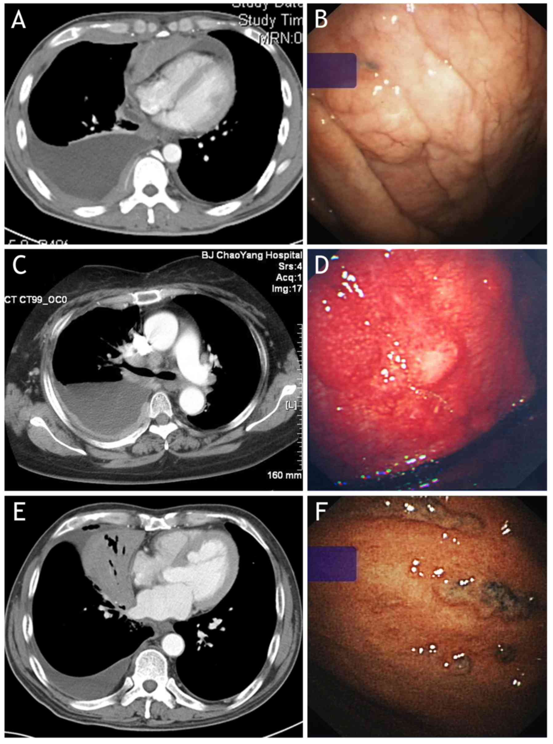
Pleural effusions were bilateral in 3 patients. In
the other 7 patients, effusions were in the right hemithorax in 4
patients and in the left hemithorax in 3 patients (Table I). The volume of a pleural effusion
was estimated as small, moderate or large based on CT imaging,
according to the techniques described by Moy et al (21). In either unilateral or bilateral
effusion, the volume of pleural effusions was small in 2 patients,
moderate in 2, and large in 6.
In addition to pleural effusion, CT scan (Table II) following administration of
intravenous contrast medium revealed diffuse pleural thickening
(n=5), focal pleural thickening (n=5), pleural nodules (n=3), and
pleural masses (n=2). Isolated pleural effusion with no
demonstrable pleural thickening or mass could also be seen. No
patients had evidence of visceral pleural abnormalities on CT scan.
Other pulmonary involvements included pulmonary consolidation or
infiltration (n=6), thoracic lymphadenopathy (n=4), lung nodule or
mass (n=3), and obstructive atelectasis (n=1).
 | Table II.Chest computed tomography scan
findings (n=10). |
Table II.
Chest computed tomography scan
findings (n=10).
|
|
Patient
no. |
|
|---|
|
|
|
|
|---|
| Finding | 1 | 2 | 3 | 4 | 5 | 6 | 7 | 8 | 9 | 10 | Total, n |
|---|
| Pleural
effusion | + | + | + | + | + | + | + | + | + | + | 10 |
| Diffuse pleural
thickening |
| + |
| + |
|
|
| + | + | + | 5 |
| Focal pleural
thickening | + |
| + | + | + |
| + |
|
|
| 5 |
| Pleural
nodules |
|
|
| + | + |
|
| + |
|
| 3 |
| Pleural masses |
|
|
| + |
|
|
| + |
|
| 2 |
| Pulmonary
consolidation or infiltration |
|
| + |
|
| + | + | + | + | + | 6 |
| Thoracic
lymphadenopathy |
|
|
| + |
|
| + | + |
| + | 4 |
| Lung nodule or
mass |
|
| + | + |
|
|
| + |
|
| 3 |
| Obstructive
atelectasis |
|
|
|
|
|
|
| + |
|
| 1 |
| Pericardial
effusion |
|
|
|
|
|
|
|
|
| + | 1 |
| Abdominal
lymphadenopathy | + |
|
| + | + |
|
| + | + | + | 6 |
| Abdominal mass |
|
|
|
|
|
|
|
|
| + | 1 |
| Hepatomegaly |
|
|
|
|
|
|
|
|
| + | 1 |
| Liver
metastases |
|
|
|
|
|
|
|
| + |
| 1 |
| Overall, n | 3 | 2 | 4 | 8 | 4 | 2 | 4 | 9 | 5 | 8 |
|
Subsequent CT examination of the abdomen revealed
other previously undetected abnormalities, including abdominal
lymphadenopathy (n=6), abdominal mass (n=1), hepatomegaly (n=1),
and liver metastasis (n=1).
Thoracoscopic results
Thoracentesis showed the appearance of pleural fluid
was bloodstained in 5 patients, and yellow in the remaining 5
patients. According to Light's criteria (22), exudative pleural effusions were
observed in all 10 patients, with 48.1±19.9 g/l total protein and
185±45 IU/l lactate dehydrogenase (Table III). The concentration of glucose in
pleural fluid was 5.69±2.33 mmol/l (corresponding serum glucose was
5.42±1.81 mmol/l). The pleural fluid leukocyte count was
(4.27±2.42)x109/l, with 80.2±5.3% lymphocytes. Cultures
of pleural fluid samples from all patients failed to grow any
infectious agents. Investigation of pleural fluid cytology for
malignancy was non-diagnostic in 9 patients, with malignant cells
found in the pleural fluid of 1 patient, but their origin and type
could not be identified.
 | Table III.Characteristics of pleural fluid. |
Table III.
Characteristics of pleural fluid.
| Patient no. | Effusion
appearance | Effusion
nature | Protein, g/l | LDH, IU/l | Glucose,
mmol/l | Cell counts
×109/l | Lymphocytes, % | Mycobacterial smear
and culture | Malignant
cells |
|---|
| 1 | Blood-stained | Exudative | 93.4 | 99 | 5.87 | 3.30 | 88 | Negative | Negative |
| 2 | Blood-stained | Exudative | 53.3 | 221 | 0.26 | 1.35 | 62 | Negative | Positive |
| 3 | Yellow | Exudative | 48.8 | 220 | 5.95 | 5.93 | 81 | Negative | Negative |
| 4 | Yellow | Exudative | 43.0 | 134 | 7.70 | 1.50 | 85 | Negative | Negative |
| 5 | Yellow | Exudative | 64.0 | 226 | 5.72 | 1.60 | 80 | Negative | Negative |
| 6 | Blood-stained | Exudative | 53.2 | 144 | 7.35 | 8.00 | 85 | Negative | Negative |
| 7 | Yellow | Exudative | 25.2 | 212 | 4.16 | 2.98 | 70 | Negative | Negative |
| 8 | Blood-stained | Exudative | 35.4 | 205 | 5.61 | 5.10 | 85 | Negative | Negative |
| 9 | Yellow | Exudative | 35.0 | 174 | 5.39 | 6.41 | 87 | Negative | Negative |
| 10 | Blood-stained | Exudative | 29.8 | 213 | 8.86 | 6.50 | 79 | Negative | Negative |
Under MT, one or more abnormalities could be
observed on the surface of parietal and/or visceral pleura in each
of the 10 patients studied. As shown in Fig. 1 and Table
IV, pleural nodules were observed in 6 patients, hyperemia in 5
patients, plaque-like lesions in 4 patients, pleural thickening in
3 patients, cellulose in 3 patients, ulcer in 2 patients, adhesion
in 2 patients and scattered hemorrhagic spots in 1 patient.
 | Table IV.Characteristics of thoracoscopic
findings (n=10). |
Table IV.
Characteristics of thoracoscopic
findings (n=10).
| Patient no. | Thoracoscopic
findings |
|---|
| 1 | Pleural nodules,
pleural plaques and pleural hyperemia |
| 2 | Pleural nodules,
pleural plaques and cellulose |
| 3 | Pleural thickening,
pleural plaques and pleural adhesion |
| 4 | Pleural nodules,
pleural thickening and pleural hyperemia |
| 5 | Scattered pleural
nodules |
| 6 | Pleural adhesion,
hyperemia, ulcer and scattered hemorrhagic spots |
| 7 | Pleural nodules,
hyperemia, ulcer and cellulose |
| 8 | Pleural thickening
and cellulose |
| 9 | Pleural hyperemia
and pleural nodules |
| 10 | Pleural
plaques |
Histological and immunophenotyping
studies
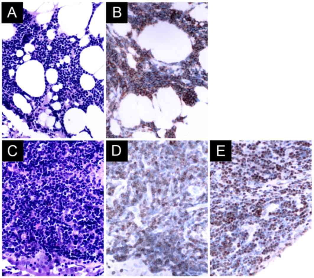
Histopathological examination revealed the presence
of the following pathological findings (Fig. 2; Table
V): i) The lymphoma cells infiltrate around reactive external
B-cell follicles to a preserved follicle mantle in a marginal zone
distribution and spread out to form larger confluent area; ii)
diffused small cell neoplasms that have slightly irregular nuclei
with moderately dispersed chromatin conspicuous or non-conspicuous
nucleoli; iii) neoplastic cells of a medium size with a high
nuclear-to-cytoplasmic ratio and with finely dispersed chromatin
and relatively prominent nucleoli, or with condensed nuclear
chromatin and no evident nucleoli; and iv) atypical lymphatic cell
infiltrates in fibrous tissue.
 | Table V.Characteristics of histopathology and
immunophenotype. |
Table V.
Characteristics of histopathology and
immunophenotype.
| Patient no. | Specimen | Histopathology | Main
immunophenotype | Final
diagnosis |
|---|
| 1 | Partial pleura | Lymphoma cells
infiltrated around reactive B-cell follicles external to a
preserved follicle mantle, in a marginal zone distribution and
spread out to form larger confluent area. | CD20+,
CD3−, CD5−, CD10−, Ki67
<5% | MALT lymphoma |
| 2 | Partial pleura | Some large atypical
cells in necrosis. | CD20+,
CD79a+, CD3−, CD10+,
BCL2+, Ki67=40% | Diffuse large
B−cell lymphoma |
| 3 | Partial pleura | Diffused small cell
neoplasm, which have slightly irregular nuclei with moderately
dispersed chromatin non−conspicuous nucleoli. | CD20+,
CD3−, CD5−, CD10−,
CD23−, CD21−, Ki67=10% | MALT lymphoma |
| 4 | Partial pleura | Diffused small cell
neoplasm, which haves lightly irregular nuclei with moderately
dispersed chromatin conspicuous nucleoli. | CD20+,
CD79a+, CD3−, CD5−, CD10
(−), Ki67=20% | MALT lymphoma |
| 5 | Partial pleura | Lymphoma cells
infiltrated around reactive B−cell follicles external to
a preserved follicle mantle, in a marginal zone distribution and
spread out to form larger confluent area. | CD20+,
CD79a+, CD3−, CD5−,
CD10−, BCL2+, Ki67=10% | MALT lymphoma |
| 6 | Partial pleura | Diffused small cell
neoplasm, which have slightly irregular nuclei with moderately
dispersed chromatin conspicuous nucleoli. | CD20+,
CD3−, CD5+, CD23+,
Calretinin−, Ki67=20% | Small lymphocytic
lymphoma |
| 7 | Partial pleura | The neoplastic
cells are of medium size with a high nuclear cytoplasmic ratio and
with finely dispersed chromatin and relatively prominent
nucleoli. | CD3+,
CD5+, CD4+, CD20−,
TdT+, CK−, PAX5−, Ki67=50% | T lymphoblastic
lymphoma |
| 8 | Partial pleura | The neoplastic
cells are of small size with a high nuclear cytoplasmic ratio and
with condensed nuclear chromatin and no evident nucleoli. | CD3+,
CD5+, CD20−, TdT+,
CK19−, TTF1−, CD56−,
Syn−, CK−, PAX5−, Ki67=90% | T lymphoblastic
lymphoma |
| 9 | Partial pleura | Atypical lymphatic
cell infiltrates in fibrous tissue. | CD20+,
CD3−, CD5−, CD10−,
BCL6−, Ki67=10% | MALT lymphoma |
| 10 | Partial pleura | Mesothelial cell
and fibroblastic proliferation with focal lymphocytes
infiltrates. | Not done | Follicular
lymphoma |
|
| Lymph nodes | Closely packed
follicles efface the nodal architecture. Neoplastic follicles are
poorly defined and absent mantle zones. | CD20+,
CD79a+, CD3−, CD5−,
CD10+, BCL2+, BCL6+, Ki67=25% |
|
Histopathological and immunophenotyping studies of
pleural biopsy samples revealed extranodal marginal zone lymphoma
of mucosa-associated lymphoid tissue (MALT lymphoma) in 5 patients,
T-lymphoblastic lymphoma in 2 patients, diffuse large B-cell
lymphoma in 1 patient, and small lymphocytic lymphoma in 1 patient.
The pleural biopsy tissue was not sufficient for immunohistological
staining in 1 patient, meaning a final diagnosis of follicular
lymphoma was established by lymph node biopsy.
Discussion
The success of MT in diagnosing pleural lymphomas
has been reported previously (13–15).
Although the present study's sample size is relatively small, the
current retrospective review is the first to specifically assess
the utility of MT in the diagnosis of MPE induced by NHL. In the
present study, the definite diagnosis of NHL in 9 out of 10
patients that presented with exudative lymphocytic pleural effusion
was finally established though MT. These patients did not have any
other indications of NHL based on medical history, physical
examination, routine blood work and CT scans of the chest and
abdomen; thoracentesis and/or closed pleural biopsy did not reach
identify the cause of their MPEs.
CT findings in pleural NHL are similar to those in
pleural metastasis or malignant mesothelioma (23–25). The
present study revealed that, in addition to pleural effusion, NHL
mainly appears as diffuse pleural thickening and localized nodules
or masses on CT scans. The pattern of pleural thickening appeared
continuous in 5 patients and discontinuous in 5 patients. In total,
5 patients had additional tumor nodules or masses of the pleura. It
appears as isolated pleural effusion with no demonstrable pleural
thickening or mass in 1 patient. A total of 6 patients had
pulmonary consolidation/infiltration and 4 patients had mediastinal
lymphadenopathy. Although thoracic CT findings are not specific and
are not informative for the diagnosis of NHL, areas of pleural
thickening or nodularity identified on CT scan can be used to guide
subsequent biopsy and thus improve the diagnostic yield (26,27).
Thoracentesis with pleural fluid cytology is usually
an early step in diagnosing MPE. In cases of MPE induced by NHL,
even experienced cytologists may not be able to make a definite
diagnosis because: i) Lymphomatous cells in pleural fluid are
always sparse (4,28); ii) lymphomatous cells in pleural fluid
are usually identical to lymphocytes in other tissues involved,
such as lymph nodes (4,29); and iii) difficulties in
morphologically distinguishing lymphomatous cells from reactive
lymphocytes arise in low-grade lymphomas, in which the involved
cells are small mature lymphocytes, as well as whenever a mixed
population of lymphomatous and reactive lymphocytes exists
(30,31).
The presence of malignant cells in the pleural fluid
of a patient with NHL does not necessarily indicate pleural
involvement with the tumor, as is invariably true in patients with
metastatic carcinoma (32). As NHL
lesions on the pleural surface are scattered, a closed pleural
biopsy adds little to the diagnosis (28,33).
Previous studies have shown that real-time image-guided pleural
biopsy is a promising technique for sampling the pleura, as it can
improve MPE diagnostic sensitivity (26,34,35). MT
has been a routine method for patients with exudative pleural
effusion that remain undiagnosed by clinical, radiologic,
laboratory or cytological investigation in Beijing Chaoyang
Hospital since June 2005. During this period, a total of 833
patients with undiagnosed pleural effusions underwent MT
successfully, and 342 of them were eventually confirmed as having
MPE (16).
Malignant neoplasm is the leading cause of death in
the adult Chinese population (36).
MPE occurs in up to 15% of patients with advanced malignancies
(37). More recently, data show that
in 342 patients with MPEs who underwent MT, the most frequent cause
of MPE was lung cancer (67.8%), followed by mesothelioma (10.2%)
and lymphoma (2.9%) (16). Although
any type of lymphoma can be involved in the pleura, diffuse large
B-cell lymphoma is most common, followed by follicular lymphoma, at
rates of 60 and ~20%, respectively (38). In the present study, histopathological
and immunohistochemical staining of pleural biopsy samples obtained
through MT confirmed MALT lymphoma in 5 patients, T lymphoblastic
lymphoma in 2 patients, diffuse large B-cell lymphoma in 1 patient,
and small lymphocytic lymphoma in 1 patient. It is possible that if
the 1 patient whose final diagnosis was based on lymph node biopsy
had provided pleural biopsy tissue suitable for immunohistochemical
study, MT would have also been able to establish a definite
diagnosis of NHL by pleural biopsy examination. Regardless, the
present study provided firm evidence to demonstrate that MT can
reach a definite diagnosis of NHL in 9 out of 10 (90%) patients
with MPE.
Although cytological investigation with
immunophenotyping of pleural fluid cells can be diagnostic of
malignancy and of the lymphoma subtype (39) and cytogenetic analysis may further
support the diagnosis and define other specific types of lymphomas
(31,39,40).
However, because a diagnosis of NHL in all 10 patients with MPE was
not initially considered, flow cytometry was not performed to
investigate the immunophenotype of pleural fluid cells in the
current study.
In NHL, lymphomatous deposits arise from lymphatic
channels and lymphoid aggregates in the subpleural connective
tissue below the visceral pleura (41). MPE may develop in patients with NHL by
three different mechanisms: i) Pleural infiltration by NHL with
shedding of cells into the pleural space; ii) lymphatic obstruction
by NHL infiltration of pulmonary and mediastinal lymph nodes; and
iii) obstruction of the thoracic duct, which results in chylothorax
(8,30). One observation of the present study is
that extensive infiltration of NHL into the pleura indicates that
the major mechanism for the development of MPE in NHL is the direct
involvement of the pleura by NHL tissue, rather than obstruction to
lymphatic flow.
The present study had three limitations: It was not
a prospective study and as such all required data were collected
and analyzed retrospectively; the sample size was quite small, with
only 10 NHL patients with MPE being included; and only data from
MPE patients with NHL, and no data from the other control group,
such as the patients with benign pleural diseases, were available,
meaning it was not possible to calculate the sensitivity and
specificity of MT in diagnosing MPE induced by NHL.
In summary, MT is safe with a high positive rate of
diagnosis of MPE induced by NHL. Its convenience and compatibility
with existing bronchoscopy means the technique has the potential to
be a more widely performed procedure. Therefore, it is worth
actively pursuing for patients with suspected MPE, if the
facilities to conduct MT are available.
Acknowledgements
The present study was supported in part by grants
from National Natural Science Foundation of China (grant nos.
91442109, 31470883 and 81270149), and in part by the Key Project Of
The Department Of Science And Technology, Beijing, China (grant no.
D141107005214003).
References
|
1
|
Marel M, Stastny B, Melínová L, Svandová E
and Light RW: Diagnosis of pleural effusions. Experience with
clinical studies, 1986 to 1990. Chest. 107:1598–1603. 1995.
View Article : Google Scholar : PubMed/NCBI
|
|
2
|
Johnston WW: The malignant pleural
effusion. A review of cytopathologic diagnoses of 584 specimens
from 472 consecutive patients. Cancer. 56:905–909. 1985. View Article : Google Scholar : PubMed/NCBI
|
|
3
|
Das DK: Serous effusions in malignant
lymphomas: A review. Diagn Cytopathol. 34:335–347. 2006. View Article : Google Scholar : PubMed/NCBI
|
|
4
|
Alexandrakis MG, Passam FH, Kyriakou DS
and Bouros D: Pleural effusions in hematologic malignancies. Chest.
125:1546–1555. 2004. View Article : Google Scholar : PubMed/NCBI
|
|
5
|
Hunter BD, Dhakal S, Voci S, Goldstein NP
and Constine LS: Pleural effusions in patients with Hodgkin
lymphoma: Clinical predictors and associations with outcome. Leuk
Lymphoma. 55:1822–1826. 2014. View Article : Google Scholar : PubMed/NCBI
|
|
6
|
Wong FM, Grace WJ and Rottino A: Pleural
effusions, ascites, pericardial effusions and Edema in Hodgkin's
disease. Am J Med Sci. 246:678–682. 1963. View Article : Google Scholar : PubMed/NCBI
|
|
7
|
Manoharan A, Pitney WR, Schonell ME and
Bader LV: Intrathoracic manifestations in non-Hodgkin's lymphoma.
Thorax. 34:29–32. 1979. View Article : Google Scholar : PubMed/NCBI
|
|
8
|
Berkman N, Breuer R, Kramer MR and
Polliack A: Pulmonary involvement in lymphoma. Leuk Lymphoma.
20:229–237. 1996. View Article : Google Scholar : PubMed/NCBI
|
|
9
|
Cordier JF, Chailleux E, Lauque D,
Reynaud-Gaubert M, Dietemann-Molard A, Dalphin JC, Blanc-Jouvan F
and Loire R: Primary pulmonary lymphomas. A clinical study of 70
cases in nonimmunocompromised patients. Chest. 103:201–208. 1993.
View Article : Google Scholar : PubMed/NCBI
|
|
10
|
Nador RG, Cesarman E, Chadburn A, Dawson
DB, Ansari MQ, Sald J and Knowles DM: Primary effusion lymphoma: A
distinct clinicopathologic entity associated with the Kaposi's
sarcoma-associated herpes virus. Blood. 88:645–656. 1996.PubMed/NCBI
|
|
11
|
Hengge UR, Ruzicka T, Tyring SK, Stuschke
M, Roggendorf M, Schwartz RA and Seeber S: Update on Kaposi's
sarcoma and other HHV8 associated diseases. Part 2: Pathogenesis,
Castleman's disease, and pleural effusion lymphoma. Lancet Infect
Dis. 2:344–352. 2002. View Article : Google Scholar : PubMed/NCBI
|
|
12
|
Aozasa K: Pyothorax-associated lymphoma. J
Clin Exp Hematop. 46:5–10. 2006. View Article : Google Scholar : PubMed/NCBI
|
|
13
|
Steiropoulos P, Kouliatsis G, Karpathiou
G, Popidou M and Froudarakis ME: Rare cases of primary pleural
Hodgkin and non-Hodgkin lymphomas. Respiration. 77:459–463. 2009.
View Article : Google Scholar : PubMed/NCBI
|
|
14
|
Oikonomou A, Giatromanolaki A, Margaritis
D, Froudarakis M and Prassopoulos P: Primary pleural lymphoma:
Plaque-like thickening of the pleura. Jpn J Radiol. 28:62–65. 2010.
View Article : Google Scholar : PubMed/NCBI
|
|
15
|
Giardino A, O'Regan KN, Hargreaves J,
Jagannathan J, Park D, Ramaiya N and Fisher D: Primary pleural
lymphoma without associated pyothorax. J Clin Oncol. 29:e413–e415.
2011. View Article : Google Scholar : PubMed/NCBI
|
|
16
|
Wang XJ, Yang Y, Wang Z, Xu LL, Wu YB,
Zhang J, Tong ZH and Shi HZ: Efficacy and safety of diagnostic
thoracoscopy in undiagnosed pleural effusions. Respiration.
90:251–255. 2015. View Article : Google Scholar : PubMed/NCBI
|
|
17
|
Wang Z, Tong ZH, Li HJ, Zhao TT, Li XY, Xu
LL, Luo J, Jin ML, Li RS and Wang C: Semi-rigid thoracoscopy for
undiagnosed exudative pleural effusions: A comparative study. Chin
Med J (Engl). 121:1384–1389. 2008.PubMed/NCBI
|
|
18
|
Wang Z, Xu LL, Wu YB, Wang XJ, Yang Y,
Zhang J, Tong ZH and Shi HZ: Diagnostic value and safety of medical
thoracoscopy in tuberculous pleural effusion. Respir Med.
109:1188–1192. 2015. View Article : Google Scholar : PubMed/NCBI
|
|
19
|
Zelenetz AD, Abramson JS, Advani RH,
Andreadis CB, Bartlett N, Bellam N, Byrd JC, Czuczman MS, Fayad LE,
Glenn MJ, et al: Non-Hodgkin's lymphomas. J Natl Compr Canc Netw.
9:484–560. 2011. View Article : Google Scholar : PubMed/NCBI
|
|
20
|
Tan D, Tan SY, Lim ST, Kim SJ, Kim WS,
Advani R and Kwong YL: Management of B-cell non-Hodgkin lymphoma in
Asia: Resource-stratified guidelines. Lancet Oncol. 14:e548–e561.
2013. View Article : Google Scholar : PubMed/NCBI
|
|
21
|
Moy MP, Levsky JM, Berko NS, Godelman A,
Jain VR and Haramati LB: A new, simple method for estimating
pleural effusion size on CT scans. Chest. 143:1054–1059. 2013.
View Article : Google Scholar : PubMed/NCBI
|
|
22
|
Light RW, Macgregor MI, Luchsinger PC and
Ball WC Jr: Pleural effusions: The diagnostic separation of
transudates and exudates. Ann Intern Med. 77:507–513. 1972.
View Article : Google Scholar : PubMed/NCBI
|
|
23
|
Parnell AP and Frew I: Case report:
Non-Hodgkin's lymphoma presenting as an encasing pleural mass. Br J
Radiol. 68:926–927. 1995. View Article : Google Scholar : PubMed/NCBI
|
|
24
|
Barahona ML, Dueñas VP, Sánchez MT and
Plaza BV: Case report. Primary mucosa-associated lymphoid tissue
lymphoma as a pleural mass. Br J Radiol. 84:e229–e231. 2011.
View Article : Google Scholar : PubMed/NCBI
|
|
25
|
Aquino SL, Chen MY, Kuo WT and Chiles C:
The CT appearance of pleural and extrapleural disease in lymphoma.
Clin Radiol. 54:647–650. 1999. View Article : Google Scholar : PubMed/NCBI
|
|
26
|
Maskell NA, Gleeson FV and Davies RJ:
Standard pleural biopsy versus CT-guided cutting-needle biopsy for
diagnosis of malignant disease in pleural effusions: A randomised
controlled trial. Lancet. 361:1326–1330. 2003. View Article : Google Scholar : PubMed/NCBI
|
|
27
|
Rahman NM, Ali NJ, Brown G, Chapman SJ,
Davies RJ, Downer NJ, Gleeson FV, Howes TQ, Treasure T, Singh S, et
al: Local anaesthetic thoracoscopy: British Thoracic Society
Pleural Disease Guideline 2010. Thorax. 65 Suppl 2:ii54–ii60. 2010.
View Article : Google Scholar : PubMed/NCBI
|
|
28
|
Celikoglu F, Teirstein AS, Krellenstein DJ
and Strauchen JA: Pleural effusion in non-Hodgkin's lymphoma.
Chest. 101:1357–1360. 1992. View Article : Google Scholar : PubMed/NCBI
|
|
29
|
Bangerter M, Hildebrand A and Griesshammer
M: Immunophenotypic analysis of simultaneous specimens from
different sites from the same patient with malignant lymphoma.
Cytopathology. 12:168–176. 2001. View Article : Google Scholar : PubMed/NCBI
|
|
30
|
Xaubet A, Diumenjo MC, Maŕin A, Montserrat
E, Estopá R, Llebaría C, Austí A and Rozman C: Characteristics and
prognostic value of pleural effusions in non-Hodgkin's lymphomas.
Eur J Respir Dis. 66:135–140. 1985.PubMed/NCBI
|
|
31
|
Pietsch JB, Whitlock JA, Ford C and Kinney
MC: Management of pleural effusions in children with malignant
lymphoma. J Pediatr Surg. 34:635–638. 1999. View Article : Google Scholar : PubMed/NCBI
|
|
32
|
Clarkson B: Relationship between cell
type, glucose concentration, and response to treatment in
neoplastic effusions. Cancer. 17:914–928. 1964. View Article : Google Scholar : PubMed/NCBI
|
|
33
|
Froudarakis ME: Diagnostic work-up of
pleural effusions. Respiration. 75:4–13. 2008. View Article : Google Scholar : PubMed/NCBI
|
|
34
|
Metintas M, Ak G, Dundar E, Yildirim H,
Ozkan R, Kurt E, Erginel S, Alatas F and Metintas S: Medical
thoracoscopy vs CT scan-guided Abrams pleural needle biopsy for
diagnosis of patients with pleural effusions: A randomized,
controlled trial. Chest. 137:1362–1368. 2010. View Article : Google Scholar : PubMed/NCBI
|
|
35
|
Koegelenberg CF, Bolliger CT, Theron J,
Walzl G, Wright CA, Louw M and Diacon AH: Direct comparison of the
diagnostic yield of ultrasound-assisted Abrams and Tru-Cut needle
biopsies for pleural tuberculosis. Thorax. 65:857–862. 2010.
View Article : Google Scholar : PubMed/NCBI
|
|
36
|
He J, Gu D, Wu X, Reynolds K, Duan X, Yao
C, Wang J, Chen CS, Chen J, Wildman RP, et al: Major causes of
death among men and women in China. N Engl J Med. 353:1124–1134.
2005. View Article : Google Scholar : PubMed/NCBI
|
|
37
|
Haas AR, Sterman DH and Musani AI:
Malignant pleural effusions: Management options with consideration
of coding, billing, and a decision approach. Chest. 132:1036–1041.
2007. View Article : Google Scholar : PubMed/NCBI
|
|
38
|
Vega F, Padula A, Valbuena JR, Stancu M,
Jones D and Medeiros LJ: Lymphomas involving the pleura: A
clinicopathologic study of 34 cases diagnosed by pleural biopsy.
Arch Pathol Lab Med. 130:1497–1502. 2006.PubMed/NCBI
|
|
39
|
Bangerter M, Hildebrand A and Griesshammer
M: Combined cytomorphologic and immunophenotypic analysis in the
diagnostic workup of lymphomatous effusions. Acta Cytol.
45:307–312. 2001. View Article : Google Scholar : PubMed/NCBI
|
|
40
|
Matolcsy A, Nádor RG, Cesarman E and
Knowles DM: Immunoglobulin VH gene mutational analysis suggests
that primary effusion lymphomas derive from different stages of B
cell maturation. Am J Pathol. 153:1609–1614. 1998. View Article : Google Scholar : PubMed/NCBI
|
|
41
|
Dynes MC, White EM, Fry WA and Ghahremani
GG: Imaging manifestations of pleural tumors. Radiographics.
12:1191–1201. 1992. View Article : Google Scholar : PubMed/NCBI
|


