Introduction
Liver cancer is a leading cause of cancer-associated
mortality and morbidity worldwide, and consists of hepatocellular
carcinoma (HCC) and hepatoblastoma (HB) (1,2). HCC and
HB are two different types of liver cancer with their own
distinctive cytological features (3).
HepG2 was previously considered to be a HCC cell line; however, in
2009, López-Terrada et al (4)
identified that HepG2 is a HB-derived cell line. The
misidentification of HepG2 cells remains widespread. The properties
of the HepG2 cell line significantly differ from those of HCC
(5); thus, HepG2 is not an
appropriate cell line to use to investigate HCC but can be used in
liver cancer research as a HB cell line (6). Thus, the aim of the present study was to
investigate the mechanism of HepG2 cells to develop a potential
novel method of treating HB.
Although the overall incidence of HB is rare, it is
the most common type of liver tumor diagnosed in children (7). Complete resection of the tumor via
surgery often leads to remission in patients with liver cancer;
however, the majority of patients are unable to undergo tumor
resection at diagnosis, as certain tumors are accompanied by
familial hepatoblastomas or adrenocortical tumors, for which
surgical resection is not the best treatment option (8). In such situations, patients undergo
chemotherapy as a first-line treatment. Cisplatin is commonly used
as a chemotherapeutic drug to treat patients with cancer, as it is
effective against cancer cells derived from solid tumors. It is a
DNA-damaging anticancer agent and induces apoptosis in cancer cells
(9). However, long-term chemotherapy
with cisplatin may induce resistance and reduce the sensitivity of
cancer cells to apoptosis, which is a major cause of the
uncontrolled progression of various types of cancer (10). Due to the development of
chemoresistance, the prognosis of patients with advanced-stage
cancer is particularly poor (11).
Current therapeutic strategies of treating intermediate and
advanced liver cancer use a combination of cisplatin and other
chemotherapy agents, including paclitaxel, gemcitabine and
cyclophosphamide. However, the mechanism underlying their
anticancer or apoptosis-promoting effects remains unclear and
numerous studies have indicated that autophagy may serve a
potential role in the anticancer effect of chemotherapeutic
reagents (12,13).
Autophagy is a process of cell self-destruction that
occurs in all eukaryotic cells. Damaged molecules and organelles
are absorbed by autophagosomes and subsequently degraded by
lysosomal hydrolases to recycle energy. Previous studies have
demonstrated that autophagy acts as a double-edged sword in the
initiation, development and metastasis of cancer, depending upon
the stage of autophagy (14,15). For example, in early stages of
autophagy, the initiation and development of cancer are stimulated,
while cancer cell death begins to occur during later stages of
autophagy. Autophagy serves an important antitumor role (16) but also protects tumor cells against
stress as an adaptive response (17,18).
Autophagy dysregulation has been associated with numerous diseases,
including cancer (19); however, the
role of autophagy in the development of cancer chemoresistance
remains unknown. The inhibition of autophagy significantly enhances
the cytocidal activity of combinatorial treatments, indicating that
the induction of autophagy is a compensatory response to
therapeutic stress (20).
The phosphoinositide 3-kinase (PI3K)/Akt/mechanistic
target of rapamycin (mTOR) signaling pathway is a key regulator of
the physiological cellular processes associated with proliferation,
differentiation, autophagy, apoptosis, motility and metabolism
(21). During energy and oxygen
deficiency, the PI3K/Akt signaling pathway negatively regulates
autophagy by mediating mTOR expression. Furthermore, AMP-activated
protein kinase (AMPK), which is a serine/threonine protein kinase,
positively regulates autophagy by phosphorylating ULK1 at specific
sites (22). By contrast, the
combination of ULK1 and mTOR inhibits ULK1 activity, which inhibits
the interaction between ULK1 and AMPK. Furthermore, it has been
demonstrated that AMPK phosphorylates the tuberous sclerosis
complex, thus inhibiting the mTOR pathway (23). However, the detailed mechanisms of
different anticancer drug treatments, all of which involve an
association between autophagy and apoptosis, remain poorly
understood.
Gingko is the only living species of the
gymnospermae group and may exhibit considerable medicinal benefits
(24). Ginkgo biloba extract (EGb) is
one of the most commonly administered therapeutic agents and is
primarily used to promote blood circulation and the dilation of
blood vessels. Chen et al (25) and the results of our previous study
(26), demonstrated that EGb
effectively inhibits cell division and induces apoptosis in the
hepatoma cell line SMMC-7721. However, to the best of our
knowledge, the effect of ginkgol on cisplatin-induced autophagy in
HB has not yet been investigated. The aim of the present study was
to investigate the effect of ginkgol C17:1 combined with
chemotherapy on autophagy and apoptosis in HepG2 cells.
Materials and methods
Reagents
NH4Cl was purchased from Sinopharm
Chemical reagent Co., Ltd. (Shanghai, China). MTT (cat. no. M5655),
Hoechst 33342 (cat. no. B2261), cisplatin (cat. no. P4394) and
3-methyladenine (3-MA; cat. no. M9281) were purchased from
Sigma-Aldrich; Merck KGaA (Darmstadt, Germany). Penicillin and
streptomycin were purchased from Harbin Pharmaceutical Group Co.,
Ltd. (Harbin, China). The Ad-mRFP-GFP-LC3 adenovirus was obtained
from the Hanheng Biotechnology Company (Shanghai, China). Mouse
monoclonal antibody (mAb) against β-actin (cat. no. sc-47778) was
purchased from Santa Cruz Biotechnology, Inc. (Dallas, TX, USA).
Rabbit mAbs against Beclin-1 (cat. no. 3495),
microtubule-associated protein 1 light chain 3 (LC3) I/II (cat. no.
12741), mTOR (cat. no. 2983), phosphorylated (p)-mTOR (Ser2448;
cat. no. 5536), p-ULK1 (Ser555; cat. no. 5869), PI3K (cat. no.
4249), p-PI3K (Tyr458; cat. no. 4228), B-cell lymphoma 2
(Bcl-2)-associated X protein (Bax; cat. no. 5023) and cleaved
caspase-3 (cat. no. 9661) were purchased from Cell Signaling
Technology, Inc. (Danvers, MA, USA). Horseradish peroxidase
(HRP)-labeledanti-mouse (cat. no. A0216) and HRP-conjugated
anti-rabbit (cat. no. A0208) secondary antibodies were purchased
from Beyotime Institute of Biotechnology (Haimen, China). Compound
C (cat. no. ab120843), rapamycin (cat. no. ab120224), rabbit mAb
SQSTM1/p62 (cat. no. ab91526) and rabbit anti-ULK1 (cat. no.
ab128859) were purchased from Abcam (Cambridge, UK). Dulbecco's
modified Eagle's medium (DMEM), fetal bovine serum (FBS),
trypsin-EDTA solution, rabbit anti-Akt (cat. no. IM001-0359),
anti-p-Akt1/2/3 (Tyr315/316/312; cat. no. IM001-0270), anti-c-JUN
N-terminal kinase (JNK; cat. no. IM001-0504), anti-p-JNK (Tyr185;
cat no. IM001-0272) and anti-Bcl-2 (cat. no. IM001-0363) were
purchased from Shanghai ExCell Biology, Inc. (Shanghai, China).
Mouse anti-AMPKα1 (cat. no. RLM3361) and anti-p-AMPKα1/2 (Thr172;
cat. no. RLM0575) were purchased from Suzhou Ruiying-Runze Trading
Co., Ltd. (Suzhou, China). Skimmed milk was purchased from Bright
Dairy & Food Co., Ltd. (Harbin, China). Ginkgol C17:1
(>96.5%, as determined by high-performance liquid
chromatography) was obtained from the Laboratory of the Food and
Biological Engineering School at Jiangsu University.
Cell line and culture
The human HB HepG2 cell line was obtained from the
Institute of Cell Biology at the Chinese Academy of Sciences
(Shanghai, China). HepG2 cells were cultured in DMEM supplemented
with 10% FBS, penicillin and streptomycin (10 mg/l) at 37°C in a
humidified atmosphere containing 5% CO2 and 95% air. The
medium was replenished every 2 days and cells were maintained at
sub-confluence.
MTT assay
HepG2 cells were seeded in a 96-well plate
(5×10³ cells/well) in a humidified atmosphere with 5%
CO2 at 37°C and treated with ginkgol C17:1 alone (0, 10,
20, 40, 80 and 160 µg/ml), cisplatin alone (0, 1, 2, 4, 8 and 16
µg/ml), or ginkgol (0, 20, 40 and 80 µg/ml) in combination with
cisplatin (2 µg/ml) for 24 h. Subsequently, 10 µl MTT (5 mg/ml) was
added to each well and cells were incubated for an additional 4–6
h. Following removal of the supernatant, dimethyl sulfoxide (100
µl/well) was added to dissolve the blue formazan crystals converted
from MTT by living cells. Cell viability was assessed using a
microplate reader at an optical density of 490 nm.
Analysis of cell autophagy
HepG2 cells were seeded in a 24-well plate
(5×10⁴ cells/well) in a humidified atmosphere with 5%
CO2 at 37°C for 12 h. Cells were infected with
Ad-mRFP-GFP-LC3 adenovirus for 12 h (MOI=50) and the DMEM medium
was replaced every 2 h. This process was performed as the weakening
of GFP expression is considered to indicate the formation of
autophagy-lysosomes and is therefore able to reflect the level of
cell autophagic flux. After 10–12 h, the cells were treated with
ginkgol C17:1 (0, 20, 40 and 80 µg/ml) and/or cisplatin (2 µg/ml)
for 24 h. Cell fixation was performed using 4% paraformaldehyde at
room temperature for 15 min. Subsequent to fixation and washing to
remove the excess water, sealing was performed using drops of
Aqueous Mounting Medium (Beyotime Institute of Biotechnology; cat.
no. P0126) were applied to each of the treated wells. The treated
cells were immobilized for 20 min at room temperature. LC3 is a
marker protein of autophagy, and LC3 proteins cluster together to
form LC3 puncta. In the present study, LC3 puncta (fluorescing
green) were observed under a fluorescence microscope at a
magnification of ×200. The numbers of LC3 puncta (green, selected
to demonstrate the intensity of autophagy, and not the autophagy
flux) from five different wells that underwent the same treatment
were calculated using ImageJ software 1.48u (National Institute of
Health, Bethesda, MD, USA) (19,27).
Hoechst 33342 staining
HepG2 cells were seeded in a 24-well plate
(5×10⁴ cells/well) in a humidified atmosphere with 5%
CO2 at 37°C. Following 24 h treatment, cells were fixed
with 4% paraformaldehyde for 2 h at room temperature. Cells were
counterstained with Hoechst 33342 reagent (5 µg/ml) for 15 min at
room temperature and stained cell nuclei were observed using a
fluorescence microscope. Subsequently, the amount of nuclei
aberration from five different wells that underwent the same
treatment was calculated using ImageJ software (National Institute
of Health).
Protein extraction
HepG2 cells (1×10⁶ cells/well) were
cultured with ginkgol C17:1 (0, 20, 40 or 80 µg/ml) or cisplatin (2
µg/ml) with/without NH4Cl (an upstream inhibitor of
autophagy; 0.535 mg/ml) or 3-MA (a downstream inhibitor of
autophagy; 0.75 mg/ml), rapamycin (an mTOR inhibitor; 100 ng/ml) or
compound C (an AMPK inhibitor; 8 µg/ml) for 24 h in 6-well plates
until they reached ~80% confluence. Subsequently, cells were lysed
in lysis buffer [50 mM Tris, 150 mM NaCl, 1 mM EDTA and 1% Triton
X-100 (pH 7.4)], washed 3 times with cold PBS and treated with 1 mM
phenylmethylsulfonyl fluoride (Shanghai Bogoo Biotechnology Co.,
Ltd., Shanghai, China) for 30 min on ice. Following transfer into
an Eppendorf tube (Corning Incorporated, Corning, NY, USA),
proteins were centrifuged at 12,000 × g for 5 min at 4°C. Finally,
the supernatant was obtained as the whole cell protein extract.
Western blot analysis
Total protein was quantified using a bicinchoninic
acid assay and proteins (5 µg/lane) underwent electrophoresis on
10% (for proteins with a molecular weight <60 kDa) or 12% (for
proteins with a molecular weight >60 kDa) SDS polyacrylamide
gels. Subsequently, the proteins were transferred onto PVDF
membranes (Bio-Rad Laboratories, Inc.). PVDF membranes were
initially blocked with 5% skimmed milk for 1 h at room temperature.
Subsequently, proteins were incubated with the following primary
antibodies: β-actin, Beclin-1, LC3I/II, p62, mTOR, p-mTOR, ULK1,
p-ULK1, PI3K, p-PI3K, Bcl-2, Bax and cleaved caspase-3 (dilution,
1:1,000), at 4°C overnight. They were subsequently incubated with
horseradish peroxidase-conjugated anti-mouse and anti-rabbit
secondary antibodies (dilution, 1:1,000) for 1 h at room
temperature. Immobilon western chemiluminescent HRP substrates (EMD
Millipore, Billerica, MA, USA) were used as visualization reagents
and bands were imaged using a MiniChemi miniature chemiluminescence
imager (Beijing Sage Creation Science Co., Ltd., Beijing,
China).
Statistical analysis
All data are presented as the mean ± standard
deviation (n=5). Independent sample one-way analysis of variance
was used to assess the differences between experimental groups.
Dunnett's test was used to perform multiple comparisons. All data
were analyzed using SPSS 16.0 software (SPSS, Inc., Chicago, IL,
USA) and P<0.05 was considered to indicate a statistically
significant difference.
Results
Ginkgol C17:1 induces autophagy and
apoptosis in HepG2 cells
Prior to investigating the effects of ginkgol C17:1
on the viability of HepG2 cells treated with cisplatin, the
cytotoxicity of ginkgol C17:1 was initially analyzed by performing
an MTT assay. It was revealed that ginkgol C17:1 significantly
decreased the viability of HepG2 cells at doses of 40, 80 and 160
µg/ml (P<0.05; Fig. 1A). This
inhibition of HepG2 cell viability occurred in a dose-dependent
manner. Inhibition was higher following treatment with higher
concentrations of ginkgol C17:1.
 | Figure 1.Ginkgol C17:1 boosts autophagy and
apoptosis in HepG2 cells. (A) Following treatment of HepG2 cells
with the indicated concentrations of ginkgol C17:1 (0, 10, 20, 40,
80 and 160 µg/ml) for 24 h, cell viability was detected by an MTT
assay. (B) The autophagy protein LC3 was examined by an
immunofluorescence assay (magnification, ×200) following infection
with GFP-RFP-LC3 adenovirus for 36 h and treatment with ginkgol
C17:1 (40 µg/ml) for 24 h; (C) the relative rate of GFP-LC3 puncta
was then quantified. (D) Following treatment of HepG2 cells with
ginkgol C17:1 (40 µg/ml) with or without NH4Cl (0.535
mg/ml) for 24 h, the expression of Beclin-1, LC3I/II and p62 were
analyzed by western blotting. Following treatment with ginkgol
C17:1 (40 µg/ml) for 24 h, the morphology of HepG2 nuclei was
observed by (E) immunofluorescence microscopy (magnification, ×200)
and they were stained with Hoechst 33342. (F) The nucleus
aberration rate was then analyzed and (G) western blot analysis was
performed to determine the expression of cleaved caspase-3, Bax and
Bcl-2. All values are presented as the mean ± standard deviation
from three independent experiments (n=5). *P<0.05 and
**P<0.01 vs. control. GFP, green fluorescent protein; RFP, red
fluorescent protein; LC3, light chain 3; Bcl-2, B-cell lymphoma 2;
Bax, Bcl-2-associated X protein. |
Autophagy and apoptosis are two different forms of
programed cell death. To determine whether ginkgol C17:1 induces
autophagy in HepG2 cells, cells were infected with Ad-mRFP-GFP-LC3
adenovirus. The results of the LC3 punctum assay revealed that
ginkgol C17:1 enhanced autophagy as the LC3 punctarate increased
(Fig. 1B and C). Furthermore, LC3
upregulation enhanced upstream autophagy and suppressed downstream
degradation. During autophagy, NH4Cl, a downstream
inhibitor of autophagy, may stimulate the accumulation of autophagy
proteins. Following the treatment of HepG2 cells with ginkgol
C17:1, the expression of Beclin-1 and LC3I/II autophagy proteins
was increased and that of autophagy downstream protein, p62, was
decreased through autophagic degradation, consequently increasing
the level of autophagy. Following treatment with NH4Cl
in combination with ginkgol C17:1, increased the expression of
autophagy proteins compared with ginkgol C17:1 monotherapy
(Fig. 1D), indicating that
NH4Cl induces autophagy. To determine whether ginkgol
C17:1 induces apoptosis in HepG2 cells, HepG2 cells that underwent
Hoechst 33342 staining were treated with ginkgol C17:1 (40 µg/ml).
Cells treated with ginkgol C17:1 exhibited nuclear shrinkage and
rupture (Fig. 1E), along with a
significantly increased nuclear aberration rate (Fig. 1F). In addition, treatment with ginkgol
C17:1 increased the expression of the pro-apoptotic proteins Bax
and cleaved caspase-3 but reduced the expression of the
anti-apoptotic protein Bcl-2 (Fig.
1G). These results indicate that ginkgol C17:1 induced the
apoptosis of HepG2 cells.
Ginkgol C17:1 reduces
cisplatin-induced autophagy and enhances cisplatin-induced
apoptosis in HepG2 cells
Following treatment of cells with various
concentrations of cisplatin, cell viability was measured using an
MTT assay (Fig. 2A). The results
revealed that 2, 4, 8 and 16 µg/ml cisplatin significantly
decreased the viability of HepG2 cells; therefore, 2 µg/ml
cisplatin was used to treat HepG2 cells in subsequent experiments.
Combination therapy of 2 µg/ml cisplatin with various
concentrations of ginkgol C17:1 significantly inhibited the
viability of HepG2 cells in a dose-dependent manner (P<0.05;
Fig. 2B). Furthermore, the results of
the LC3 punctum assay revealed that cisplatin significantly
enhanced autophagy (P<0.01); however, treatment with all
concentrations of ginkgol C17:1 significantly reduced it
(P<0.05; Fig. 2C). Additionally,
the results of western blotting demonstrated that ginkgol C17:1
inhibits cisplatin-induced autophagy (Fig. 2D). Fig.
2D demonstrates that the expression of autophagy-associated
proteins is decreased following treatment with ginkgol C17:1,
thereby indicating that this drug inhibited cisplatin-induced
autophagy. Hoechst 33342 staining revealed that ginkgol C17:1
significantly enhanced the nucleus aberration rate associated with
cisplatin-induced apoptosis in a dose-dependent manner (P<0.05;
Fig. 2E) and the results of the
western blot analysis revealed that ginkgol C17:1 and cisplatin
increased the expression of cleaved caspase-3 and the ratio of
Bax/Bcl-2 (Fig. 2F), indicating that
ginkgol C17:1 increases the rate of HepG2 cell apoptosis.
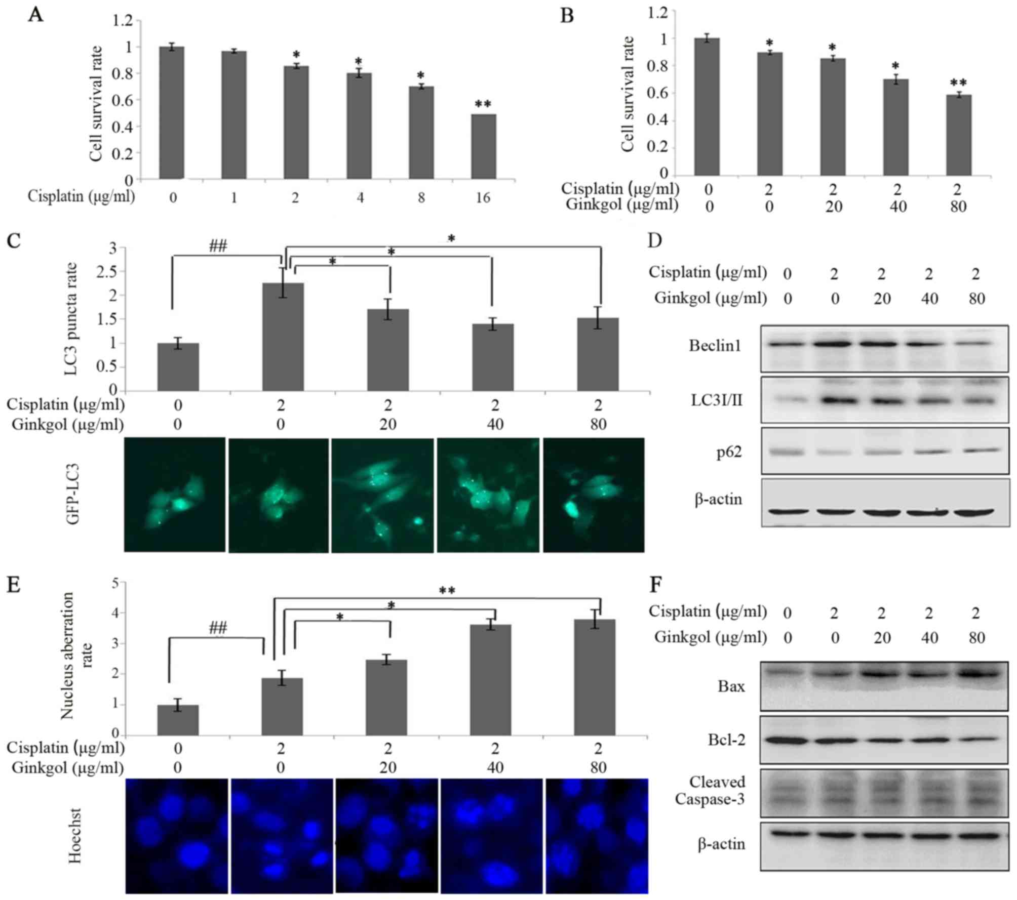 | Figure 2.Effects of ginkgol C17:1 on
cisplatin-induced autophagy and apoptosis in HepG2 cells. (A)
Following treatment with cisplatin (0, 1, 2, 4, 8 and 16 µg/ml) for
24 h, HepG2 cell viability was determined via an MTT assay. (B)
Cell viability was detected by MTT assay following treatment with
cisplatin (2 µg/ml) and ginkgol C17:1 (0, 20, 40 and 80 µg/ml) for
24 h. Following co-treatment with cisplatin and ginkgol C17:1, LC3
autophagosomes were detected by an (C) immunofluorescence assay
(magnification, ×200) and the expression of Beclin-1, LC3I/II and
p62 were analyzed by (D) a western blot assay. Under the same
conditions, the morphology of HepG2 nuclei was observed by (E)
immunofluorescence microscopy (magnification, ×200) staining with
Hoechst 33342 and the expression of cleaved caspase-3, Bax and
Bcl-2 were analyzed by (F) western blot analysis. Data are
presented as the mean ± standard deviation from three independent
experiments. *P<0.05 and **P<0.01 vs. control group, and
##P<0.01. LC3, light chain 3; Bcl-2, B-cell lymphoma
2; Bax, Bcl-2-associated X protein; GFP, green fluorescent
protein. |
Inhibition of autophagy can enhance
apoptosis during chemotherapy
To determine the association between autophagy and
apoptosis in HepG2 cells treated with cisplatin, cells were
incubated with cisplatin, 3-MA (an upstream inhibitor of autophagy)
or ginkgol C17:1. LC3 punctum and western blot assays revealed that
3-MA significantly inhibited the induction of autophagy by
cisplatin (P<0.05; Fig. 3A and B).
Furthermore, Hoechst 33342 staining indicated that 3-MA
significantly enhanced the nuclear aberration rate of cells treated
with cisplatin (P<0.05; Fig. 3C)
and the ratio of Bax/Bcl-2 (Fig. 3D).
The effects of ginkgol C17:1 on cisplatin-induced autophagy and
apoptosis were similar to 3-MA (Fig. 3E
and F). The present study revealed that ginkgol C17:1 has a
similar function to that of 3-MA in regulating cisplatin-induced
autophagy and apoptosis in HepG2 cells; ginkgol C17:1 and 3-MA
enhance cisplatin-induced apoptosis and inhibit cisplatin-induced
autophagy.
 | Figure 3.Ginkgol C17:1 enhances apoptosis and
inhibits the autophagy of cells, similar to 3MA. Following
infection of HepG2 cells with the GFP-LC3 virus and treatment with
or without cisplatin and 3-MA for 24 h, (A) LC3 autophagosomes were
detected by an immunofluorescence assay and the average rate of LC3
puncta was valued (magnification, ×200). (B) Western blot analysis
measuring the expression of Beclin-1 and p62 was performed.
Following treatment with or without cisplatin and 3MA for 24 h, (C)
nuclei morphology were observed by immunofluorescence microscopy
(magnification, ×200) following staining with Hoechst 33342 and (D)
the expression of Beclin-1, LC3 and p62 were measured by western
blotting. Following treatment of HepG2 with cisplatin with or
without ginkgol C17:1 and 3MA for 24 h, western blot analysis
investigating the expression of (E) Beclin-1 and p62 and (F) Bax
and Bcl-2 was performed. Data are presented as the mean ± standard
deviation from five independent experiments. *P<0.05 and
##P<0.01. Cisplatin, 2 µg/ml; 3MA, 0.75 mg/ml and
ginkgol C17:1, 40 µg/ml. GFP, green fluorescent protein; LC3, light
chain 3; Bcl-2, B-cell lymphoma 2; Bax, Bcl-2-associated X protein;
3-MA, 3-methyladenine. |
Ginkgol C17:1 regulates
cisplatin-induced autophagy by inhibiting the AMPK/ULK1
pathway
AMPK is one of the primary stress-sensing enzymes
and actively regulates cell metabolism and proliferation. The
AMPK/ULK1 pathway serves an important role in regulating autophagy.
A western blot assay was therefore performed to investigate the
effect of ginkgol C17:1 on the AMPK/ULK1 pathway in HepG2 cells.
The expression of p-AMPK was increased in cells treated with
cisplatin, indicating that cisplatin induces autophagy (Fig. 4A). However, treatment with ginkgol
C17:1 decreased the expression of p-AMPK (Thr172) and p-ULK1
(Ser555) in cells treated with cisplatin in a dose-dependent
manner. In addition, the AMPK inhibitor compound C reduced the
expression of p-AMPK and p-ULK1 (Fig.
4B). The expression of Beclin-1 and LC3 I/II were increased
following treatment with cisplatin and this was inhibited following
treatment with ginkgol C17:1 (Fig.
4B). However, the effects of ginkgol C17:1 were reversed
following the inhibition of AMPK by compound C. By contrast, the
expression of Bax and Bcl-2 remained unchanged following treatment
with compound C, indicating that ginkgol C17:1 still induced a
pro-apoptotic effect in cells treated with compound C and ginkgol
C17:1 (Fig. 4B). These data imply
that the AMPK/ULK1 signaling pathway is a vital regulatory pathway
involved in cisplatin-induced autophagy on HepG2 cells.
 | Figure 4.Effect of ginkgol C17:1 on the
AMPK/ULK1 pathway. (A) Following co-treatment of HepG2 cells with
cisplatin (2 µg/ml) and ginkgol C17:1 (0, 20, 40 and 80 µg/ml) for
24 h, western blot analysis was performed to determine the
expression of AMPK, p-AMPK (Thr172), ULK1 and p-ULK1 (Ser555). (B)
Following treatment of cells with cisplatin (2 µg/ml) with or
without ginkgol C17:1 (40 µg/ml) and compound C (8 µg/ml) for 24 h,
western blot analysis was performed to measure the expression of
p-AMPK (Thr172), p-ULK1 (Ser555), Beclin-1, LC3I/II, Bax and Bcl-2.
AMPK, AMP-activated protein kinase; p-, phosphorylated; LC3, light
chain 3; Bcl-2, B-cell lymphoma 2; Bax, Bcl-2-associated X
protein. |
Ginkgol C17:1 regulates
cisplatin-induced apoptosis by inhibiting the PI3K/Akt/mTOR
pathway
The PI3K/Akt/mTOR signaling pathway is involved in
the molecular biological mechanisms by which autophagy or apoptosis
occur (21). The expression of p-PI3K
(Tyr458), p-Akt (Tyr315) and p-mTOR (Ser2448) decreased in a
dose-dependent manner following the co-culture of HepG2 cells with
cisplatin and ginkgol C17:1 (Fig.
5A). To determine whether ginkgol C17:1 affects the autophagy
and apoptosis induced by cisplatin via the PI3K/Akt/mTOR signaling
pathway, HepG2 cells were treated with rapamycin (an mTOR
inhibitor). The effect of ginkgol C17:1 on the expression of Bax
and Bcl-2 activated by cisplatin was attenuated following rapamycin
treatment, however, the expression of the autophagy proteins
Beclin-1 and LC3I/II remained evidently unchanged following
co-treatment with or without ginkgol C17:1, and following the
inhibition of mTOR signaling by rapamycin (Fig. 5B). These data imply that ginkgol C17:1
stimulated cisplatin-induced apoptosis by inhibiting the
PI3K/Akt/mTOR signaling pathway in HepG2 cells, but did not affect
cisplatin-induced autophagy.
 | Figure 5.Influence of ginkgol C17:1 on the
PI3K/Akt/mTOR pathway. (A) Following co-treatment of HepG2 cells
with cisplatin (2 µg/ml) and ginkgol C17:1 (0, 20, 40 and 80 µg/ml)
for 24 h, western blot analysis was performed to measure the
expression of proteins in the upstream pathway: PI3K and p-PI3K
(Tyr458), Akt and p-Akt (Tyr315), mTOR and p-mTOR (Ser2448), JNK
and p-JNK (Tyr185). (B) Following treatment of cells were with
cisplatin (2 µg/ml), with or without ginkgol C17:1 (40 µg/ml) and
rapamycin (100 ng/ml) for 24 h, western blotting was performed to
measure the expression of p-mTOR (Ser2448), Beclin-1, LC3I/II, Bax
and Bcl-2. p-, phosphorylated; PI3K, phosphoinositide 3-kinase;
mTOR, mechanistic target of rapamycin; LC3, light chain 3; Bcl-2,
B-cell lymphoma 2; Bax, Bcl-2-associated X protein. |
Discussion
The theory of substrate selectivity in autophagy has
received increasing recognition. This differs from the original
understanding of autophagy, which was that autophagy was a
non-specific process of self-destruction (28). In its ‘active form’, LC3II assists in
elongating the phagophore membrane and in the recruitment of
damaged or degraded organelles to the phagophore (29). Beclin-1 is sometimes essential for
autophagy (27). However, it has been
suggested that Beclin-1 expression is only necessary for autophagy
occurring in certain types of cells and that it may not be required
for autophagy induced by cytotoxic compounds, including
resveratrol, staurosporin or gossypol (30). To the best of our knowledge, the
impact of autophagy on antitumor therapy has not yet been
demonstrated. It has been demonstrated that autophagy and apoptosis
are distinctive; however, the two processes are associated and
cross-talk also occurs between them, where by individual proteins
may serve functions in both processes (31). The cross-talk that occurs between them
is partly complicated by the fact that they share numerous common
regulatory molecules, including Bcl-2 and Beclin-1, as well as the
PI3K/Akt/mTOR signaling pathway (32,33).
Cisplatin is commonly used in chemotherapeutic
regimes to treat patients with cancer. However, the efficacy of
cisplatin is limited due to the development of drug resistance in
many patients and the fact that it causes multiple side effects;
therefore, it is important to improve the pharmacological effect of
cisplatin (34). It has been
demonstrated that cisplatin induces autophagy and apoptosis in
cancer cells (35,36), which has also been determined in the
present study. Apoptosis directly causes the death of cancer cells;
by contrast, autophagy may have two effects on tumors; an antitumor
or protective effect (14,37). Therefore, autophagy may beinvolved in
the regulation of tumor growth. A previous study demonstrated that
ginkgol ultimately induced apoptosis by activating the expression
of caspases via inhibition ofthe PI3K/Akt pathway (38). In the present study, ginkgol C17:1 not
only induced apoptosis in HepG2 cells but also induced autophagy.
Following co-treatment with cisplatin, ginkgol C17:1 stimulated
cisplatin-induced apoptosis, however, it also inhibited
cisplatin-induced autophagy. Previous studies have demonstrated
that inhibitors of autophagy administered in combination with
anti-cancer agents may enhance chemosensitization in human cancer
cells (39–41). The resultsof the present study
indicating the effects of ginkgol C17:1 on cisplatin-induced
autophagy are in accordance with the results of a study by Bao
et al (33); they suggest that
ginkgol C17:1 may overcome cisplatin resistance.
The present study revealed that ginkgol C17:1 and
cisplatin may affect the PI3K/Akt/mTOR and AMPK/ULK1 signaling
pathways, which are two primary methods of regulating autophagy and
apoptosis (42–44). Co-treatment with ginkgol C17:1 and
cisplatin inhibited these pathways, while treatment with ginkgol
C17:1 or cisplatin alone caused the AMPK/ULK1 pathway to become
activated. However, monotherapy with either drug resulted in the
inhibition of the PI3K/Akt/mTOR pathway.
AMPK is a multi-functional protein kinase; thus, its
activation may promote apoptosis by inducing the phosphorylation of
p53, activating the nuclear factor-κB and c-myc, or the c-Jun
pathway (45–47). Based on this function, activation of
the AMPK pathway may block tumor cell proliferation and regulate
tumor development. Thus activating the AMPK pathway may be a novel
method of treating patients with cancer.
It has been demonstrated that AMPK activates
autophagy by directly and indirectly activating ULK1 (48). AMPK indirectly activates ULK1 by
inhibiting mTOR, which phosphorylates and inhibits ULK1 to disrupt
the interaction between AMPK and ULK1 (49). However, as feedback, activated ULK1
also phosphorylates AMPK and inhibits its activation, providing a
potential negative-feedback loop resulting in autophagy induction
(50). Following co-treatment with
ginkgol C17:1 and cisplatin, the AMPK/ULK1 pathway was inhibited
and the negative-feedback loop was activated by ginkgol C17:1.
Due to the multifaceted role of autophagy in cancer
cells, manipulating the activation and inhibition of AMPK may be
developed as an additional therapeutic strategy for cancer
treatment. Enhancing AMPK activity may be a novel method of
sensitizing tumor cells to radiotherapy and chemotherapy (51). However, previous studies have
demonstrated that autophagy also provides a survival advantage
against cancer therapies by inducing AMPK and suppressing the
apoptosis pathway (52,53). Thus, suppressing autophagy enhances
cell death by inhibiting AMPK. In the present study, ginkgol C17:1
reduced autophagy by inhibiting the activity of AMPK to induce cell
death.
When the two signaling pathways were interrupted, it
was revealed that rapamycin (mTOR inhibitor) blocked the action of
ginkgol C17:1 in cisplatin-induced apoptosis rather than autophagy.
This indicates that ginkgol C17:1 enhances the apoptosis induced by
cisplatin via the PI3K/Akt/mTOR signaling pathway. Additionally,
compound C (an AMPK inhibitor) blocked the action of ginkgol C17:1
in cisplatin-induced autophagy rather than apoptosis, indicating
that the AMPK/ULK1 pathway is a vital regulatory pathway involved
in cisplatin-induced autophagy in HepG2 cells. These signaling
pathways are associated with autophagy and apoptosis; however,
studies highlight that different stimuli may induce autophagy via
different mechanisms under different conditions (41,54). This
may have therapeutic implications in the treatment of HB.
In conclusion, autophagy and apoptosis are two
important mechanisms involved in cell regulation. In the present
study, treatment with ginkgol C17:1 affected cancer cells primarily
through autophagy and apoptosis. Furthermore, the present study
indicated that ginkgol C17:1 inhibits cisplatin-induced autophagy
via AMPK/ULK1 signaling and increases cisplatin-induced apoptosis
via the PI3K/Akt/mTOR pathway. Ultimately, ginkgol C17:1
significantly inhibited human liver cancer cells and enhanced the
anticancer activity of cisplatin. Furthermore, the present study
provides an important theoretical basis for the future of antitumor
research. To further assess the antitumor effect of ginkgol C17:1
and potentially develop it as a novel cancer treatment, further
studies are required to identify the antitumor effects of ginkgol
C17:1.
Acknowledgements
The present study was supported by the National
Natural Science Foundation of China (grant no. 81372404); the
College Students' Scientific Research Project of Jiangsu University
(grant no. 14A056) and the Zhenjiang Social Development Project
(grant no. SH2015072).
References
|
1
|
Farazi PA and Depinho RA: Hepatocellular
carcinoma pathogenesis: From genes to environment. Nat Rev Cancer.
6:674–687. 2006. View
Article : Google Scholar : PubMed/NCBI
|
|
2
|
Khaderi S, Guiteau J, Cotton RT, O'Mahony
C, Rana A and Goss JA: Role of liver transplantation in the
management of hepatoblastoma in the pediatric population. World J
Transplant. 4:294–298. 2014. View Article : Google Scholar : PubMed/NCBI
|
|
3
|
Abdul HA, Lai ML and Phaik LC: Tissue
microarray immunohistochemical profiles of p53 and pRB in
hepatocellular carcinoma and hepatoblastoma. Asian Pac J Cancer
Prev. 15:3959–3963. 2014. View Article : Google Scholar : PubMed/NCBI
|
|
4
|
López-Terrada D, Cheung SW, Finegold MJ
and Knowles BB: Hep G2 is a hepatoblastoma-derived cell line. Hum
Pathol. 40:1512–1517. 2009. View Article : Google Scholar
|
|
5
|
Pang RTK, Poon TCW, Wong N, Lai PSB, Wong
NLY, Chan CML, Yu JWS, Chan ATC and Sung JJY: Comparison of protein
expression patterns between hepatocellular carcinoma cell lines and
a hepatoblastoma cell line. Clin Proteomics. 1:313–331. 2004.
View Article : Google Scholar
|
|
6
|
Rishi RR, Kimberlee KS, Ruth IH, Michael
SB, Max B and Lisa EH: Hepatoblastoma: A need for cell lines and
tissue banks to develop targeted drug therapies. Front Pediatr.
4:222016.PubMed/NCBI
|
|
7
|
Pateva IB, Egler RA and Stearns DS:
Hepatoblastoma in an 11-year-old: Case report and a review of the
literature. Medicine (Baltimore). 96:e58582017. View Article : Google Scholar : PubMed/NCBI
|
|
8
|
Tomlinson GE and Kappler R: Genetics and
epigenetics of hepatoblastoma. Pediatr Blood Cancer. 59:785–792.
2012. View Article : Google Scholar : PubMed/NCBI
|
|
9
|
Boulikas T and Vougiouka M: Cisplatin and
platinum drugs at the molecular level (Review). Oncol Rep.
10:1663–1682. 2003.PubMed/NCBI
|
|
10
|
Amable L: Cisplatin resistance and
opportunities for precision medicine. Pharmacol Res. 106:27–36.
2016. View Article : Google Scholar : PubMed/NCBI
|
|
11
|
Muggia F: Platinum compounds 30 years
after the introduction of cisplatin: Implications for the treatment
of ovarian cancer. Gynecol Oncol. 112:275–281. 2009. View Article : Google Scholar : PubMed/NCBI
|
|
12
|
Zhang HQ, Fang N, Liu XM, Xiong SP, Liao
YQ, Jin WJ, Song RF and Wan YY: Antitumor activity of chloroquine
in combination with Cisplatin in human gastric cancer xenografts.
Asian Pac J Cancer Prev. 16:3907–3912. 2015. View Article : Google Scholar : PubMed/NCBI
|
|
13
|
García-Cano J, Ambroise G, Pascual-Serra
R, Carrión MC, Serrano-Oviedo L, Ortega-Muelas M, Cimas FJ, Sabater
S, Ruiz-Hidalgo MJ, Sanchez Perez I, et al: Exploiting the
potential of autophagy in cisplatin therapy: A new strategy to
overcome resistance. Oncotarget. 6:15551–15565. 2015. View Article : Google Scholar : PubMed/NCBI
|
|
14
|
Lu SZ and Harrison-Findik DD: Autophagy
and cancer. World J Biol Chem. 4:64–70. 2013. View Article : Google Scholar : PubMed/NCBI
|
|
15
|
Mizushima N and Komatsu M: Autophagy:
Renovation of cells and tissues. Cell. 147:728–741. 2011.
View Article : Google Scholar : PubMed/NCBI
|
|
16
|
Mathew R, Karp CM, Beaudoin B, Vuong N,
Chen G, Chen HY, Bray K, Reddy A, Bhanot G, Gelinas C, et al:
Autophagy suppresses tumorigenesis through elimination of p62.
Cell. 137:1062–1075. 2009. View Article : Google Scholar : PubMed/NCBI
|
|
17
|
Liu M, Ma S, Liu M, Hou Y, Liang B, Su X
and Liu X: Synergistic killing of lung cancer cells by cisplatin
and radiation via autophagy and apoptosis. Oncol Lett. 7:1903–1910.
2014.PubMed/NCBI
|
|
18
|
Sakamoto A and Iwamoto Y: Current status
and perspectives regarding the treatment of osteo-sarcoma:
Chemotherapy. Rev Recent Clin Trials. 3:228–231. 2008. View Article : Google Scholar : PubMed/NCBI
|
|
19
|
Choi AM, Ryter SW and Levine B: Autophagy
in human health and disease. N Engl J Med. 368:651–662. 2013.
View Article : Google Scholar : PubMed/NCBI
|
|
20
|
Chen J, Wang Q, Yin FQ, Zhang W, Yan LH
and Li L: MTRR silencing inhibits growth and cisplatin resistance
of ovarian carcinoma via inducing apoptosis and reducing autophagy.
Am J Transl Res. 7:1510–1527. 2015.PubMed/NCBI
|
|
21
|
Vanhaesebroeck B, Stephens L and Hawkins
P: PI3K signalling: The path to discovery and understanding. Nat
Rev Mol Cell Biol. 13:195–203. 2012. View
Article : Google Scholar : PubMed/NCBI
|
|
22
|
Tsai JP, Lee CH, Ying TH, Lin CL, Lin CL,
Hsueh JT and Hsieh YH: Licochalcone A induces autophagy through
PI3K/Akt/mTOR inactivation and autophagy suppression enhances
Licochalcone A-induced apoptosis of human cervical cancer cells.
Oncotarget. 6:28851–28866. 2015. View Article : Google Scholar : PubMed/NCBI
|
|
23
|
Laplante M and Sabatini DM: mTOR signaling
in growth control and disease. Cell. 149:274–293. 2012. View Article : Google Scholar : PubMed/NCBI
|
|
24
|
Nash KM and Shah ZA: Current perspectives
on the beneficial role of ginkgo biloba in neurological and
cerebrovascular disorders. Integr Med InsIghts. 10:1–9. 2015.
View Article : Google Scholar : PubMed/NCBI
|
|
25
|
Chen Q, Yang GW and An LG: Apoptosis of
hepatoma cells SMMC-7721 induced by ginkgo biloba seed
polysaccharide. World J Gastroenterol. 8:832–836. 2002. View Article : Google Scholar : PubMed/NCBI
|
|
26
|
Yang XM, Wang YF, Li YY and Ma HL: Thermal
stability of ginkgolic acids from ginkgo biloba and the effects of
ginkgol C17:1 on the apoptosis and migration of SMMMC7721 cells.
Fitoterapia. 98:66–76. 2014. View Article : Google Scholar : PubMed/NCBI
|
|
27
|
Ma X, Liu H, Foyi SR, Godar RJ, Weinheimer
CJ, Hill JA and Diwan A: Impaired autophagosome clearance
contributes to cardiomyocyte death in ischemia/reperfusion injury.
Circulation. 125:3170–3181. 2012. View Article : Google Scholar : PubMed/NCBI
|
|
28
|
Reggiori F, Komatsu M, Finley K and
Simonsen A: Autophagy: More than a nonselective pathway. Int J Cell
Biol. 2012:2196252012. View Article : Google Scholar : PubMed/NCBI
|
|
29
|
Kabeya Y, Mizushima N, Ueno T, Yamamoto A,
Kirisako T, Noda T, Kominami E, Ohsumi Y and Yoshimori T: LC3, a
mammalian homologue of yeast Apg8p, is localized in autophagosome
membranes after processing. EMBO J. 19:5720–5728. 2000. View Article : Google Scholar : PubMed/NCBI
|
|
30
|
Lindqvist LM, Simon AK and Baehrecke EH:
Current questions and possible controversies in autophagy. Cell
Death Discov. 1:pii: 150362015. View Article : Google Scholar
|
|
31
|
Zhang Z, Shao Z, Xiong L and Yang S:
Inhibition of autophagy enhances cisplatin-induced apoptosis in the
MG63 human osteosarcoma cell line. Oncol Lett. 10:2941–2946.
2015.PubMed/NCBI
|
|
32
|
Amaravadi RK and Thompson CB: The roles of
therapy-induced autophagy and necrosis in cancer treatment. Clin
Cancer Res. 13:7271–7279. 2007. View Article : Google Scholar : PubMed/NCBI
|
|
33
|
Bao L, Jaramillo MC, Zhang Z, Zheng Y, Yao
M, Zhang DD and Yi X: Induction of autophagy contributes to
cisplatin resistance in human ovarian cancer cells. Mol Med Rep.
11:91–98. 2015. View Article : Google Scholar : PubMed/NCBI
|
|
34
|
Le Grazie M, Biagini MR, Tarocchi M,
Polvani S and Galli A: Chemotherapy for hepatocellular carcinoma:
The present and the future. World J Hepatol. 9:907–920. 2017.
View Article : Google Scholar : PubMed/NCBI
|
|
35
|
Cavallo F, Feldman DR and Barchi M:
Revisiting DNA damage repair, p53-mediated apoptosis and cisplatin
sensitivity in germ cell tumors. Int J Dev Biol. 57:273–280. 2013.
View Article : Google Scholar : PubMed/NCBI
|
|
36
|
Zhang HQ, He B, Fang N, Lu S, Liao YQ and
Wan YY: Autophagy inhibition sensitizes cisplatin cytotoxicity in
human gastric cancer cell line SGC7901. Asian Pac J Cancer Prev.
14:4685–4688. 2013. View Article : Google Scholar : PubMed/NCBI
|
|
37
|
Kimura T, Takabatake Y, Takahashi A and
Isaka Y: Chloroquine in cancer therapy: A double-edged sword of
autophagy. Cancer Res. 73:3–7. 2013. View Article : Google Scholar : PubMed/NCBI
|
|
38
|
Li YY, Liu J, Yang XM, Dong Y, Liu YL and
Chen M: Ginkgol C17:1 inhibits tumor growth by blunting the
EGF-PI3K/Akt signaling pathway. J Biomed Res. 31:232–239.
2017.PubMed/NCBI
|
|
39
|
Tang JY, Dai T, Zhang H, Xiong WJ, Xu MZ,
Wang XJ, Tang QH, Chen B and Xu M: GDC-0980-induced apoptosis is
enhanced by autophagy inhibition in human pancreatic cancer cells.
Biochem Biophys Res Commun. 453:533–538. 2014. View Article : Google Scholar : PubMed/NCBI
|
|
40
|
Wu HM, Jiang ZF, Ding PS, Shao LJ and Liu
RY: Hypoxia-induced autophagy mediates cisplatin resistance in lung
cancer cells. Sci Rep. 5:122912015. View Article : Google Scholar : PubMed/NCBI
|
|
41
|
Yan MM, Ni JD, Song D, Ding M and Huang J:
Interplay between unfolded protein response and autophagy promotes
tumor drug resistance. Oncol Lett. 10:1959–1969. 2015.PubMed/NCBI
|
|
42
|
Luo J, Manning BD and Cantley LC:
Targeting the PI3K-Akt pathway in human cancer: Rationale and
promise. Cancer Cell. 4:257–262. 2003. View Article : Google Scholar : PubMed/NCBI
|
|
43
|
Li F, Zeng J, Gao Y, Guan Z, Ma Z, Shi Q,
Du C, Jia J, Xu S, Wang X, et al: G9a inhibition induces autophagic
cell death via AMPK/mTOR pathway in bladder transitional cell
carcinoma. PLoS One. 10:e01383902015. View Article : Google Scholar : PubMed/NCBI
|
|
44
|
Nikoletopoulou V, Markaki M, Palikaras K
and Tavernarakis N: Crosstalk between apoptosis, necrosis and
autophagy. Biochim Biophys Acta. 1833:3448–3459. 2013. View Article : Google Scholar : PubMed/NCBI
|
|
45
|
Okoshi R, Ozaki T, Yamamoto H, Ando K,
Koida N, Ono S, Koda T, Kamijo T, Nakagawara A and Kizaki H:
Activation of AMP-activated protein kinase induces p53-dependent
apoptotic cell death in response to energetic stress. J Biol Chem.
283:3979–3987. 2008. View Article : Google Scholar : PubMed/NCBI
|
|
46
|
Kefas BA, Cai Y, Ling Z, Heimberg H, Hue
L, Pipeleers D and Van de Casteele M: AMP-activated protein kinase
can induce apoptosis of insulin-producing MIN6 cells through
stimulation of c-Jun-N-terminal kinase. J Mol Endocrinol.
30:151–161. 2003. View Article : Google Scholar : PubMed/NCBI
|
|
47
|
Nieminen AI, Eskelinen VM, Haikala HM,
Tervonen TA, Yan Y, Partanen JI and Klefström J: Myc-induced
AMPK-phospho p53 pathway activates Bak to sensitize mitochondrial
apoptosis. Proc Natl AcadSci USA. 110:pp. E1839–E1848. 2013;
View Article : Google Scholar
|
|
48
|
Kim J, Kundu M, Viollet B and Guan KL:
AMPK and mTOR regulate autophagy through direct phosphorylation of
Ulk1. Nat Cell Biol. 13:132–141. 2011. View Article : Google Scholar : PubMed/NCBI
|
|
49
|
Jeon SM: Regulation and function of AMPK
in physiology and diseases. Exp Mol Med. 48:e2452016. View Article : Google Scholar : PubMed/NCBI
|
|
50
|
Löffler AS, Alers S, Dieterle AM, Keppeler
H, Franz-Wachtel M, Kundu M, Campbell DG, Wesselborg S, Alessi DR
and Stork B: Ulk1-mediated phosphorylation of AMPK constitutes a
negative regulatory feedback loop. Autophagy. 7:696–706. 2011.
View Article : Google Scholar : PubMed/NCBI
|
|
51
|
Sanli T, Steinberg GR, Singh G and
Tsakiridis T: AMP-activated protein kinase (AMPK) beyond
metabolism: A novel genomic stress sensor participating in the DNA
damage response pathway. Cancer Biol Ther. 15:156–169. 2014.
View Article : Google Scholar : PubMed/NCBI
|
|
52
|
Kondo Y, Kanzawa T, Sawaya R and Kondo S:
The role of autophagy in cancer development and response to
therapy. Nat Rev Cancer. 5:726–734. 2005. View Article : Google Scholar : PubMed/NCBI
|
|
53
|
Xie BS, Zhao HC, Yao SK, Zhuo DX, Jin B,
Lv DC, Wu CL, Ma DL, Gao C, Shu XM and Ai ZL: Autophagy inhibition
enhances etoposide-induced cell death in human hepatoma G2 cells.
Int J Mol Med. 27:599–606. 2011.PubMed/NCBI
|
|
54
|
Wu T, Wang MC, Jing L, Liu ZY, Guo H, Liu
Y, Bai YY, Cheng YZ, Nan KJ and Liang X: Autophagy facilitates lung
adenocarcinoma resistance to cisplatin treatment by activation of
AMPK/mTOR signaling pathway. Drug Des Devel Ther. 9:6421–6431.
2015. View Article : Google Scholar : PubMed/NCBI
|


