Introduction
Colorectal cancer (CRC) is one of the leading causes
of cancer-associated mortality in the USA and East Asia (1–3). While
segmental surgical resection of the colon or rectum is the standard
treatment, surgery alone is not sufficient to cure the majority of
patients with CRC (4). Patients who
experience local or distant recurrences following surgery require
chemotherapy (4). Furthermore,
patients with stage III CRC, as well as selected patients with
stage II, also require adjuvant chemotherapy to reduce the risk of
recurrence (5). 5-Fluorouracil (5-FU)
has been used as a standard chemotherapy regimen in the past few
decades and remains a key drug in several recently introduced
combination chemotherapies (6).
FOLFOX [folinic acid (LV), 5-FU and oxaliplatin] and FORFIRI (LV,
5-FU and irinotecan) provide a high response rate, and their
therapeutic effect is evident in numerous patients with CRC
(7–10). However, drug resistance to these
combination chemotherapies has finally occurred and may be a major
problem in the treatment of CRC (11).
One of the mechanisms of drug resistance is the
transportation of anticancer drugs out of cells, and the major drug
transporters are the adenosine triphosphate (ATP)-binding cassette
(ABC) and solute carrier (SLC) superfamilies (12). ABC transporters contain membrane
proteins that translocate a variety of substrates across extra- and
intracellular membranes, and use the energy from ATP
binding/hydrolysis to transport multiple types of drugs, including
anticancer agents (13). In
vitro studies have revealed that more than half of ABC
transporters are associated with chemotherapy failure (14). The most characterized ABC transporter
is ABCB1 (also known as multidrug resistance protein 1), the
overexpression of which in tumor cells has been associated with
drug resistance to molecularly targeted and chemotherapy drugs
(15,16). By contrast, SLC drug transporters do
not rely on ATP and uptake small molecules into cells (12). While cancer cells express several
different types of ABC transporters to gain drug resistance
(17), the expression of SLC
transporters in malignant tumors remains unknown. SLC transporters
include almost 400 members organized into 52 families, and it has
been reported that certain transporters, including SLC22A6 and
SLC22A8 from the SLC22 family, transport methotrexate (18). The present study suggested that SLC
transporters may also be associated with drug resistance (18). However, the molecular functions of the
majority of SLC transporters in cancer remain to be elucidated.
The SLC37 family consists of four proteins, namely
A1, A2, A3 and A4, which are involved in sugar-phosphate exchange
(19). SLC37A4 is known as a
glucose-6-phosphate (G6P) transporter that performs an important
role in glucose production (19);
however, the functional role of other SLC37 family genes is not yet
fully understood. However, since this transporter functions in
glycolipid metabolism, it has been hypothesized that abundant
expression of SLC37 family genes is involved in cancer development
(19). It is known that oncogenes and
tumor-suppressor genes dysregulate glucose transport and energy
metabolism pathways due to satisfy the requirement of a large
amount of glucose for cancer cells (20,21).
Therefore, a previous study investigated the roles of SLC
transporters and expected them to be a useful therapeutic target
(12).
In the current study, SLC37 family gene expression
was investigated in patients with CRC. Furthermore, to clarify the
functional role of SLC37 genes, the association between SLC37 gene
expression and glycolipid metabolism was investigated.
Materials and methods
Clinical samples of patients
A total of 231 surgical specimens obtained from 231
patients with CRC who had undergone surgical resection at Fukushima
Medical University Hospital (Fukushima, Japan) between January 1991
and December 2007 were included in the present study. In 10 cases,
messenger RNAs (mRNAs) were extracted from cancer tissues and
adjacent non-tumor tissues. Information regarding age, sex,
tumor-node-metastasis stage and pathological diagnosis, including
lymphatic and venous invasion, was retrospectively collected
(Tables III and IV). The carcinomas at the time of primary
tumor resection were staged according to the Union for
International Cancer Control classification (7th classification)
(22,23). Written informed consent was obtained
from all patients. The present study was approved by the ethics
committee of Fukushima Medical University (approval no. 1476).
 | Table III.Clinicopathological factors and
SLC37A1 IHC expression. |
Table III.
Clinicopathological factors and
SLC37A1 IHC expression.
|
| SLC37A1 IHC |
|---|
|
|
|
|---|
| Variables | Total (n) | Positive (n) | Negative (n) | P-value |
|---|
| Total | 231 | 157 | 74 |
|
| Age, (years) |
|
|
| 0.542 |
|
≥60 | 162 | 108 | 54 |
|
|
<60 | 69 | 49 | 20 |
|
| Sex |
|
|
| 1 |
|
Male | 136 | 92 | 44 |
|
|
Female | 95 | 65 | 30 |
|
| Stage |
|
|
| 0.479 |
| 0 |
5 |
4 | 1 |
|
| I | 36 | 23 | 13 |
|
| II | 85 | 58 | 27 |
|
|
III | 67 | 42 | 25 |
|
| IV | 38 | 30 | 8 |
|
| Tumor location |
|
|
| 0.179 |
|
Right | 78 | 58 | 20 |
|
|
Left | 133 | 99 | 54 |
|
| Histology |
|
|
| 0.479 |
|
Well-differentiated | 100 | 64 | 36 |
|
|
Moderately-differentiated | 102 | 74 | 31 |
|
|
Poorly-differentiated |
7 |
4 | 3 |
|
|
Mucinous | 19 | 15 | 4 |
|
| Depth |
|
|
| 0.888 |
| T1 | 28 | 18 | 10 |
|
| T2 | 27 | 18 | 9 |
|
| T3 | 159 | 108 | 51 |
|
| T4 | 17 | 13 | 4 |
|
| Lymphatic
invasion |
|
|
| 0.729 |
|
Absent | 47 | 31 | 16 |
|
|
Present | 184 | 126 | 58 |
|
| Venous
invasion |
|
|
| 0.034 |
|
Absent | 46 | 25 | 21 |
|
|
Present | 185 | 132 | 53 |
|
| Lymph node
metastasis |
|
|
| 0.315 |
|
Negative | 127 | 97 | 40 |
|
|
Positive | 94 | 60 | 34 |
|
| Liver
metastasis |
|
|
| 0.013 |
|
Negative | 200 | 130 | 70 |
|
|
Positive | 31 | 27 | 4 |
|
| Table IV.Univariate and multivariate Cox
regression analyses of SLC37A1 expression levels and other clinical
covariates. |
Table IV.
Univariate and multivariate Cox
regression analyses of SLC37A1 expression levels and other clinical
covariates.
|
| Univariate
analysis | Multivariate
analysisa |
|---|
|
|
|
|
|---|
| Clinical
covariates | HR (95% CI) | P-value | HR (95% CI) | P-value |
|---|
| SLC37A1 expression
(positive vs. negative) | 2.25
(1.04–5.59) | 0.04 | 2.02
(0.91–5.14) | 0.09 |
| Age (≥65 years vs.
65< years) | 1.18
(0.60–2.36) | 0.63 |
|
|
| Sex (male vs.
female) | 0.89
(0.46–1.77) | 0.74 |
|
|
| Location (right vs.
left) | 0.97
(0.44–1.98) | 0.94 |
|
|
| Stage (II–IV vs.
0–I) | 8.83
(1.91–157) |
0.18×10⁻² |
0.66×10¹⁰ (1.07–37.0) | 0.04 |
| Histology (others
vs. well)b | 3.53
(1.63–8.79) |
0.09×10⁻² | 2.90
(1.33–7.26) | 0.01 |
| ly (present vs.
absent) | 3.25
(1.16–13.6) | 0.02 | 1.11
(0.38–4.72) | 0.87 |
| v (present vs.
absent) | 5.16
(1.56–31.8) |
0.04×10⁻¹ | 2.21
(0.64–14.0) | 0.24 |
| n (positive vs.
negative) | 4.47
(2.24–9.51) |
<0.01×10⁻² | 3.79
(1.81–8.53) |
0.03×10⁻² |
Cell line culture
The colon cancer cell lines (SW620, LoVo, SW480,
HCT116, SW48, HCT15, RKO, SW837, COLO-201, COLO-205, LS174T and
LS180) used in the present study were originally obtained from the
American Type Culture Collection (Manassas, VA, USA). The cells
were cultured in the recommended media (Dulbecco's modified Eagle's
medium (Thermo Fisher Scientific, Inc., Waltham, MA, USA) for RKO,
LS174T and LS180 and RPMI-1640 medium (Sigma-Aldrich; Merck KGaA,
Darmstadt, Germany) for SW480, Lovo, HCT15, SW48, SW620 and HCT116
supplemented with 10% fetal bovine serum (Thermo Fisher Scientific,
Inc.). The monolayer cells were maintained in a 37°C incubator with
5% CO2, observed regularly under a light microscope
(magnification, ×40) and subcultured when they reached 80–90%
confluency.
Reverse transcription-quantitative
polymerase chain reaction (RT-qPCR)
Total RNA was extracted from the tissues and cells
using TRIzol reagent (Thermo Fisher Scientific, Inc., Waltham, MA,
USA) according to the manufacturer's protocol, as previously
described (24). Complementary DNA
(cDNA) was synthesized from 5 µg of total RNA with a random hexamer
using the SuperScript III First-Strand Synthesis System (Thermo
Fisher Scientific, Inc.). The cDNAs from the CRC tissues were used
for the measurement of four SLC37 family genes. β-actin was used as
an internal control. Primer sequences are shown in Table I. To quantify the gene expression, the
band was scanned, and a densitometry analysis was performed using
the software ImageJ version 1.51k (National Institutes of Health,
Bethesda, MD, USA). The experiment was repeated at least two
times.
| Table I.Primers used for reverse
transcription-quantitative polymerase chain reaction analysis. |
Table I.
Primers used for reverse
transcription-quantitative polymerase chain reaction analysis.
| Gene | Forward primer
(5′-3′) | Reverse primer
(5′-3′) | Size (bp) |
|---|
| SLC37A1 |
TCATTGATCGCTGGCTACTG |
CAAGGTGACCACATTCGTG | 388 |
| SLC37A2 |
CCTTTCACCTCGCTCTTTG |
ATTTGGCCACAGTCTCAAGG | 446 |
| SLC37A3 |
CTCGGGTATTGAGGCAGAAG |
AGCCTGTCTCCTTAGCACGA | 445 |
| SLC37A4 |
TCAATCGCAAGACCTTCTCC |
CATCAGCAGGTTCATTGTGG | 495 |
| β-actin |
GCTCGTCGTCGACAACGGCTC |
CAAACATGATCTGGGTCATCTTCTC | 353 |
RT-qPCR amplification of the SLC37A1 gene was
performed with the GeneAmp PCR System 9700 (Thermo Fisher
Scientific, Inc.) at 94°C initial denaturation for 5 min, followed
by 25–35 cycles of 94°C for 1 min, 55–60°C for 45 sec and 72°C for
45 sec. These cDNAs were used for the measurement of gene
expression by real-time PCR with an Applied Biosystems®
7500 Real-time PCR system (Thermo Fisher Scientific, Inc.) using
TaqMan® probes that were purchased from Thermo Fisher
Scientific, Inc., and β-actin was used as an internal control:
SLC37A1 (catalog no., Hs00375251_m1) and β-actin (catalog no.,
Hs99999903_m1). Relative SLC37A1 gene expression was calculated
using the 2−ΔΔCq method (25), according to the supplier's protocol
(Thermo Fisher Scientific, Inc.). This sets the average control
expression at 1. This experiment was repeated this three times.
Immunohistochemical (IHC) staining and
evaluation
IHC staining was carried out on paraffin-embedded
histological sections (4-µm thick) using a polymer peroxidase
method in which colon cancer specimens were fixed in 20%
phosphate-buffered formalin (pH 7.4) at room temperature overnight.
Following deparaffinization with xylene and rehydration using
alcohol-water mixtures, prior to heat treatment in 10 mM citric
acid (pH 6.0) for antigen retrieval, the sections were treated with
0.3% hydrogen peroxide in methanol for 30 min at room temperature
to block endogenous peroxidase activity. Subsequent to rinsing in
PBS, the sections were incubated with anti-SLC37A1 antibody
(catalog no., HPA030418; dilution, 1:200; Atlas Antibodies, Bromma,
Sweden) at 4°C overnight. An additional wash in PBS was followed by
treatment with a peroxidase-labeled polymer conjugated to goat
anti-rabbit immunoglobulins (Dako EnVision+ System-HRP Labelled
Polymer; ready-to-use; catalog no., K4003; Dako; Agilent
Technologies, Inc., Santa Clara, CA, USA), as the secondary
antibody for 30 min at room temperature. The staining was
visualized with 3,3′-diaminobenzidine, followed by counterstaining
with hematoxylin.
Expression of SLC37A1 proteins was evaluated using
optical microscopy (BX43; Olympus Corporation, Tokyo, Japan) as
positive when detected in the nucleus of the tumorous tissue, but
not in the nucleoli, cytoplasm or endoplasmic reticulum, and the
total field of view was observed at ×400 magnification. Blinded to
the origin of the features and clinical outcomes, the staining of
each specimen was evaluated by two independent researches (D.K. and
K.S.). Rates were classified as follows: 0–5%, 0; 6–20%, 1; 21–50%,
2; and 51–100%, 3. Staining intensity was scored as 0 (negative), 1
(weak), 2 (moderate) and 3 (strong). Positive staining was defined
by a score of >2, while negative staining was a score of 0 or
1.
Small interfering RNA (siRNA)
transfection
A knockdown experiment was performed using the siRNA
method with SLC37A1 Stealth RNAi oligos HSS1227622 (SLC37A1-siRNA1)
and HSS182427 (SLC37A1-siRNA2) (Thermo Fisher Scientific, Inc.),
and Lipofectamine RNAiMAX (Thermo Fisher Scientific, Inc.)
according to the manufacturer's protocol. Stealth RNAi siRNA
negative control (NC-siRNA) (catalog no. 12935-300; Thermo Fisher
Scientific, Inc.) was used as control. One day prior to
transfection, a CRC cell line, LS180, was seeded at
5×10⁵ cells per well on a 6-cm plate. Transfection with
a final concentration of 40 nM siRNA was conducted when the cell
density was 30–50% on the plates, and the plates were then
incubated for 48 h at 37°C.
Fluorescence-activated cell sorting
(FACS)
FACS was employed to characterize sialyl Lewis A and
sialyl Lewis X protein expression levels. The anti-sialyl Lewis A
antibody (catalog no., 2D3; Seikagaku Kogyo Co., Ltd., Tokyo,
Japan) and anti-sialyl Lewis X (catalog no., 2H5; Seikagaku Kogyo
Co., Ltd.) were prepared as previously described (26). For flow-cytometric analysis, the cells
were harvested at a semi-confluent stage and stained with a
fluorescein isothiocyanate-conjugated secondary antibody for 30 min
at 25°C (diliution, 1:250; Chemicon; EMD Millipore, Billerica, MA,
USA) prior to analysis using FACSCanto II (BD Biosciences, San
Jose, CA, USA).
Statistical analysis
Statistical analysis was carried out with an
unpaired Student's t-test using GraphPad Prism v5.0
(GraphPad Software, Inc., La Jolla, CA, USA). Survival curves were
generated by the Kaplan-Meier method and compared by log-rank
tests. Univariate and multivariate Cox regression analyses were
performed by JMP 10 software (SAS Institute, Inc., Cary, NC, USA).
P<0.05 was considered to indicate a statistically significant
difference.
Results
Upregulated SLC37A1 expression is
associated with worse patient outcomes
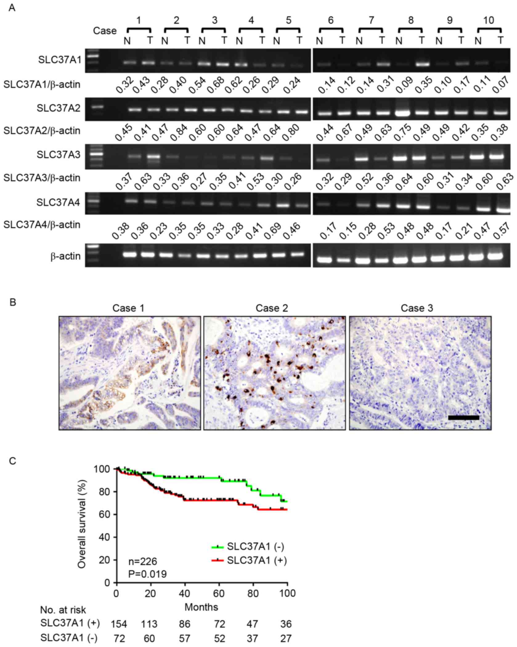
To investigate SLC37A family gene expression in CRC,
the mRNA expression of each cluster gene was investigated in 10
representative cases using RT-qPCR (Fig.
1A and Table I). While all SLC37A
family genes were detected in tumor and adjacent non-cancerous
tissue, SLC37A1 expression appeared to be markedly increased in
tumor tissue compared with its expression in non-tumor tissue
(Table II). To investigate these
comparisons, SLC37A1 protein expression was evaluated by IHC
staining in 231 patients with CRC (Fig.
1B). SLC37A1 expression was positive in 157 cases (68.0%) and
negative in 74 cases (32.0%). SLC37A1 expression was observed in
the nucleus of cancer cells of patients with CRC.
| Table II.Tumor/non-tumor ratioa of SLC37A gene expression. |
Table II.
Tumor/non-tumor ratioa of SLC37A gene expression.
|
|
Case |
|---|
|
|
|
|---|
| Gene | 1 | 2 | 3 | 4 | 5 | 6 | 7 | 8 | 9 | 10 | Mean |
|---|
| SLC37A1 | 1.35 | 1.43 | 1.27 | 0.42 | 0.84 | 0.84 | 2.23 | 3.98 | 1.70 | 0.67 | 1.48 |
| SLC37A2 | 0.91 | 1.79 | 1.01 | 0.74 | 1.24 | 1.54 | 1.28 | 0.65 | 0.86 | 1.08 | 1.11 |
| SLC37A3 | 1.74 | 1.11 | 1.29 | 1.32 | 0.87 | 0.89 | 0.69 | 0.93 | 1.10 | 1.06 | 1.10 |
| SLC37A4 | 0.95 | 1.56 | 0.95 | 1.45 | 0.67 | 0.89 | 1.92 | 1.01 | 1.24 | 1.22 | 1.19 |
SLC37A1 expression level was then compared with
clinicopathological factors in patients with CRC (Table III). The SLC37A1 expression level
was not associated with age, sex, stage, tumor location, histology,
depth of invasion, lymphatic invasion or lymph node metastasis.
However, positive expression of SLC37A1 was significantly
associated with positive venous invasion (P=0.034) and liver
metastasis (P=0.013). Based on the Kaplan-Meier analysis, positive
expression of SLC37A1 was significantly associated with worse
overall survival (P=0.019) (Fig. 1C).
This association was marginally significant (P=0.085) and
independent of all clinical covariates in multivariate models
(Table IV), demonstrating the
potential for SLC37A1 as a useful prognostic biomarker for CRC.
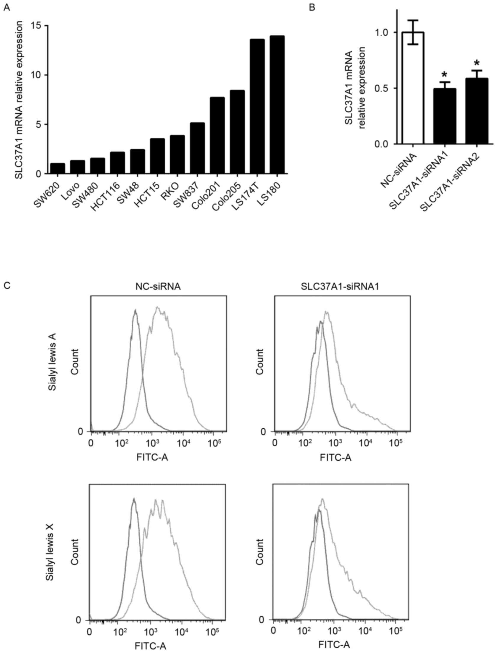
Knockdown of SLC37A1 decreases sialyl
Lewis A and X expression in colon cancer cells
To evaluate the role of SLC37A1 in glycolipid
metabolism, gene knockdown technology was used to investigate
sialyl Lewis A and X expression. SLC37A1 expression in the 12 colon
cancer cell lines (SW620, LoVo, SW480, HCT116, SW48, HCT15, RKO,
SW837, COLO-201, COLO-205, LS174T and LS180) used in the present
study was evaluated in order to select appropriate cells for the
experiment (Fig. 2A). SLC37A1 was
then knocked down by siRNA oligonucleotide in LS180 cells
(SLC37A1-siRNA), which originally expressed upregulated SLC37A1
(Fig. 2B). While no morphological
changes were observed in the SLC37A1-knockdown cells, the
expression of sialyl Lewis A and sialyl Lewis X was markedly
reduced compared with that in NC-siRNA cells (Fig. 2C).
Discussion
In the present study, increased SLC37A1 expression
was observed in patients with CRC, which was associated with
positive venous invasion and liver metastasis. The present study
confirmed that the tumor expression of SLC37A1 is upregulated at
the mRNA and protein levels in patients with CRC, whereas other
SLC37A family genes are not upregulated. Furthermore, positive
expression of SLC37A1 is associated with poor patient survival,
indicating an oncogenic role of SLC37A1 in CRC.
SLC37A1, located at chromosome 21q22.3, is one of
the SLC37 family genes, and is abundantly expressed in the adult
kidney, bone marrow, intestine, spleen and liver (27). While the functional role of SLC37A1
has not yet been characterized, an association with glycolipid
metabolism has been suggested (28).
SLC37A1 shares 30% of sequence similarity with the
glycerol-3-phophate (G3P) transporter; therefore, SLC37A1 may be a
G3P transporter (19,27). By contrast, SLC37A1 also appears to
function as a G6P transporter (29),
indicating that even its fundamental role as a transporter remains
controversial. It has been reported that SLC37A1 appears to be
involved in phospholipid biosynthesis, indicating that SLC37A1 may
accelerate cell proliferation (30).
It appears that cells express SLC37A1 transporters to gain drug
resistance, but the cell viability did not significantly differ
between LS180 cells with high SLC37A1 expression and SW480 cells
with low SLC37A1 expression (data not shown). In vitro
experiments revealed that the expression of SLC37A1 was upregulated
by epidermal growth factor (EGF) via the EGF receptor
(EGFR)/mitogen-activated protein kinase/Fos transduction pathway in
breast cancer cell lines (30). Since
SLC37A1 expression was not associated with EGFR expression by IHC
evaluation in the present cohort (data not shown), SLC37A1 may be
upregulated by the downstream factor of EGFR signaling. The EGFR
signaling pathway is also activated by KRAS mutations in CRC,
indicating that SLC37A1 upregulation is involved in CRC tumor
development. Approximately 50% of CRC cases have KRAS mutations
(31), and they do not respond to
anti-EGFR antibody therapy (32).
This means that upstream inhibition of EGFR has no therapeutic
effect in CRC cases with KRAS mutations (32). In addition, thus far, KRAS mutation
has not served as a direct druggable target (33,34).
Therefore, to identify a candidate therapeutic target downstream of
KRAS is reasonable.
Sialyl Lewis A and sialyl Lewis X are ligands for
the adhesion molecule E-selectin, which is expressed in vascular
endothelial cells and is associated with tumor metastasis,
recurrence and overall survival in various patients with cancer
(35). Our previous study reported
that sialyl Lewis A and sialyl Lewis X were significantly induced
by upregulation of uridine diphosphate-galactose transporter and
hypoxia (26,36). These carbohydrate antigens are
responsible for the adhesion of cancer cells to the endothelium
during metastasis (37). In the
present study, positive SLC37A1 expression in CRC tumor tissue was
associated with the presence of liver metastasis, indicating that
SLC37A1 performs an important role in the hematogenous metastasis
of CRC. Therefore, SLC37A1 was knocked down in colon cancer cells,
and a significant reduction of sialyl Lewis A and sialyl Lewis X
expression was observed. These results indicated that induced
SLC37A1 expression in CRC tumor tissue accelerates cancer
metastasis and may be a therapeutic target in patients with
CRC.
In conclusion, to the best of our knowledge, the
present study is the first to report that SLC37A1 is upregulated in
CRC and is associated with poor patient outcomes. Additional
studies are required to investigate the possibility of SLC37A as a
candidate therapeutic target or biomarker to detect high-risk
patients.
Acknowledgements
The present study was supported by the Japan Society
for the Promotion of Science via the Grants-in-Aid for Scientific
Research KAKENHI program (grant no. 15k10143).
References
|
1
|
Siegel RL, Miller KD and Jemal A: Cancer
statistics, 2016. CA Cancer J Clin. 66:7–30. 2016. View Article : Google Scholar : PubMed/NCBI
|
|
2
|
Chen W, Zheng R, Zhang S, Zhao P, Li G, Wu
L and He J: Report of incidence and mortality in China cancer
registries, 2009. Chin J Cancer Res. 25:10–21. 2013.PubMed/NCBI
|
|
3
|
Katanoda K, Hori M, Matsuda T, Shibata A,
Nishino Y, Hattori M, Soda M, Ioka A, Sobue T and Nishimoto H: An
updated report on the trends in cancer incidence and mortality in
Japan, 1958–2013. Jpn J Clin Oncol. 45:390–401. 2015. View Article : Google Scholar : PubMed/NCBI
|
|
4
|
Venook AP, Weiser MR and Tepper JE:
Colorectal cancer: All hands on deck. Am Soc Clin Oncol Educ Book
83–89. 2014. View Article : Google Scholar
|
|
5
|
Carrato A: Adjuvant treatment of
colorectal cancer. Gastrointest Cancer Res. 2 4 Suppl:S42–S46.
2008.PubMed/NCBI
|
|
6
|
Longley DB, Harkin DP and Johnston PG:
5-fluorouracil: Mechanisms of action and clinical strategies. Nat
Rev Cancer. 3:330–338. 2003. View
Article : Google Scholar : PubMed/NCBI
|
|
7
|
de Gramont A, Figer A, Seymour M, Homerin
M, Hmissi A, Cassidy J, Boni C, Cortes-Funes H, Cervantes A, Freyer
G, et al: Leucovorin and fluorouracil with or without oxaliplatin
as first-line treatment in advanced colorectal cancer. J Clin
Oncol. 18:2938–2947. 2000. View Article : Google Scholar : PubMed/NCBI
|
|
8
|
Douillard JY, Cunningham D, Roth AD,
Navarro M, James RD, Karasek P, Jandik P, Iveson T, Carmichael J,
Alakl M, et al: Irinotecan combined with fluorouracil compared with
fluorouracil alone as first-line treatment for metastatic
colorectal cancer: A multicentre randomised trial. Lancet.
355:1041–1047. 2000. View Article : Google Scholar : PubMed/NCBI
|
|
9
|
Goldberg RM, Sargent DJ, Morton RF, Fuchs
CS, Ramanathan RK, Williamson SK, Findlay BP, Pitot HC and Alberts
SR: A randomized controlled trial of fluorouracil plus leucovorin,
irinotecan, and oxaliplatin combinations in patients with
previously untreated metastatic colorectal cancer. J Clin Oncol.
22:23–30. 2004. View Article : Google Scholar : PubMed/NCBI
|
|
10
|
Tournigand C, André T, Achille E, Lledo G,
Flesh M, Mery-Mignard D, Quinaux E, Couteau C, Buyse M, Ganem G, et
al: FOLFIRI followed by FOLFOX6 or the reverse sequence in advanced
colorectal cancer: A randomized GERCOR study. J Clin Oncol.
22:229–237. 2004. View Article : Google Scholar : PubMed/NCBI
|
|
11
|
Hu T, Li Z, Gao CY and Cho CH: Mechanisms
of drug resistance in colon cancer and its therapeutic strategies.
World J Gastroenterol. 22:6876–6889. 2016. View Article : Google Scholar : PubMed/NCBI
|
|
12
|
Lin L, Yee SW, Kim RB and Giacomini KM:
SLC transporters as therapeutic targets: Emerging opportunities.
Nat Rev Drug Discov. 14:543–560. 2015. View
Article : Google Scholar : PubMed/NCBI
|
|
13
|
Dean M, Rzhetsky A and Allikmets R: The
human ATP-binding cassette (ABC) transporter superfamily. Genome
Res. 11:1156–1166. 2001. View Article : Google Scholar : PubMed/NCBI
|
|
14
|
Szakács G, Annereau JP, Lababidi S,
Shankavaram U, Arciello A, Bussey KJ, Reinhold W, Guo Y, Kruh GD,
Reimers M, et al: Predicting drug sensitivity and resistance:
Profiling ABC transporter genes in cancer cells. Cancer Cell.
6:129–137. 2004. View Article : Google Scholar : PubMed/NCBI
|
|
15
|
Higgins CF: Multiple molecular mechanisms
for multidrug resistance transporters. Nature. 446:749–757. 2007.
View Article : Google Scholar : PubMed/NCBI
|
|
16
|
Shukla S, Chen ZS and Ambudkar SV:
Tyrosine kinase inhibitors as modulators of ABC
transporter-mediated drug resistance. Drug Resist Updat. 15:70–80.
2012. View Article : Google Scholar : PubMed/NCBI
|
|
17
|
Gottesman MM, Fojo T and Bates SE:
Multidrug resistance in cancer: Role of ATP-dependent transporters.
Nat Rev Cancer. 2:48–58. 2002. View
Article : Google Scholar : PubMed/NCBI
|
|
18
|
Lopez-Lopez E, Ballesteros J, Piñan MA, de
Toledo J Sanchez, de Andoin N Garcia, Garcia-Miguel P, Navajas A
and Garcia-Orad A: Polymorphisms in the methotrexate transport
pathway: A new tool for MTX plasma level prediction in pediatric
acute lymphoblastic leukemia. Pharmacogenet Genomics. 23:53–61.
2013. View Article : Google Scholar : PubMed/NCBI
|
|
19
|
Chou JY and Mansfield BC: The SLC37 family
of sugar-phosphate/phosphate exchangers. Curr Top Membr.
73:357–382. 2014. View Article : Google Scholar : PubMed/NCBI
|
|
20
|
Levine AJ and Puzio-Kuter AM: The control
of the metabolic switch in cancers by oncogenes and tumor
suppressor genes. Science. 330:1340–1344. 2010. View Article : Google Scholar : PubMed/NCBI
|
|
21
|
Chen JQ and Russo J: Dysregulation of
glucose transport, glycolysis, TCA cycle and glutaminolysis by
oncogenes and tumor suppressors in cancer cells. Biochim Biophys
Acta. 1826:370–384. 2012.PubMed/NCBI
|
|
22
|
Sobin LH and Compton CC: TNM seventh
edition: What's new, what's changed: Communication from the
international union against cancer and the American joint Committee
on cancer. Cancer. 116:5336–5339. 2010. View Article : Google Scholar : PubMed/NCBI
|
|
23
|
Sobin LH, Gospodarowicz M and Wittekind
Ch: International Union Against Cancer TNM Classification of
Malignant Tumors. 7th. Wiley-Blackwell; Oxford, UK: 2009
|
|
24
|
Okano M, Kumamoto K, Saito M, Onozawa H,
Saito K, Abe N, Ohtake T and Takenoshita S: Upregulated Annexin A1
promotes cellular invasion in triple-negative breast cancer. Oncol
Rep. 33:1064–1070. 2015. View Article : Google Scholar : PubMed/NCBI
|
|
25
|
Livak KJ and Schmittgen TD: Analysis of
relative gene expression data using real-time quantitative PCR and
the 2(-Delta Delta C(T)) Method. Methods. 25:402–408. 2001.
View Article : Google Scholar : PubMed/NCBI
|
|
26
|
Koike T, Kimura N, Miyazaki K, Yabuta T,
Kumamoto K, Takenoshita S, Chen J, Kobayashi M, Hosokawa M,
Taniguchi A, et al: Hypoxia induces adhesion molecules on cancer
cells: A missing link between Warburg effect and induction of
selectin-ligand carbohydrates. Proc Natl Acad Sci USA. 101:pp.
8132–8137. 2004; View Article : Google Scholar : PubMed/NCBI
|
|
27
|
Bartoloni L, Wattenhofer M, Kudoh J, Berry
A, Shibuya K, Kawasaki K, Wang J, Asakawa S, Talior I, Bonne-Tamir
B, et al: Cloning and characterization of a putative human glycerol
3-phosphate permease gene (SLC37A1 or G3PP) on 21q22.3: Mutation
analysis in two candidate phenotypes, DFNB10 and a glycerol kinase
deficiency. Genomics. 70:190–200. 2000. View Article : Google Scholar : PubMed/NCBI
|
|
28
|
Bartoloni L and Antonarakis SE: The human
sugar-phosphate/phosphate exchanger family SLC37. Pflugers Arch.
447:780–783. 2004. View Article : Google Scholar : PubMed/NCBI
|
|
29
|
Pan CJ, Chen SY, Jun HS, Lin SR, Mansfield
BC and Chou JY: SLC37A1 and SLC37A2 are phosphate-linked,
glucose-6-phosphate antiporters. PLoS One. 6:e231572011. View Article : Google Scholar : PubMed/NCBI
|
|
30
|
Iacopetta D, Lappano R, Cappello AR, Madeo
M, De Francesco EM, Santoro A, Curcio R, Capobianco L, Pezzi V,
Maggiolini M and Dolce V: SLC37A1 gene expression is up-regulated
by epidermal growth factor in breast cancer cells. Breast Cancer
Res Treat. 122:755–764. 2010. View Article : Google Scholar : PubMed/NCBI
|
|
31
|
Cancer Genome Atlas N, . Comprehensive
molecular characterization of human colon and rectal cancer.
Nature. 487:330–337. 2012. View Article : Google Scholar : PubMed/NCBI
|
|
32
|
Siddiqui AD and Piperdi B: KRAS mutation
in colon cancer: A marker of resistance to EGFR-I therapy. Ann Surg
Oncol. 17:1168–1176. 2010. View Article : Google Scholar : PubMed/NCBI
|
|
33
|
Dietlein F, Kalb B, Jokic M, Noll EM,
Strong A, Tharun L, Ozretic L, Künstlinger H, Kambartel K,
Randerath WJ, et al: A Synergistic Interaction between Chk1- and
MK2 Inhibitors in KRAS-Mutant Cancer. Cell. 162:146–159. 2015.
View Article : Google Scholar : PubMed/NCBI
|
|
34
|
Zhu Z, Aref AR, Cohoon TJ, Barbie TU,
Imamura Y, Yang S, Moody SE, Shen RR, Schinzel AC, Thai TC, et al:
Inhibition of KRAS-driven tumorigenicity by interruption of an
autocrine cytokine circuit. Cancer Discov. 4:452–465. 2014.
View Article : Google Scholar : PubMed/NCBI
|
|
35
|
Liang JX, Liang Y and Gao W:
Clinicopathological and prognostic significance of sialyl Lewis X
overexpression in patients with cancer: A meta-analysis. Onco
Targets Ther. 9:3113–3125. 2016.PubMed/NCBI
|
|
36
|
Kumamoto K, Goto Y, Sekikawa K,
Takenoshita S, Ishida N, Kawakita M and Kannagi R: Increased
expression of UDP-galactose transporter messenger RNA in human
colon cancer tissues and its implication in synthesis of
Thomsen-Friedenreich antigen and sialyl Lewis A/X determinants.
Cancer Res. 61:4620–4627. 2001.PubMed/NCBI
|
|
37
|
Takada A, Ohmori K, Yoneda T, Tsuyuoka K,
Hasegawa A, Kiso M and Kannagi R: Contribution of carbohydrate
antigens sialyl Lewis A and sialyl Lewis X to adhesion of human
cancer cells to vascular endothelium. Cancer Res. 53:354–361.
1993.PubMed/NCBI
|