Introduction
Alzheimer's disease (AD), a severe age-related
neurodegenerative disorder, is characterized by the accumulation of
amyloid-β (Aβ) plaques and neurofibrillary tangles, synaptic and
neuronal loss, and cognitive decline (1,2). An
estimated 5.3 million Americans have AD; 5.1 million of these are
aged 65 and over, and approximately 200,000 are <65 years-of-age
and have younger onset AD (3). In
2013, official death certificates recorded 84,767 deaths from AD,
making AD the sixth leading cause of death in the United States and
the fifth leading cause of death in Americans aged 65 years
(3). There is currently no effective
treatment capable of slowing down the progression of AD.
Among the genes associated with AD, β-site
APP-cleaving enzyme 1 (BACE1) is a rate-limiting enzyme for Aβ
production and has been demonstrated to be significantly
upregulated in patients with AD (4).
Furthermore, BACE1 has been implicated as a potential target for
therapies against AD (4). Therefore,
understanding the mechanism underlying BACE1 upregulation in AD may
further the development of therapeutic strategies for the treatment
of AD.
MicroRNAs (miRs/miRNAs), a class non-coding RNAs
18–25 nucleotides in length, have been reported to cause mRNA
degradation or inhibition of protein translation through directly
binding to the 3′-untranslated region (UTR) of their target mRNAs
(5). Recently, several studies have
implicated certain miRs in neuronal survival, proliferation,
differentiation and migration (6,7). Yang
et al revealed that miR-29c negatively mediates the
expression of DNA methyltransferase 3, which contributes to
neuronal proliferation, by regulating the expression of
brain-derived neurotrophic factor (6). Furthermore, the dysfunction of certain
miRs has been suggested to be involved in the development of AD
(8–10). Denk et al investigated the
expression profiling of 1,178 miRs in cerebrospinal fluid samples
from patients with AD and normal controls, and discrimination
analysis using a combination of miR-100, miR-103 and miR-375 was
able to detect AD by positively classifying controls and AD cases
with 96.4 and 95.5% accuracy, respectively (8). Furthermore, Lei et al reported
that the downregulation of miR-29c was correlated with increased
BACE1 expression levels in sporadic Alzheimer's disease (4). Recently, Liu et al used miR
microarrays to analyze the miR expression profiles of amyloid
precursor protein (APP)/presenilin 1 (PS1) in the hippocampi of
transgenic and wild-type mice, and identified that miR-135a was
significantly downregulated in the hippocampi of APP/PS1 transgenic
mice compared with the wild-type control, suggesting that
downregulation of miR-135a may have a role in the development of AD
(11). However, the exact role of
miR-135b in AD still remains largely unclear.
The primary aim of the present study was to
investigate the expression levels and role of miR-135b in AD. The
underlying mechanism involving BACE1 was also investigated.
Materials and methods
Collection of blood samples
The present study was approved by the ethics boards
of Xinxiang Medical School (Weihui, China). Blood samples from
patients from The First Affiliated Hospital of Xinxiang Medical
University, (Weihui, China) with AD (n=25; 12 male, 13 female) aged
between 65 and 81 years old and age-matched normal subjects (n=25)
were collected from our hospital between April 2013 and March 2014.
Blood samples were stored in anticoagulation tubes at −80°C.
Patients with diabetes, heart disease, stroke and cancer were
excluded from the study. Written informed consent was obtained from
all participants.
Cell culture
Primary hippocampal cells (purchased from Amspring,
Changsha, China), obtained from the embryonic hippocampi of
senescence-accelerated mouse resistant 1 (SAMR1) mice, were
cultured in Dulbecco's modified Eagle medium (Thermo Fisher
Scientific, Inc., Waltham, MA, USA) with 10% fetal bovine serum
(FBS; Thermo Fisher Scientific, Inc.), and 100 kU/l of penicillin
and streptomycin (Thermo Fisher Scientific, Inc.). Hippocampal
cells were cultured in a humidified atmosphere of 95% air and 5%
CO2.
Reverse transcription-quantitative
polymerase chain reaction (RT-qPCR) analysis
Total RNA was extracted from the human tissue and
mouse hippocampal cells using TRIzol reagent (Thermo Fisher
Scientific, Inc.). A Taqman miRNA Reverse Transcription kit (Thermo
Fisher Scientific, Inc.) was used to convert RNA into cDNA. A
miScript SYBR-Green PCR kit (Guangzhou RiboBio Co., Ltd. Guangzhou,
China) was used to determine the miRNA expression levels, according
to the manufacturer's protocol. U6 was used as an endogenous
control. Expression levels of mRNA were determined using the SYBR
green qPCR assay (CWBio, Beijing, China) following the
manufacturer's protocol. An Applied Biosystems 7500 Thermocycler
(Applied Biosystems; Thermo Fisher Scientific, Inc.) The specific
primers were as follows: Forward, 5′-TCTGTCGGAGGGAGCATGAT-3′ and
reverse, 5′-GCAAACGAAGGTTGGTGGT-3′ for BACE1; forward,
5′-ACAACTTTGGTATCGTGGAAGG-3′ and reverse, 5′-GCCATCACGCCACAGTTTC-3′
for GAPDH. Expression of GAPDH was used as an endogenous control.
The PCR cycling conditions were as follows: 95°C for 5 min, and 40
cycles of denaturation at 95°C for 15 sec and annealing/elongation
step at 60°C for 30 sec. Data were analyzed using the
2−ΔΔqt method (12).
Dual luciferase reporter assay
The seed sequences of miR-135b (5′-AAGCCAUA-3′)
within the BACE1 3′-UTR, or the mutant binding sequences of
miR-135b within the BACE1 3′-UTR, were cloned downstream of the
luciferase gene driven by the cytomegalovirus (CMV) promoter,
generating Luc-BACE1 and Luc-mutant BACE1 vectors (GeneChem,
Shanghai, China), respectively. The vectors used in the luciferase
reporter assay were directly purchased from Amspring. Lipofectamine
2000 (Invitrogen; Thermo Fisher Scientific, Inc., Waltham, MA, USA)
was used to co-transfect (for 24 h at 37°C) hippocampal cells with
Luc-BACE1 or Luc-mutant BACE1 vectors and miR-135b mimics or
scramble miR-negative control (miR-NC) mimics (GeneChem Co., Ltd.),
respectively. Following a transfection period of 24 h, luciferase
activity was determined using an LD400 luminometer (Beckman Coulter
Inc., Brea, CA, USA).
Western blot analysis
Tissues and cells were solubilized in cold RIPA
lysis and extraction buffer (Thermo Fisher Scientific, Inc.). The
concentration of protein was determined using a BCA kit (Pierce
Biotechnology, Inc., Rockford, IL, USA). Proteins were separated
with 10% sodium dodecyl sulfate-polyacrylamide gel electrophoresis
and transferred onto a polyvinylidene difluoride membrane (PVDF;
Thermo Fisher Scientific, Inc.). The PVDF membrane was incubated
with phosphate-buffered saline (PBS) containing 5% non-fat milk
overnight at 4°C, then incubated with mouse monoclonal anti-BACE1
(cat no. ab201946; 1:50; Abcam, Cambridge, MA, USA) or anti-GAPDH
antibody (cat no. ab8245; 1:100; Abcam) at room temperature for 3
h. Subsequent incubation with rabbit anti-mouse secondary antibody
(cat no. ab6728; 1:10,000; Abcam) at room temperature for 1 h was
then performed. An enhanced chemiluminescence kit (Pierce
Biotechnology, Inc.) was utilized to perform chemiluminescent
detection. The relative protein expression levels were analyzed
with Image Pro Plus software (version 6.0; Media Cybernetics,
Rockville, MD, USA), represented as the density ratio versus
GAPDH.
Cell proliferation assay
An MTT assay was performed to investigate cell
proliferation. For each group, 5,000 hippocampal cells per well
were seeded in 96-well plates and incubated for 6, 12, 24 or 48 h
at 37°C and 5% CO2. Following this, MTT (5 mg/ml) was
added to each well and cells were then incubated at 37°C for 4 h.
The medium containing MTT was then removed and 100 µl dimethyl
sulfoxide was added. Absorbance was detected at 492 nm using a
microplate reader (Multiskan FC Microplate Photometer; Thermo
Fisher Scientific, Inc.).
Animal treatment
The experimental protocol was approved by the Animal
Care and Use Committee of Xinxiang Medical School, in compliance
with the National Institutes of Health Guide for the Care and Use
of Laboratory Animals (13). Male
senescence accelerated mouse prone 8 (SAMP8) mice (n=18; 8 months
old) were purchased from the Animal Center of Xinxiang Medical
School. Mice were housed at 22±1°C in a 12-h light/dark cycle. The
third ventricle of anesthetized mice (0.5 ml/100g 10% chloral
hydrate; Tanghua, Changsha, China) were injected with 3 µl PBS
containing 0.5 nM of miR-135b mimic or scramble miR-NC mimic
(GeneChem Co., Ltd. Shanghai, China). In the control group, mice
received 3 µl of PBS. Each group was composed of 6 mice. At 3 h
after injection, the mice were sacrificed using cervical
dislocation under anesthetization using 10% chloral hydrate. The
heads of mice were anatomized, and the hippocampal tissues were
obtained.
Y-maze test
The maze apparatus (DOiT, Shanghai, China) was
constructed of wood painted in black with three arms. The mice were
placed at the end of one arm and allowed to move freely for 10 min.
Spontaneous alternation was defined as successive entries into the
three arms in overlapping triplet sets. The alternation percentage
was determined as the ratio of actual alternations to maximum
alternations multiplied by 100.
Statistical analysis
Data are expressed as mean ± standard deviation.
Differences between two groups were determined by Student's t-test.
Statistical analyses were performed using GraphPad Prism software
(version 5; Graphpad Software, Inc., La Jolla, CA, USA). P<0.05
was considered to indicate a statistically significant
difference.
Results
miR-135b levels are reduced in the
blood of patients with AD
To reveal the role of miR-135b in AD, RT-qPCR was
conducted to determine the miR-135b expression levels in the
peripheral blood of patients with AD and age-matched normal
controls. The data revealed that the expression levels of miR-135b
were significantly reduced in the peripheral blood of AD patients
compared with the normal controls (P<0.01; Fig. 1), suggesting that the downregulation
of miR-135b may have a role in the pathogenesis of AD.
miR-135b promotes the proliferation of
hippocampal cells
The role of miR-135b in the regulation of
hippocampal cell proliferation was subsequently investigated.
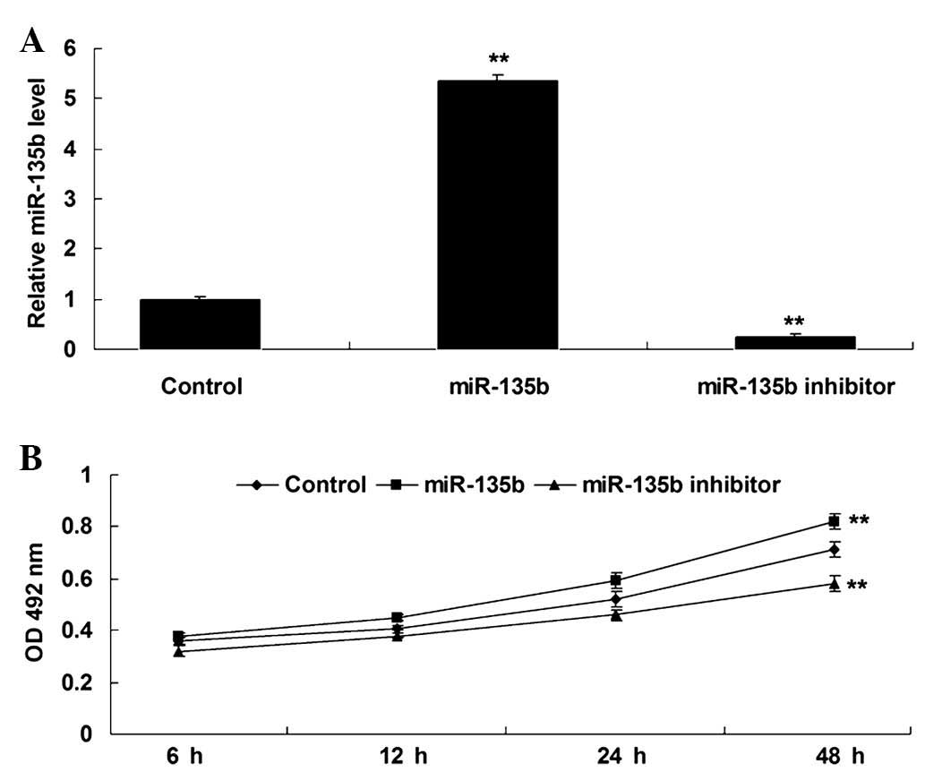
Hippocampal cells were transfected with miR-135b mimics or a
miR-135b inhibitor, respectively. Following transfection, RT-qPCR
was performed to detect the expression levels of miR-135b in each
group. As displayed in Fig. 2A,
transfection with miR-135b mimics led to a significant increase in
miR-135b expression levels in hippocampal cells (P<0.01), while
transfection with the miR-135b inhibitor caused a significant
downregulation of miR-135b (P<0.01), compared with the control,
indicating that the transfection was successful. Following this, an
MTT assay was conducted to examine the proliferating capacity of
hippocampal cells in each group. As demonstrated in Fig. 2B, upregulation of miR-135b
significantly enhanced the proliferation of hippocampal cells,
while knockdown of miR-135b significantly inhibited hippocampal
cell proliferation, compared with the control group (P<0.01).
These findings indicate that miR-135b promotes the proliferation of
hippocampal cells.
miR-135b has a neuroprotective role in
SAMP8 mice
SAMP8 mice are senescence-accelerated and thus have
been widely used to investigate AD (14,15). In
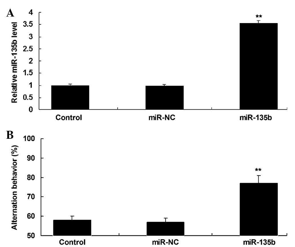
the present study, miR-135b mimics were injected into the third
ventricle of SAMP8 mice to reveal the role of miR-135b in
vivo. Following injection, RT-qPCR was conducted to detect the
expression of miR-135b in the hippocampal tissue. As demonstrated
in Fig. 3A, miR-135b was
significantly upregulated in the hippocampal tissue following
injection with miR-135b mimics, when compared with the control
group (P<0.01). A Y-maze test was then performed to examine the
learning and memory capacities of mice in each group. As shown in
Fig. 3B, injection with miR-135b
mimics into the hippocampi significantly enhanced the learning and
memory behaviors of SAMP8 mice compared with the control group
(P<0.01; Fig. 3B) and indicated
that miR-135b has a neuroprotective role in vivo.
BACE1 is a target gene of
miR-135b
The putative targets of miR-135b that had been
suggested to be associated with AD were also investigated.
Bioinformatic analysis predicated that BACE1 is a putative target
gene of miR-135b (Fig. 4A). To
confirm this predication, the putative miR-135b target sequence
within the BACE1 3′-UTR, in addition to a mutant lacking
complementarity with the miR-135b seed sequence, were cloned
downstream of the luciferase gene driven by the CMV promoter,
generating Luc-BACE1 and Luc-mutant BACE1 vectors, respectively
(Fig. 4B). Hippocampal cells were
then co-transfected with Luc-BACE1 or Luc-mutant BACE1 vectors and
miR-135b mimics or scramble miR mimics (miR-NC), respectively. The
luciferase reporter assay data revealed that luciferase activity
was only significantly decreased in hippocampal cells
co-transfected with the Luc-BACE1 vector and miR-135b mimics
compared with the control group (P<0.01; Fig. 4C). However, the luciferase activity
revealed no difference in other groups when compared with the
control group (Fig. 4C). Therefore,
the results of the present study indicate that BACE1 is a target
gene of miR-135b.
 | Figure 4.(A) Targetscan software predicted that
BACE1 was a target gene of miR-135b. (B) The predicted miR-135b
target sequence within the BACE1 3′-UTR and a mutant lacking
complimentarity with miR-135b seed sequence are indicated. The seed
sequences of miR-135b within the BACE1 3′-UTR, or the mutant
binding sequences of miR-135b within the BACE1 3′-UTR were cloned
downstream of the luciferase gene, generating Luc-BACE1 and
Luc-mutant BACE1 vectors, respectively. Hippocampal cells were
transfected with Luc-BACE1 or Luc-mutant BACE1 vector and miR-135b
mimics or scramble miR mimics (miR-NC), respectively. (C) The
luciferase activity was determined after transfection for 24 h.
Control: Hippocampal cells transfected with Luc-BACE1 or Luc-mutant
BACE1 vectors alone. **P<0.01 vs. control. BACE1, β-site APP
cleaving enzyme 1; miR, microRNA; NC, negative control; UTR,
untranslated region; Luc, luciferase; PCT, probability
of conserved targeting. |
BACE1 is negatively regulated by
miR-135b in vitro and in vivo
As miRs negatively mediate their target genes at a
post-transcriptional level, the effect of miR-135b on the protein
expression levels of BACE1 in hippocampal cells of SAMR1 mice, as
well as in the hippocampal tissues of SAMP8 mice, were
investigated. Western blot analysis data revealed that the
overexpression of miR-135b led to a significant decrease in the
protein expression levels of BACE1, while knockdown of miR-135b
caused a significant increase in the protein expression levels of
BACE1 in hippocampal cells (P<0.01; Fig. 5A). Subsequently, the protein
expression levels of BACE1 were examined in the hippocampal tissue
of SAMP8 mice. As displayed in Fig.
5B, injection with miR-135b mimics caused a significant
decrease in the protein expression levels of BACE1 in the
hippocampal tissue of SAMP8 mice when compared with the control
group (P<0.01). On the basis of the aforementioned data, we
propose that BACE1 is negatively regulated by miR-135b in
vitro and in vivo.
Discussion
It has been suggested that the dysfunction of
certain miRs are involved in the development of AD. However, the
exact role of miR-135b in AD, in addition to its underlying
mechanism, have yet to be elucidated. The present study revealed
that miR-135b was significantly downregulated in the peripheral
blood of patients with AD compared with normal controls. The
present study also indicated that overexpression of miR-135b
significantly enhanced the proliferation of hippocampal cells, and
injection with miR-135b mimics into the third ventricle of
anesthetized SAMP8 mice enhanced their learning and memory
capacities. Furthermore, BACE1 was identified as a target gene of
miR-135b, and miR-135b negatively mediated the protein expression
levels of BACE1 in hippocampal cells of SAMR1 mice, as well as in
hippocampal tissues, of SAMP8 mice.
Previous studies have indicated that certain genes
or miRs are significantly downregulated or upregulated in the
peripheral blood or cerebrospinal fluid of patients with AD,
including BACE1 (16–18). The present study revealed that the
expression levels of miR-135b were significantly decreased in the
peripheral blood of AD patients compared with those of the
age-matched normal controls, suggesting that serum miR-135b levels
may be used for the clinical diagnosis of AD. In agreement with the
findings of the present study, Liu et al analyzed the miR
hippocampi expression profiles of APP/PS1 transgenic and wild-type
mice, and observed that miR-135a was significantly downregulated in
the hippocampi of the transgenic mice compared with those of the
wild type mice (11). Therefore,
miR-135a and miR-135b may both participate in the development and
progression of AD. Hébert et al also identified that
miR-29a/b-1 was significantly downregulated in the brains of
patients with sporadic AD and correlated with increased BACE1
expression (19). In addition,
Müller et al revealed that miR-16, miR-34c, miR-107,
miR-128a and miR-146a were differentially regulated in the
hippocampi of patients with AD and age-matched normal controls,
while only miR-16 and miR-146a were reliably detected in the
cerebrospinal fluid of patients with AD (20).
The survival and proliferation of hippocampal cells
in patients with AD is significantly suppressed; thus, promoting
their survival and proliferation is critical for the treatment of
AD (21). In the present in
vitro study, it was revealed that the overexpression of
miR-135b significantly promotes the proliferation of hippocampal
cells. Conversely, the inhibition of miR-135b expression levels
significantly suppressed the proliferation of hippocampal cells,
indicating that miR-135b may have a neuroprotective role.
Furthermore, several other miRs have also been revealed to mediate
the proliferation of neural cells. The overexpression of miR-125b,
for example, inhibited the proliferation of neural stem/progenitor
cells (22), and miR-34a was
revealed to enhance cell proliferation and function of newly
generated neurons, as well as improve behavioral outcomes (23). Furthermore, as patients with AD are
characterized by learning and memory deficits (24), SAMP8 mice were used to investigate
the effect of miR-135b on learning and memory behaviors in the
present study. It was identified that injection with miR-135b
mimics into the third ventricle significantly upregulated miR-135b
expression levels in the hippocampal tissues of SAMP8 mice. This
was accompanied by the upregulation of the learning and memory
behaviors. Accordingly, miR-135a also appears to demonstrate a
neuroprotective role in vivo.
Typically, Aβ peptide is deposited in the brain of
AD patients (25). BACE1 is a type I
integral membrane glycoprotein and aspartic protease that is
responsible for the proteolytic cleavage of APP, generating Aβ
peptide (25). Therefore, BACE1 has
a critical role in AD. The expression levels of BACE1 have been
revealed to be significantly increased in patients with AD
(4); thus, BACE 1 is a potential
therapeutic target for novel AD therapies (26). Several BACE1 inhibitors have
demonstrated promising results for the treatment of AD, which have
recently been used in the human clinical trials (26). In the present study, BACE1 was
identified as a direct target gene of miR-135b, and miR-135b was
revealed to negatively mediate the protein expression levels of
BACE1 in hippocampal cells of SAMR1 mice, as well as in the
hippocampal tissues of SAMP8 mice. Therefore, miR-135b may be a
potential candidate for the treatment of AD through inhibiting
BACE1. In addition, several other miRs have been observed to
directly target BACE1, including the miR-29 family, miR-124,
miR-195 and miR-339-5p, and thus may contribute to the treatment of
AD (4,27–30).
In conclusion, the present study indicates that
miR-135b has a neuroprotective role via directly inhibiting BACE1
protein expression. Consequently, we propose that miR-135b may be
utilized in the treatment of AD. Further studies are required that
focus on investigating the downstream signaling pathways of
miR-135b/BACE1 in the pathogenesis of AD.
References
|
1
|
Zlomuzica A, Dere D, Binder S, De Souza
Silva MA, Huston JP and Dere E: Neuronal histamine and cognitive
symptoms in Alzheimer's disease. Neuropharmacology. May
27–2015.(Epub ahead of print). View Article : Google Scholar : PubMed/NCBI
|
|
2
|
Dezsi L, Tuka B, Martos D and Vecsei L:
Alzheimer's disease, astrocytes and kynurenines. Curr Alzheimer
Res. 12:462–480. 2015. View Article : Google Scholar : PubMed/NCBI
|
|
3
|
Alzheimer's Association: 2015 Alzheimer's
disease facts and figures. Alzheimers Dement. 11:332–384. 2015.
View Article : Google Scholar : PubMed/NCBI
|
|
4
|
Lei X, Lei L, Zhang Z, Zhang Z and Cheng
Y: Downregulated miR-29c correlates with increased BACE1 expression
in sporadic Alzheimer's disease. Int J Clin Exp Pathol.
8:1565–1574. 2015.PubMed/NCBI
|
|
5
|
Ambros V: The functions of animal
microRNAs. Nature. 431:350–355. 2004. View Article : Google Scholar : PubMed/NCBI
|
|
6
|
Yang G, Song Y, Zhou X, Deng Y, Liu T,
Weng G, Yu D and Pan S: DNA methyltransferase 3, a target of
microRNA-29c, contributes to neuronal proliferation by regulating
the expression of brain-derived neurotrophic factor. Mol Med Rep.
12:1435–1442. 2015.PubMed/NCBI
|
|
7
|
Stary CM, Xu L, Sun X, Ouyang YB, White
RE, Leong J, Li J, Xiong X and Giffard RG: MicroRNA-200c
contributes to injury from transient focal cerebral ischemia by
targeting Reelin. Stroke. 46:551–556. 2015. View Article : Google Scholar : PubMed/NCBI
|
|
8
|
Denk J, Boelmans K, Siegismund C, Lassner
D, Arlt S and Jahn H: MicroRNA Profiling of CSF reveals potential
biomarkers to detect Alzheimer's disease. PLoS One.
10:e01264232015. View Article : Google Scholar : PubMed/NCBI
|
|
9
|
Yang G, Song Y, Zhou X, Deng Y, Liu T,
Weng G, Yu D and Pan S: MicroRNA-29c targets β-site amyloid
precursor protein-cleaving enzyme 1 and has a neuroprotective role
in vitro and in vivo. Mol Med Rep. 2015. View Article : Google Scholar
|
|
10
|
Zhu Y, Li C, Sun A, Wang Y and Zhou S:
Quantification of microRNA-210 in the cerebrospinal fluid and
serum: Implications for Alzheimer's disease. Exp Ther Med.
9:1013–1017. 2015.PubMed/NCBI
|
|
11
|
Liu CG, Wang JL, Li L, Xue LX, Zhang YQ
and Wang PC: MicroRNA-135a and −200b, potential Biomarkers for
Alzheimer's disease, regulate β secretase and amyloid precursor
protein. Brain Res. 1583:55–64. 2014. View Article : Google Scholar : PubMed/NCBI
|
|
12
|
Livak KJ and Schmittgen TD: Analysis of
relative gene expression data using real-time quantitative PCR and
the 2(−Delta Delta C(T)) method. Methods. 25:402–408. 2001.
View Article : Google Scholar : PubMed/NCBI
|
|
13
|
National Research Council (US) Committee
for the Update of the Guide for the Care and Use of Laboratory
Animals: Washington (DC) (8th). National Academies Press. US:
2011.
|
|
14
|
Hansen HH, Fabricius K, Barkholt P,
Niehoff ML, Morley JE, Jelsing J, Pyke C, Knudsen LB, Farr SA and
Vrang N: The GLP-1 receptor agonist liraglutide improves memory
function and increases hippocampal CA1 neuronal numbers in a
senescence-accelerated mouse model of Alzheimer's disease. J
Alzheimers Dis. 46:877–888. 2015. View Article : Google Scholar : PubMed/NCBI
|
|
15
|
Takagane K, Nojima J, Mitsuhashi H, Suo S,
Yanagihara D, Takaiwa F, Urano Y, Noguchi N and Ishiura S: Aβ
induces oxidative stress in senescence-accelerated (SAMP8) mice.
Biosci Biotechnol Biochem. 79:912–918. 2015. View Article : Google Scholar : PubMed/NCBI
|
|
16
|
Decourt B, Walker A, Gonzales A,
Malek-Ahmadi M, Liesback C, Davis KJ, Belden CM, Jacobson SA and
Sabbagh MN: Can platelet BACE1 levels be used as a biomarker for
Alzheimer's disease? Proof-of-concept study. Platelets. 24:235–238.
2013. View Article : Google Scholar : PubMed/NCBI
|
|
17
|
Shaw LM, Vanderstichele H, Knapik-Czajka
M, Clark CM, Aisen PS, Petersen RC, Blennow K, Soares H, Simon A,
Lewczuk P, et al: Alzheimer's Disease Neuroimaging Initiative:
Cerebrospinal fluid biomarker signature in Alzheimer's disease
neuroimaging initiative subjects. Ann Neurol. 65:403–413. 2009.
View Article : Google Scholar : PubMed/NCBI
|
|
18
|
Bibl M, Esselmann H, Lewczuk P,
Trenkwalder CF, Otto M, Kornhuber J, Wiltfang J and Mollenhauer B:
Combined analysis of CSF Tau, Aβ42, Aβ1–42% and Aβ1–40% in
Alzheimer's disease, dementia with Lewy bodies and Parkinson's
disease dementia. Int J Alzheimers Dis 2010. Article ID 761571.
2010.
|
|
19
|
Hébert SS, Horré K, Nicolaï L,
Papadopoulou AS, Mandemakers W, Silahtaroglu AN, Kauppinen S,
Delacourte A and De Strooper B: Loss of microRNA cluster
miR-29a/b-1 in sporadic Alzheimer's disease correlates with
increased BACE1/beta-secretase expression. Proc Natl Acad Sci USA.
105:6415–6420. 2008. View Article : Google Scholar : PubMed/NCBI
|
|
20
|
Müller M, Kuiperij HB, Claassen JA,
Küsters B and Verbeek MM: MicroRNAs in Alzheimer's disease:
Differential expression in hippocampus and cell-free cerebrospinal
fluid. Neurobiol Aging. 35:152–158. 2014. View Article : Google Scholar : PubMed/NCBI
|
|
21
|
Moon M, Cha MY and Mook-Jung I: Impaired
hippocampal neurogenesis and its enhancement with ghrelin in 5XFAD
mice. J Alzheimers Dis. 41:233–241. 2014.PubMed/NCBI
|
|
22
|
Cui Y, Xiao Z, Han J, Sun J, Ding W, Zhao
Y, Chen B, Li X and Dai J: MiR-125b orchestrates cell
proliferation, differentiation and migration in neural
stem/progenitor cells by targeting Nestin. BMC Neurosci.
13:1162012. View Article : Google Scholar : PubMed/NCBI
|
|
23
|
Mollinari C, Racaniello M, Berry A, Pieri
M, de Stefano MC, Cardinale A, Zona C, Cirulli F, Garaci E and
Merlo D: miR-34a regulates cell proliferation, morphology and
function of newborn neurons resulting in improved behavioural
outcomes. Cell Death Dis. 6:e16222015. View Article : Google Scholar : PubMed/NCBI
|
|
24
|
Buckley RF, Ellis KA, Ames D, Rowe CC,
Lautenschlager NT, Maruff P, Villemagne VL, Macaulay SL, Szoeke C,
Martins RN, et al: Phenomenological characterization of memory
complaints in preclinical and prodromal Alzheimer's disease.
Neuropsychology. 29:571–581. 2015. View Article : Google Scholar : PubMed/NCBI
|
|
25
|
Vassar R, Kuhn PH, Haass C, Kennedy ME,
Rajendran L, Wong PC and Lichtenthaler SF: Function, therapeutic
potential and cell biology of BACE proteases: Current status and
future prospects. J Neurochem. 130:4–28. 2014. View Article : Google Scholar : PubMed/NCBI
|
|
26
|
Yan R and Vassar R: Targeting the β
secretase BACE1 for Alzheimer's disease therapy. Lancet Neurol.
13:319–329. 2014. View Article : Google Scholar : PubMed/NCBI
|
|
27
|
Long JM, Ray B and Lahiri DK:
MicroRNA-339-5p down-regulates protein expression of β-site amyloid
precursor protein-cleaving enzyme 1 (BACE1) in human primary brain
cultures and is reduced in brain tissue specimens of Alzheimer
disease subjects. J Biol Chem. 289:5184–5198. 2014. View Article : Google Scholar : PubMed/NCBI
|
|
28
|
Zhu HC, Wang LM, Wang M, Song B, Tan S,
Teng JF and Duan DX: MicroRNA-195 downregulates Alzheimer's disease
amyloid-beta production by targeting BACE1. Brain Res Bull.
88:596–601. 2012. View Article : Google Scholar : PubMed/NCBI
|
|
29
|
Roshan R, Ghosh T, Gadgil M and Pillai B:
Regulation of BACE1 by miR-29a/b in a cellular model of
Spinocerebellar Ataxia 17. RNA Biol. 9:891–899. 2012. View Article : Google Scholar : PubMed/NCBI
|
|
30
|
Fang M, Wang J, Zhang X, Geng Y, Hu Z,
Rudd JA, Ling S, Chen W and Han S: The miR-124 regulates the
expression of BACE1/β-secretase correlated with cell death in
Alzheimer's disease. Toxicol Lett. 209:94–105. 2012. View Article : Google Scholar : PubMed/NCBI
|


