Introduction
Human benign and malignant breast diseases,
including atypical hyperplasia, papilloma, fibroma and ductal
carcinoma, are usually associated with inflammation (1,2). Benign
breast conditions are common and the majority of breast changes are
not cancer; however, certain benign diseases increase the risk of
breast cancer, including atypical hyperplasia, which is a potential
precursor of cancer (3). Cells
involved in the inflammatory response are attracted by cytokines
and chemokines and may promote the onset and progression of breast
diseases (4–7). Inflammation mediates the initiation and
growth of breast cancer and cytokines serve a very important role
during this process (5,8).
Previous studies have indicated that
pro-inflammatory cytokines, including interleukin (IL) 17A and
transforming growth factor α (TGF-α) are involved in the initiation
and progression of mammary diseases (5,9,10). IL-17A is produced by activated T
cells (9). The number of
IL-17A-producing tumor-infiltrating lymphocytes (TILs) is increased
in patients with breast cancer, particularly in estrogen
receptor-negative (ER−) tumors, these cells are
indicative of a poor prognosis for the patient (9,11,12).
TGF-α is secreted by various types of cells and stimulates their
proliferation, differentiation and development (10). Previous studies have demonstrated
that TGF-α levels are correlated with different types of tumor,
including mammary, squamous and renal tumors (10,13).
Anti-inflammatory cytokines are also very important in tumor
development; for example IL-10, which is a potent anti-inflammatory
cytokine, promotes the formation of a microenvironment, which
inhibits anti-tumor immune response and promotes the growth of
cancer cells (14–16). It has been demonstrated that serum
levels of IL-10 are higher in patients with breast cancer than in
healthy subjects and IL-10 is also overexpressed in ER−
breast cancer, compared with ER+ breast cancer (17). Several studies have identified the
presence of these cytokines in breast, liver and gastric cancer
(13,17–19);
however, it remains unclear whether serum levels of these cytokines
are correlated with clinical tumor stage. Additionally, it is
unclear whether the expression of human epidermal growth factor
receptor 2 (HER2) in tumors is correlated with the serum levels of
these cytokines. HER2 is a tumor antigen that is overexpressed in
breast cancer and is a very important biomarker for predicting the
prognosis of patients with breast cancer.
To fully understand the significance of IL-10,
IL-17A and TGF-α during the progression of breast cancer, the serum
levels of these cytokines in patients with mammary diseases,
including atypical hyperplasia, papilloma, fibroma and ductal
carcinoma, were compared to those in healthy women. It was also
determined whether there was a correlation between levels of these
cytokines and clinical tumor stage, as well as with ER,
progesterone receptor (PR) and HER2 antigen expression.
Materials and methods
Samples
Serum samples were collected from 378 patients with
benign (atypical hyperplasia, papilloma, fibroma and ductal
carcinoma) and malignant breast diseases (mean age, 45±11 years)
and 70 healthy subjects (mean age, 38±9 years), all female, who
were recruited from the Huai'an First People's Hospital (Huai'an,
China; Table I). All patients were
diagnosed by histopathology as blood samples were collected from
the elbow vein of each participant. The expression of ER, PR and
HER2 were analyzed if patients were suspected of having cancer. The
blood was placed in a glass container, left to clot at room
temperature for 1 h and stored overnight at 4°C. Serum was
collected and centrifuged at 150 × g for 5 min at room temperature
(to sediment erythrocytes) and then at 350 × g for 15 min at room
temperature. The serum (straw-colored supernatant) was transferred
to containers suitable for long-term storage and heated at 56°C for
30 min to destroy the heat-labile components of the complement.
 | Table I.Demographic data of recruited
patients with mammary diseases and healthy subjects. |
Table I.
Demographic data of recruited
patients with mammary diseases and healthy subjects.
| Demographic | Ductal cancer
(n=110) | Atypical
hyperplasia (n=95) | Papilloma
(n=85) | Fibroma (n=88) | Healthy subjects
(n=70) | P-value |
|---|
| Mean age,
years | 46±19 | 40±16 | 39±13 | 37±17 | 38±9 |
P>0.05a |
Patients were excluded if breast cancer was present
alongside other malignancies, or if they had other serious
conditions, including advanced organ failure or active infections.
The control group consisted of 70 healthy women. Preoperative serum
samples were collected from the patients with ductal breast cancer
and stored at −80°C prior to further analysis. The protocol of the
present study conformed to the ethical standards of the Declaration
of Helsinki and was approved by the Ethics Committee of Huai'an
First People's Hospital Faculty of Medicine. Informed consent was
obtained from each patient, according to the committee's
regulations.
Classification of ductal carcinoma
patients
Data regarding the tumor node metastasis (TNM)
classification and ER, PR, and HER2 expression in cancer tissues of
the ductal carcinoma patients were acquired from the patient's
medical records. The patients were classified by TNM classification
and the status of ER, PR and HER2 (Table II).
 | Table II.Demographic data of patients with
ductal carcinoma. |
Table II.
Demographic data of patients with
ductal carcinoma.
| Variable | Classification | No. of patients
with ductal carcinoma (n=110) |
|---|
| ER | Negative | 55 |
|
| Positive | 55 |
| PR | Negative | 50 |
|
| Positive | 60 |
| HER-2/Neu | Overexpressed | 45 |
|
| Not
overexpressed | 65 |
| TNM | I | 25 |
|
| II | 50 |
|
| III | 35 |
Measurement of serum cytokine
levels
Human IL-10 (cat. no. S1000B), IL-17A (cat. no.
DY5194-05) and TGF-α (cat. no. DTGA00) ELISA kits were purchased
from R&D Systems, Inc. (Minneapolis, MN, USA). Levels of serum
IL-10, IL-17A and TGF-α from patients and healthy subjects were
determined by ELISA following the manufacturer's protocol.
Statistical analysis
Mean ± standard error of the mean were calculated
from data. Statistical analysis was performed using GraphPad Prism
6.0 software (GraphPad, Inc., La Jolla, CA, USA). Differences among
groups were evaluated using the one-way analysis of variance,
followed by a post hoc multiple comparisons test (Tukey's test).
Pearson's correlation coefficient was used to assess correlations
between IL-10, IL-17A and TGF-α levels in patients with ductal
carcinoma. P<0.05 was considered to indicate a statistically
significant difference.
Results
Characteristics of recruited patients
with mammary diseases and healthy subjects
The age difference between patients and healthy
groups was compared to exclude the factor of age on the production
of cytokines. There were no significant differences in the mean age
of all recruited patients with mammary diseases (ductal cancer,
atypical hyperplasia, papilloma, and fibroma) and healthy subjects
(Table I).
Serum IL-10 levels in benign and
malignant breast diseases
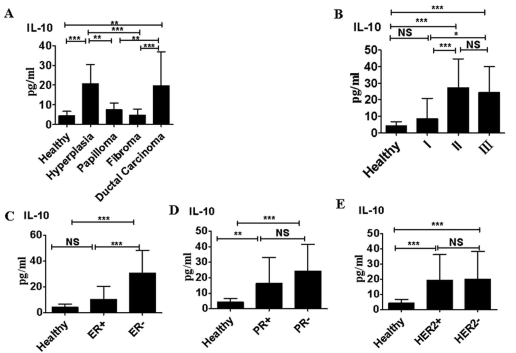
Serum levels of IL-10 in subjects with atypical
hyperplasia and ductal carcinoma were significantly higher than
those in healthy women, and in patients with papilloma or fibroma
(P<0.01; Fig. 1A). Patients with
TNM stage II and III ductal carcinoma exhibited significantly
higher serum IL-10 levels than those with stage I ductal carcinoma
(P<0.0001; Fig. 1B). Furthermore
serum IL-10 levels were significantly higher in patients with
ductal carcinoma that were ER− compared with those that
were ER+ (P<0.0001; Fig.
1C). IL-10 levels were significantly higher in sera collected
from patients with ductal carcinoma that were PR+ than
in the sera of healthy controls (P<0.01); however, IL-10 levels
did not differ significantly between patients with ductal carcinoma
that were PR+ and those that were PR−
(Fig. 1D). Furthermore, there was no
significant difference between IL-10 levels in patients with ductal
carcinoma that were HER2+ compared with those that were
HER2− (Fig. 1E).
Serum IL-17A levels in benign and
malignant breast diseases
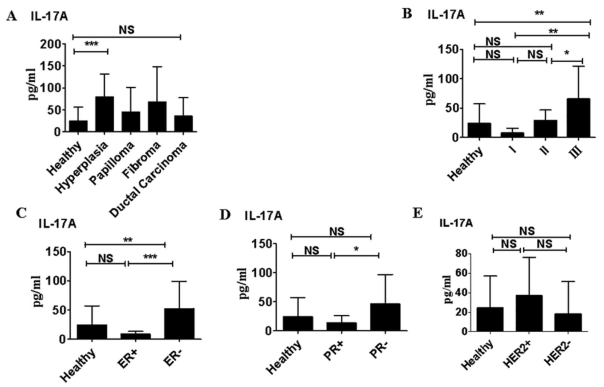
Only in patients with atypical hyperplasia were the
serum IL-17A levels significantly higher than those in healthy
women (P<0.05). IL-17A levels were slightly higher in patients
with papilloma and fibroma compared with healthy volunteers;
however, this difference was not significant (Fig. 2A). Although IL-17A levels did not
differ significantly between patients with ductal carcinoma and
healthy subjects, IL-17A levels in patients with stage III ductal
carcinoma were significantly higher than those in healthy
volunteers (P<0.01; Fig. 2B).
IL-17A levels were significantly higher in patients with ductal
carcinoma that were ER− compared with those that were
ER+ (P<0.0001; Fig.
2C). This was also the case in patients that were
PR− compared with those that were PR+
(P<0.05; Fig. 2D). However,
IL-17A levels did not differ significantly in between patients with
ductal carcinoma that were HER+ and those that were
HER− (Fig. 2E).
Serum TGF-α levels in benign and malignant breast
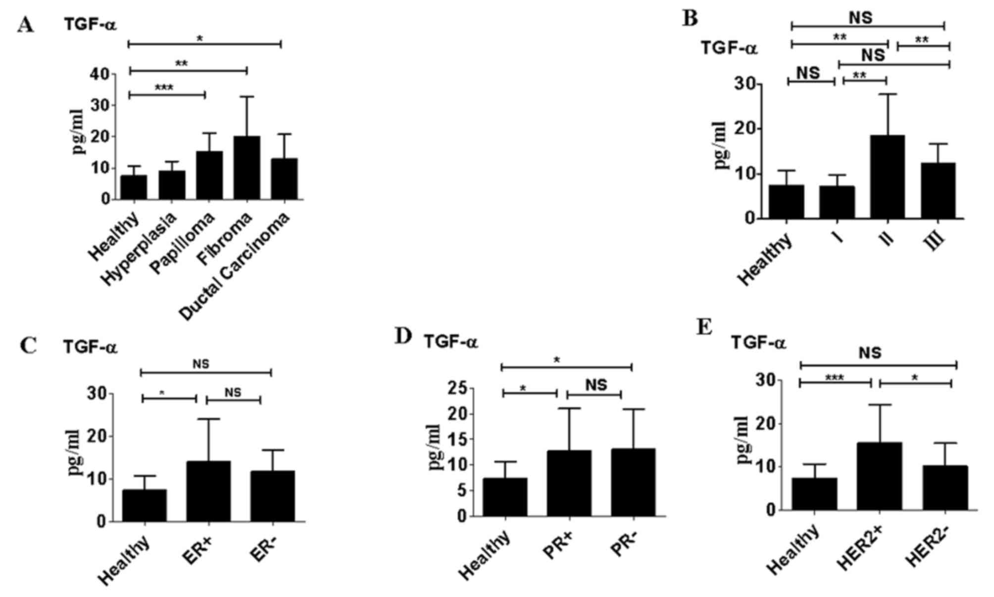
diseases. Serum levels of TGF-α were significantly higher in
patients with papilloma, fibroma and ductal carcinoma than in
healthy women (P<0.05; Fig. 3A).
However, there were no significant differences in levels of TGF-α
between patients with atypical hyperplasia and healthy women.
Patients with ductal carcinoma classified as TNM stage II also
exhibited significantly higher serum TGF-α levels than those
classed as TNM stage I (P<0.01; Fig.
3B); however there were no significant differences in TGF-α
levels between patients with stage III ductal carcinoma and those
with stage I ductal carcinoma (Fig.
3B). TGF-α levels did not differ significantly between patients
that were ER− compared with those that were
ER+ and there were no significant differences between
TGF-α levels between patients that were PR+ and those
that were PR− (Fig. 3C and
D). However, TGF-α levels were significantly higher in patients
that were HER2+ compared with those that were
HER2− (P<0.05; Fig.
3E).
Correlations between IL-10, IL-17A,
and TGF-α levels in patients with ductal carcinoma
In samples isolated from ductal carcinoma patients,
a significant positive correlation was identified between TGF-α and
IL-17A levels (P<0.001; Fig. 4A).
However, there was no correlation between TGF-α and IL-10 levels
(Fig. 4B) and no correlation between
IL-17A and IL-10 levels in patients with ductal carcinoma (Fig. 4C).
Discussion
Despite wide interest in the development of novel
diagnostic biomarkers for breast cancer (8,10,20),
limited data have been collected regarding whether IL-10, IL-17A
and TGF-α levels are associated with the clinical stage of breast
cancer, as well as ER, PR and HER2 status. The results of the
present study revealed that elevated serum levels of IL-10, IL-17A
and TGF-α are strongly associated with ductal carcinoma and are
associated with a more severe clinical stage of this disease. IL-10
and IL-17A levels were also associated with ER and PR status in
ductal carcinoma. Thus, these biomarkers may be used to diagnose
women with breast cancer and to identify patients with a poor
prognosis that may benefit from more aggressive forms of treatment
and management.
IL-10 is a pleiotropic cytokine able to suppress and
stimulate the immune response (16,21). It
has been suggested that patients with atypical hyperplasia may have
an increased risk of tumorigenesis (22). In the current study, serum IL-10
levels in patients with atypical hyperplasia were significantly
higher compared with healthy subjects and those with papilloma or
fibroma. It is therefore possible that these increased IL-10 levels
in patients with atypical hyperplasia may promote the development
of breast cancer. The results also demonstrated that IL-10 levels
were higher in sera collected from patients with stage II and III
ductal carcinoma than in the sera of patients with stage I
carcinoma. These results were not consistent with those reported by
Ikeguchi et al (19), who
reported that serum IL-10 levels were not correlated with tumor
stage in patients with gastric cancer. Therefore, it was speculated
that the intensity of the immune response varies among patients
with different types of cancer. For example, the antigens of ER and
HER2 are highly expressed on breast cancer cells but not gastric
cancer cells, and these antigens may promote or suppress cytokine
production (23). The results of the
current study indicated that high serum IL-10 levels were
associated with negative ER expression, but not with PR and HER2
expression.
IL-17A-producing TILs are found in greater numbers
in breast tumor tissue than in healthy mammary tissue (9,12). The
results of the current study revealed that serum IL-17A levels did
not differ significantly between healthy women and patients with
ductal carcinoma; however, IL-17A levels in patients with stage III
ductal carcinoma were significantly higher than those in patients
with stages I and II ductal carcinoma and healthy volunteers. This
suggests that IL-17A serves a critical role in promoting the
progression of cancer (9,14) The results of the current study
demonstrated that serum IL-10 and IL-17A levels are increased in
patients with atypical hyperplasia. These results may not actually
be contradictory, as IL-10 and IL-17A may promote tumorigenesis.
Furthermore, in the current study, serum IL-17A levels were
associated with the absence of ER in tumor tissue. Specifically,
IL-17A levels were significantly elevated in patients that were
ER− and PR−. The mechanisms underlying this
association between IL-17A and ER− remain unknown. One
possibility is that ER deficiency in breast cancer may directly
promote the expansion of IL-17A-producing TILs (9).
In breast cancer, TGF-α may promote the growth and
progression of tumors via an autocrine/paracrine loop involving the
epidermal growth factor receptor (10,13,24). In
the current study, serum TGF-α levels in patients with papilloma,
fibroma and ductal carcinoma were significantly higher than those
in healthy subjects; this suggests that TGF-α may also promote
tumor development. High serum TGF-α levels were also associated
with a more severe tumor stage and were significantly increased in
patients that were HER2+; however there was no
association between serum TGF-α levels and ER or PR expression.
Since mammary gland epithelial cells are able to secrete TGF-α, it
was hypothesized that HER2 expression in tumor cells may enhance
TGF-α production.
It was then assessed whether there were correlations
between serum IL-17A and TGF-α, IL-10 and TGF-α, and between IL-10
and IL-17A. The results demonstrated that IL-10 levels increased as
IL-17A levels increased; however, there appeared to be no
significant association between IL-10 and IL-17/TGF-α levels.
However, a strong positive correlation between TGF-α and IL-17A was
identified. Therefore, TGF-α and IL-17A may function
synergistically during the initiation and development of
tumors.
In conclusion, the results of the present study
suggest that increased serum levels of IL-10, IL-17 and TGF-α are
associated with ductal carcinoma. Increased levels of these
cytokines are also associated with a more severe clinical stage of
ductal carcinoma, and with the negative expression of ER and HER2.
Thus, these cytokines may be developed as potential diagnostic and
prognostic cancer biomarkers.
Acknowledgements
Not applicable.
Funding
The current study was supported by grants from the
National Natural Science Foundation of China (grant nos. 81472822,
and 81501377), Natural Science Foundation of Shaanxi Province
(grant no. 2015JM8385), China Postdoctoral Science Foundation
(grant no. 2014M560787), Shanxi Postdoctoral Science Foundation and
Fundamental Research Funds for the Central Universities (grant no.
2015gjhz16).
Availability of data and materials
The datasets used and/or analyzed during the current
study are available from the corresponding author on reasonable
request.
Authors' contributions
ZL and ML performed the immunoassays. JS collected
the patient and control samples. DX performed the data analysis. YM
drafted the manuscript. YM and YJ designed the study and revised
the manuscript. All authors read and approved the final
manuscript.
Ethics approval and consent to
participate
The protocol of the present study conformed to the
ethical standards of the Declaration of Helsinki. All procedures
were approved by the Ethics Committee of Huai'an First People's
Hospital Faculty of Medicine (Huai'an, China). Informed consent was
obtained from each patient, according to the committee's
regulations.
Consent for publication
Not applicable.
Competing interests
The authors declare that they have no competing
interests.
Glossary
Abbreviations
Abbreviations:
|
IL
|
interleukin
|
|
TGF-α
|
transforming growth factor α
|
|
ER
|
estrogen receptor
|
|
PR
|
progesterone receptor
|
|
HER2
|
human epidermal growth factor receptor
2
|
|
TILs
|
tumor-infiltrating lymphocytes
|
References
|
1
|
Amin AL, Purdy AC, Mattingly JD, Kong AL
and Termuhlen PM: Benign breast disease. Surg Clin North Am.
93:299–308. 2013. View Article : Google Scholar : PubMed/NCBI
|
|
2
|
Jiang X and Shapiro DJ: The immune system
and inflammation in breast cancer. Mol Cell Endocrinol.
382:673–682. 2014. View Article : Google Scholar : PubMed/NCBI
|
|
3
|
McEvoy MP, Coopey SB, Mazzola E, Buckley
J, Belli A, Polubriaginof F, Merrill AL, Tang R, Garber JE, Smith
BL, et al: Breast cancer risk and follow-up recommendations for
young women diagnosed with atypical hyperplasia and Lobular
Carcinoma In Situ (LCIS). Ann Surg Oncol. 22:3346–3349. 2015.
View Article : Google Scholar : PubMed/NCBI
|
|
4
|
Standish LJ, Sweet ES, Novack J, Wenner
CA, Bridge C, Nelson A, Martzen M and Torkelson C: Breast cancer
and the immune system. J Soc Integr Oncol. 6:158–168.
2008.PubMed/NCBI
|
|
5
|
DeNardo DG and Coussens LM: Inflammation
and breast cancer. Balancing immune response: Crosstalk between
adaptive and innate immune cells during breast cancer progression.
Breast Cancer Res. 9:2122007. View
Article : Google Scholar : PubMed/NCBI
|
|
6
|
Robertson FM, Ross MS, Tober KL, Long BW
and Oberyszyn TM: Inhibition of pro-inflammatory cytokine gene
expression and papilloma growth during murine multistage
carcinogenesis by pentoxifylline. Carcinogenesis. 17:1719–1728.
1996. View Article : Google Scholar : PubMed/NCBI
|
|
7
|
Schmid BC, Rudas M, Rezniczek GA,
Leodolter S and Zeillinger R: CXCR4 is expressed in ductal
carcinoma in situ of the breast and in atypical ductal hyperplasia.
Breast Cancer Res Treat. 84:247–250. 2004. View Article : Google Scholar : PubMed/NCBI
|
|
8
|
Chin AR and Wang SE: Cytokines driving
breast cancer stemness. Mol Cell Endocrinol. 382:598–602. 2014.
View Article : Google Scholar : PubMed/NCBI
|
|
9
|
Cochaud S, Giustiniani J, Thomas C,
Laprevotte E, Garbar C, Savoye AM, Curé H, Mascaux C, Alberici G,
Bonnefoy N, et al: IL-17A is produced by breast cancer TILs and
promotes chemoresistance and proliferation through ERK1/2. Sci Rep.
3:34562013. View Article : Google Scholar : PubMed/NCBI
|
|
10
|
Booth BW and Smith GH: Roles of
transforming growth factor-alpha in mammary development and
disease. Growth Factors. 25:227–235. 2007. View Article : Google Scholar : PubMed/NCBI
|
|
11
|
Bastid J, Bonnefoy N, Eliaou JF and
Bensussan A: Lymphocyte-derived interleukin-17A adds another brick
in the wall of inflammation-induced breast carcinogenesis.
Oncoimmunology. 3:e282732014. View Article : Google Scholar : PubMed/NCBI
|
|
12
|
Chen WC, Lai YH, Chen HY, Guo HR, Su IJ
and Chen HH: Interleukin-17-producing cell infiltration in the
breast cancer tumour microenvironment is a poor prognostic factor.
Histopathology. 63:225–233. 2013. View Article : Google Scholar : PubMed/NCBI
|
|
13
|
Kornasiewicz O, Grąt M, Dudek K,
Lewandowski Z, Gorski Z, Zieniewicz K and Krawczyk M: Serum levels
of HGF, IL-6, and TGF-α after benign liver tumor resection. Adv Med
Sci. 60:173–177. 2015. View Article : Google Scholar : PubMed/NCBI
|
|
14
|
Hamidullah, Changkija B and Konwar R: Role
of interleukin-10 in breast cancer. Breast Cancer Res Treat.
133:11–21. 2012. View Article : Google Scholar : PubMed/NCBI
|
|
15
|
Mittal SK and Roche PA: Suppression of
antigen presentation by IL-10. Curr Opin Immunol. 34:22–27. 2015.
View Article : Google Scholar : PubMed/NCBI
|
|
16
|
Igietseme JU, Ananaba GA, Bolier J, Bowers
S, Moore T, Belay T, Eko FO, Lyn D and Black CM: Suppression of
endogenous IL-10 gene expression in dendritic cells enhances
antigen presentation for specific Th1 induction: Potential for
cellular vaccine development. J Immunol. 164:4212–4219. 2000.
View Article : Google Scholar : PubMed/NCBI
|
|
17
|
Kozlowski L, Zakrzewska I, Tokajuk P and
Wojtukiewicz MZ: Concentration of interleukin-6 (IL-6),
interleukin-8 (IL-8) and interleukin-10 (IL-10) in blood serum of
breast cancer patients. Rocz Akad Med Bialymst. 48:82–84.
2003.PubMed/NCBI
|
|
18
|
Esquivel-Velázquez M, Ostoa-Saloma P,
Palacios-Arreola MI, Nava-Castro KE, Castro JI and Morales-Montor
J: The role of cytokines in breast cancer development and
progression. J Interferon Cytokine Res. 35:1–16. 2015. View Article : Google Scholar : PubMed/NCBI
|
|
19
|
Ikeguchi M, Hatada T, Yamamoto M, Miyake
T, Matsunaga T, Fukumoto Y, Yamada Y, Fukuda K, Saito H and Tatebe
S: Serum interleukin-6 and −10 levels in patients with gastric
cancer. Gastric Cancer. 12:95–100. 2009. View Article : Google Scholar : PubMed/NCBI
|
|
20
|
Goldberg JE and Schwertfeger KL:
Proinflammatory cytokines in breast cancer: Mechanisms of action
and potential targets for therapeutics. Curr Drug Targets.
11:1133–1146. 2010. View Article : Google Scholar : PubMed/NCBI
|
|
21
|
Jarnicki AG, Lysaght J, Todryk S and Mills
KH: Suppression of antitumor immunity by IL-10 and
TGF-beta-producing T cells infiltrating the growing tumor:
Influence of tumor environment on the induction of CD4+ and CD8+
regulatory T cells. J Immunol. 177:896–904. 2006. View Article : Google Scholar : PubMed/NCBI
|
|
22
|
Zhang HZ, Li XH, Zhang X, Zhang ZY, Meng
YL, Xu SW, Zheng Y, Zhu ZL, Cui DS, Huang LX, et al: PINCH protein
expression in normal endometrium, atypical endometrial hyperplasia
and endometrioid endometrial carcinoma. Chemotherapy. 56:291–297.
2010. View Article : Google Scholar : PubMed/NCBI
|
|
23
|
Chavey C, Bibeau F, Gourgou-Bourgade S,
Burlinchon S, Boissière F, Laune D, Roques S and Lazennec G:
Oestrogen receptor negative breast cancers exhibit high cytokine
content. Breast Cancer Res. 9:R152007. View
Article : Google Scholar : PubMed/NCBI
|
|
24
|
Suzuki M, Matsushima-Nishiwaki R,
Kuroyanagi G, Suzuki N, Takamatsu R, Furui T, Yoshimi N, Kozawa O
and Morishige K: Regulation by heat shock protein 22 (HSPB8) of
transforming growth factor-α-induced ovary cancer cell migration.
Arch Biochem Biophys. 571:40–49. 2015. View Article : Google Scholar : PubMed/NCBI
|


