Introduction
Dysregulation of antioxidant mechanisms along with
excessive accumulation of reactive oxygen species (ROS) is causally
linked to various health issues, including muscular dystrophy and
sarcopenia (1,2). Within the healthy muscle cell, ROS
are generated as both a by-product of metabolism and as effectors
of signaling cascades (3).
However, excessive ROS generation in skeletal muscles can influence
their contractile function by causing fatigue and increasing the
oxidative damage to cells (4,5).
Therefore, increasing the antioxidant capacity of skeletal muscles
can be beneficial for muscle performance, disease prevention, and
improved quality of life.
Many flavonoids, which are polyphenolic compounds
commonly found in a variety of fruits and vegetables and as
components of dietary supplements containing herbs, have been shown
to protect cells against oxidative stress-induced damage by virtue
of their antioxidant properties (6–8).
Morin (3,5,7,2′,4′-pentahydroxyflavone) is a naturally occurring
flavonoid that consists of a yellowish pigment found in many fruits
and herbs (9,10). It has been reported that this
compound may possess strong antioxidant properties that protect
cells against oxidative-induced damage (10–13). Morin has also been shown to induce
the activity of phase II enzymes, such as quinone reductase,
glutathione S-transferase, and glutathione reductase (14–17). In particular, morin has been
reported to protect human lens epithelial cells against oxidative
stress through the activation of nuclear factor erythroid 2-related
factor 2 (Nrf2)-dependent heme oxygenase-1 (HO-1) expression
(18). However, the inhibitory
mechanisms of morin vis-à-vis the beneficial effect of morin
against oxidative stress have not been fully studied to date.
Under normal conditions, Nrf2, a transcription
factor ubiquitously expressed in most tissues, is bound to
Kelch-like ECH-associated protein 1 (Keap1) in the cytoplasm
(19,20). Phase II enzyme inducers can
disrupt the Nrf2/Keap1 complex, resulting in the release of Nrf2
and its subsequent translocation to the nucleus (20,21). In the nucleus, Nrf2 activates the
antioxidant response element (ARE), which transcriptionally
activates many antioxidative genes (20,22). It has recently been reported that
activation of the Nrf2/HO-1 pathway can be increased by the
stabilization and/or phosphorylation of the Nrf2 protein, and
several signaling molecules, including
phosphatidylinositol-3-kinase (PI3K)/Akt and mitogen-activated
protein kinases (MAPKs), have been shown to participate in the
processes in response to a variety of phase II gene inducers
(23,24). Therefore, targeted activation of
Nrf2/HO-1 signaling may be considered an important therapeutic
strategy for protection against oxidative damage (19,20). In the present study, we
investigated the cytoprotective effect of morin against oxidative
stress damage in a mouse myoblast C2C12 cell line and the possible
protective mechanisms involved.
Materials and methods
Reagents and antibodies
Dulbecco's modified Eagle's medium (DMEM), fetal
bovine serum (FBS), and penicillin/streptomycin antibiotics were
purchased from WelGENE Inc. (Daegu, Republic of Korea). Morin,
3-[4,5-dimethylthiazol-2-yl)-2,5-diphenyltetrazolium bromide (MTT),
N-acetyl-L- cysteine (NAC), 4,6-diamidino-2-phenyllindile
(DAPI), and PD98059 (2′-amino-3′-methoxyflavone) were obtained from
Sigma-Aldrich Chemical Co. (St. Louis, MO, USA).
2′,7′-Dichlorodihydrofluorescein diacetate (DCF-DA) and a
fluorescein-conjugated Annexin V (Annexin V-FITC) staining assay
kit were purchased from Molecular Probes (Eugene, OR, USA) and BD
Biosciences (San Jose, CA, USA), respectively. An enhanced
chemiluminescence (ECL) detection kit and FITC-conjugated donkey
anti-rabbit IgG were purchased from Amersham Co. (Arlington
Heights, IL, USA) and Jackson ImmunoResearch Laboratories Inc.
(West Grove, PA, USA), respectively. Various primary antibodies
(Table I) for western blot
analysis were obtained from Cell Signaling Technology, Inc.
(Boston, MA, USA), Santa Cruz Biotechnology, Inc. (Santa Cruz, CA,
USA) and Abcam, Inc. (Cambridge, MA, USA). Horseradish
peroxidase-conjugated anti-rabbit (SC-2004), anti-mouse (SC-2005)
and anti-goat (SC-2350) antibodies were used as the secondary
antibodies, which were obtained from Santa Cruz Biotechnology, Inc.
All other chemicals were purchased from Sigma-Aldrich Chemical
Co.
 | Table IAntibodies used in the present
study. |
Table I
Antibodies used in the present
study.
| Antibody | Origin | Company | Catalogue no. |
|---|
| Actin | Mouse
monoclonal | Santa Cruz
Biotechnology, Inc. | SC-47778 |
| p-γH2AX | Rabbit
monoclonal | Cell Signaling
Technology, Inc. | 9718 |
| γH2AX | Rabbit
monoclonal | Cell Signaling
Technology, Inc. | 7631 |
| p-Nrf2 | Rabbit
monoclonal | Abcam, Inc. | ab76026 |
| Nrf2 | Rabbit
polyclonal | Santa Cruz
Biotechnology, Inc. | SC-13032 |
| Keap1 | Goat
polyclonal | Santa Cruz
Biotechnology, Inc. | SC-15246 |
| HO-1 | Rabbit
polyclonal | Santa Cruz
Biotechnology, Inc. | SC-10789 |
| NQO1 | Goat
polyclonal | Santa Cruz
Biotechnology, Inc. | SC-16464 |
| TrxR1 | Mouse
monoclonal | Santa Cruz
Biotechnology, Inc. | SC-28321 |
| Lamin B | Goat
polyclonal | Santa Cruz
Biotechnology, Inc. | SC-6216 |
| p-PI3K | Rabbit
polyclonal | Cell Signaling
Technology, Inc. | 4228 |
| PI3K | Rabbit
polyclonal | Cell Signaling
Technology, Inc. | 4249 |
| p-Akt | Rabbit
polyclonal | Santa Cruz
Biotechnology, Inc. | SC-101629 |
| Akt | Rabbit
polyclonal | Santa Cruz
Biotechnology, Inc. | SC-8312 |
| p-ERK | Mouse
monoclonal | Cell Signaling
Technology, Inc. | 9106 |
| ERK | Rabbit
polyclonal | Santa Cruz
Biotechnology, Inc. | SC-154 |
| p-JNK | Mouse
monoclonal | Cell Signaling
Technology, Inc. | 9255 |
| JNK | Rabbit
monoclonal | Cell Signaling
Technology, Inc. | 9252 |
| p-p38 MAPK | Rabbit
monoclonal | Cell Signaling
Technology, Inc. | 9211 |
| p38 MAPK | Rabbit
polyclonal | Santa Cruz
Biotechnology, Inc. | SC-535 |
Cell culture and MTT assay
C2C12 myoblasts obtained from the American Type
Culture Collection (ATCC, Manassas, VA, USA) were grown in DMEM
supplemented with 10% FBS and 100 μg/ml
penicillin/streptomycin antibiotics in a humidified 5%
CO2 atmosphere at 37°C. Morin was dissolved in dimethyl
sulfoxide (DMSO) and adjusted to final concentrations using
complete DMEM prior to use; the final DMSO concentration was
<0.1% in all experiments. In order to evaluate the degree to
which a single morin treatment affects C2C12 cell viability, the
C2C12 cells were seeded at a density of 1×10⁴ cells/well
in a 96-well plate, incubated at 37°C for 24 h, and treated with
morin at different concentrations (100–10,000 μM).
Additional cell cultivation occurred for 6 h in media where
H2O2 and NAC were simultaneously
administered, singularly administered, or were not administered.
For the cell viability assay using a colorimetric MTT assay, the
medium was discarded, MTT solution was added to each well, and the
cells were further incubated for 3 h at 37°C. The medium was
discarded and DMSO was added to dissolve the formazan product. The
optical density was then read at 450 nm using an enzyme-linked
immunosorbent assay (ELISA) plate reader (Dynatech MR-7000;
Dynatech Laboratories, Chantilly, VA, USA). Relative cell
cytotoxicity was evaluated according to the quantity of MTT
converted to insoluble formazan salt.
Intracellular ROS measurement
In order to monitor ROS generation, the cells were
incubated with 10 μM DCF-DA for 20 min at room temperature
in the dark. The ROS production in the cells was monitored with a
flow cytometer (Becton-Dickinson, San Jose, CA, USA) using
CellQuest Pro software (25).
Determination of apoptotic cells by flow
cytometry
To quantitatively assess the induced cell apoptosis
rate, an Annexin V-FITC staining assay was performed as previously
described (26). The cells were
washed twice with ice-cold phosphate-buffered saline (PBS) and
stained with Annexin V-FITC and propidium iodide (PI) in each
sample for 15 min at room temperature in the dark. The degree of
apoptosis was quantified as a percentage of Annexin V-positive and
PI-negative (Annexin V+/PI− cells) cells
using a flow cytometer.
Comet assay
To assess oxidative DNA damage, the cell suspension
was mixed with 0.5% low melting agarose (LMA) at 37°C, and the
mixture was spread on a fully frosted microscopic slide precoated
with 1% normal melting agarose. After the solidification of the
agarose, the slide was covered with 0.5% LMA and then immersed in a
lysis solution (2.5 M NaCl, 100 mM Na-ethylenediaminetetraacetic
acid (Na-EDTA), 10 mM Tris, 1% Triton X-100, and 10% DMSO, pH 10.0)
for 1 h at 4°C. The slides were then placed in a gel
electrophoresis apparatus containing 300 mM NaOH and 10 mM Na-EDTA
(pH 13.0) for 40 min to allow for DNA unwinding and the expression
of alkali-labile damage. Next, an electrical field was applied (300
mA, 25 V) for 20 min at 4°C to draw the negatively charged DNA
toward the anode. After electrophoresis, the slides were washed
three times for 5 min at 4°C in a neutralizing buffer (0.4 M Tris,
pH 7.5), followed by staining with 20 μg/ml PI. The cells
were washed twice with PBS, and images were then captured using a
fluorescence microscope (Carl Zeiss, Jena, Germany) (27).
Protein extraction and western blot
analysis
Whole-cell protein extracts from C2C12 myoblasts
were prepared with cell lysis buffer [25 mM of Tris-Cl (pH 7.5),
250 mM of NaCl, 5 mM of EDTA, 1% Nonidet P-40, 0.1 mM of sodium
orthovanadate, 2 μg/ml of leupeptin, and 100 μg/ml of
phenylmethylsulfonyl flouride] containing protease inhibitor
cocktail tablets (Roche Diagnostics, Mannheim, Germany) for 30 min.
In a parallel experiment, nuclear and cytosolic proteins were
prepared using nuclear extraction reagents (Pierce Biotechnology,
Rockford, IL, USA) according to the manufacturer's protocol. After
cell debris was discarded following centrifugation at 13,000 × g
for 15 min, the protein concentration was determined using the
Bio-Rad protein assay kit (Bio-Rad Laboratories, Hercules, CA,
USA). For western blot analysis, equal amounts of protein extracts
were subjected to electrophoresis on sodium dodecyl sulfate (SDS)
polyacrylamide gels and transferred onto nitrocellulose membranes
(Schleicher and Schuell Bioscience, Inc., Keene, NH, USA) by
electroblotting. The blots were probed with the desired primary
antibodies for 1 h, incubated with the diluted enzyme-linked
secondary antibodies, and visualized using an ECL method according
to the recommended procedure.
Small interfering RNA (siRNA)
transfection
Nrf2 siRNA and control siRNA were purchased from
Santa Cruz Biotechnology, Inc. The siRNAs were transfected into
cells according to the manufacturer's instructions using
Lipofectamine 2000 Transfection Reagent (Life Technologies,
Carlsbad, CA, USA). For transfection, the cells were seeded in
6-well culture plates and incubated with the control siRNA or Nrf2
siRNA at 50 nM for 6 h in serum-free Opti-MEM media (Life
Technologies). After incubation, the transfected cells were
subjected to treatment, as previously described (28).
Immunofluorescent staining for Nrf2
C2C12 cells were seeded on glass coverslips in
6-well plates for 24 h, and the cells were treated with morin for 6
h. Next, the cells were rinsed twice with PBS and fixed with 3.7%
paraformaldehyde in PBS for 10 min at 4°C. The cells were incubated
with 0.4% Triton X-100 for 10 min and then blocked with 5% bovine
serum albumin for 1 h, followed by probing with the anti-Nrf2
antibody overnight at 4°C and incubation with FITC-conjugated
donkey anti-rabbit IgG for 2 h at room temperature. After washing
with PBS, nuclei were counterstained with DAPI solution (1 mg/ml)
for 15 min in the dark. Images were collected using a fluorescence
microscope (Carl Zeiss, Oberkochen, Germany).
Statistical analysis
Data are expressed as mean ± standard deviation (SD)
from at least three independent experiments. Statistical
comparisons between different groups were performed using one-way
ANOVA, followed by Student's t-tests after comparing each treated
group to the negative control. Probability values P<0.01 were
considered to indicate statistically significant differences.
Results
Effects of morin on
H2O2-induced cytotoxicity in C2C12
myoblasts
To evaluate the protective effect of morin on
H2O2-induced cytotoxicity, the cells were
treated with various concentrations of morin for 24 h, and the cell
viability was evaluated for primary dose selection by determining
the percentage of MTT reduction. Morin alone at 100–500 μM
showed no cytotoxic effects, but significant cytotoxicity was noted
at 1,000 μM morin (Fig.
1A). Thus, 500 μM morin was chosen as the optimal dose
for studying the cytoprotective effect of this flavonoid.
The viability of the C2C12 cells when exposed to
H2O2 at the concentration of 1 mM for 6 h was
significantly decreased as compared with the control group, and the
survival rate was ~60% that of the control group. When the C2C12
cells were pretreated with 500 μM morin or 10 mM NAC, an ROS
scavenger that was used as positive control, for 1 h and exposed to
H2O2 for an additional 6 h, the cell
viability was significantly increased compared to the
H2O2 group, and the survival rate was 81.00
and 99.08% that of the control group, respectively (Fig. 1B).
Inhibition of
H2O2-induced ROS production by morin in C2C12
myoblasts
As the cytotoxicity of H2O2 is
mainly mediated by oxidative stress, we investigated the effect of
morin on H2O2-induced ROS accumulation using
DCF-DA reagent. Compared with the non-treated control cells, the
level of intracellular ROS was markedly increased in the C2C12
cells treated with 1 mM H2O2 for 6 h, as
indicated by the increase in the DCF-liberated fluorescent signal
(Fig. 2A). However, when the
C2C12 cells were pretreated with morin or NAC, the ROS formation
was significantly decreased, suggesting that morin pretreatment
induced a cellular antioxidative response.
Protection against
H2O2-induced C2C12 cell apoptosis by
morin
The effects of morin on C2C12 cell apoptosis induced
by H2O2 were observed using an Annexin
V-FITC/PI assay. As illustrated in Fig. 2B, the percentage of apoptotic
cells treated with 1 mM H2O2 was ~49.54%;
however, morin or NAC pretreatment effectively decreased cell
apoptosis induced by H2O2 to 7.99 and 7.86%,
respectively. The results indicate that the
H2O2-induced apoptosis was mediated by ROS
generation and that morin exerted a potent ROS scavenging effect,
preventing H2O2-induced apoptosis.
Prevention of
H2O2-induced DNA damage by morin in C2C12
myoblasts
We next examined the effects of morin on
H2O2-mediated DNA damage in the C2C12 cells
using the comet assay (single cell gel electrophoresis) and western
blot analysis. Exposure to H2O2 alone induced
significant DNA breaks, resulting in an increase in fluorescence
intensity in the tails of the comet-like structures (Fig. 3A); however, this adverse effect
was markedly reduced by pretreatment with morin or NAC. In
addition, the exposure of the C2C12 cells to
H2O2 resulted in upregulation in the level of
the phosphorylated histone variant H2AX (p-γH2AX) at serine 139, a
sensitive marker of DNA double strand breaks (29); however, pretreatment with morin or
NAC resulted in a significant decrease in p-γH2AX expression.
Effects of morin on the expression of
Nrf2 and HO-1 in C2C12 myoblasts
As Nrf2 signaling regulates cellular antioxidant
response (20,22), we examined whether morin protects
cells from oxidative stress by activating the Nrf2 signaling
pathway. Our immunoblotting results indicated that treatment of
C2C12 cells with morin induced the expression of HO-1 protein in a
time-dependent manner, but other antioxidant enzymes, NADPH-quinone
oxidoreductase 1 (NQO1) and thioredoxin reductase 1 (TrxR1), were
unaffected by morin treatment, which was associated with the
upregulation of Nrf2 expression and the downregulation of Keap1
(Fig. 4A).
Since the phosphorylation of Nrf2 at Ser40 by
several kinases is also a critical process in its stabilization and
nuclear translocation (20,23,24), we examined the phosphorylation of
Nrf2 under morin treatment to further confirm the Nrf2 activating
property of morin and observed that the treatment of cells with
morin time-dependently increased the expression levels of
phosphorylated Nrf2 (Fig. 4A).
Additionally, western blot analysis was carried out using the
nuclear and cytosolic fractions of the C2C12 cells. The results
shown in Fig. 5A and B indicate
that the amounts of total and phosphorylated Nrf2 proteins in the
nucleus were markedly increased following treatment with morin. The
immunofluorescence images also revealed that the nuclear
localization and accumulation of Nrf2 in the C2C12 cells was
significantly increased after stimulation with morin (Fig. 5C).
Nrf2-dependent induction of HO-1 by morin
in C2C12 myoblasts
In order to provide evidence for the involvement of
Nrf2 in the induction of HO-1 by morin, we transiently transfected
C2C12 cells with Nrf2 siRNA. Western blot analysis results revealed
that the silencing of Nrf2 using specific siRNA abrogated the
morin-induced increase and phosphorylation in Nrf2 expression
(Fig. 4B). Therefore, we
investigated whether Nrf2 siRNA transfection attenuated
morin-induced upregulation of HO-1 and downregulation of Keap1 and
found that Nrf2 siRNA reversed these effects, which is evidence
that the augmentation of HO-1 by morin was mediated by Nrf2.
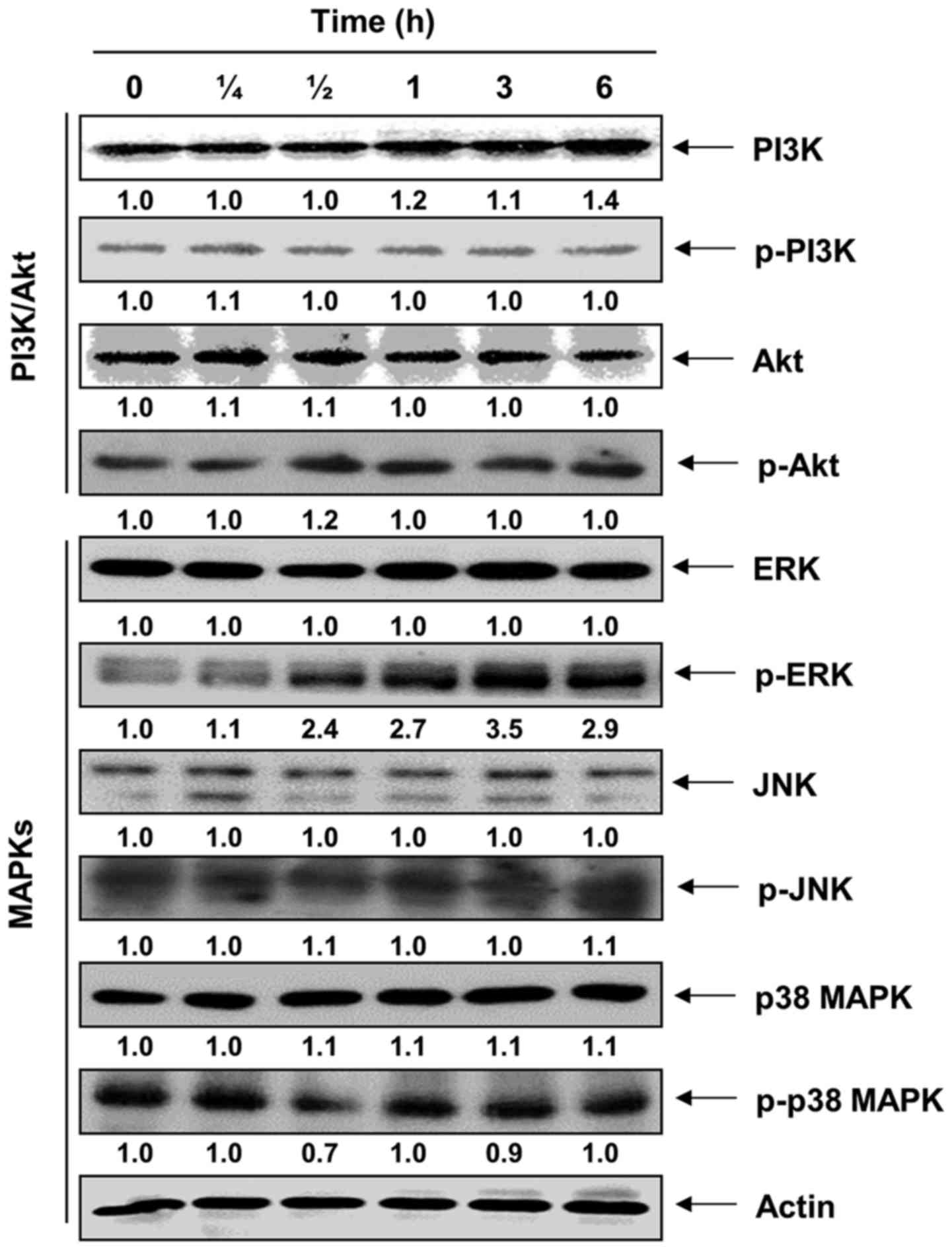
Effect of extracellular signal-regulated
kinase (ERK) signaling pathway on Nrf2-mediated HO-1 induction by
morin in C2C12 myoblasts
To identify the upstream signaling events involved
in morin-mediated Nrf2-dependent HO-1 induction, the potential
involvement of the PI3K/Akt and MAPK signaling pathways was
investigated. The total and phosphorylated levels of PI3K and Akt,
a downstream target of PI3K, did not show a notable change in the
morin-treated C2C12 cells as compared with levels in the untreated
control cells (Fig. 6).
Subsequently, although there were no notable changes observed in
the phosphorylated levels of c-Jun N-terminal kinase (JNK) and p38
MAPK, the activation of extracellular signal-regulated kinase (ERK)
was noted as early as 30 min after morin treatment, and it lasted
at least for 6 h. However, treatment with a selective inhibitor of
ERK, PD98059, blocked the morin-triggered phosphorylation of Nrf2,
and HO-1 induction was accordingly diminished without the blockade
of Nrf2 induction (Fig. 7). By
contrast, when an inhibitor of ERK was utilized, the induction of
the total protein level of Nrf2 in the morin-treated C2C12 cells
was not attenuated.
Discussion
Although there have been few studies on how
oxidative stress may quantitatively affect the load-carrying
capacity of muscle cells, oxidative stress on myoblasts should
accompany the dysfunction of muscles (3,4).
In this investigation, as part of the screening program for
therapeutic antioxidative agents from phytochemicals, we attempted
to determine whether morin offers protection from oxidative
stress-induced cytotoxicity using a C2C12 myoblast cell model. We
first observed that when C2C12 myoblasts were treated with morin in
the presence of H2O2, cell viability was
significantly recovered by inhibiting
H2O2-induced ROS generation, compared with
exposure to H2O2 alone. Our data also showed
that H2O2 exposure increased tail length and
the expression of p-γH2AX, whereas each event was mitigated in
C2C12 cells that had been treated with morin prior to
H2O2 exposure. As a result, these findings
suggest that morin may be useful for the prevention of
H2O2-induced cytotoxicity due to its strong
antioxidant effect.
There is mounting evidence that oxidative stress is
implicated in the pathogenesis of muscle dysfunction (4,5).
Oxidative stress occurs when the production of ROS exceeds its
catabolism. ROS, including superoxide radical anions, hydroxyl
radicals, singlet oxygen and hydrogen peroxide, are a class of
endogenous signaling molecules with specific functions depending on
their subcellular localization, local concentration and duration of
production. Although a moderate level of ROS transiently oxidizes
the cysteine sulfhydryl that contributes to the active sites of
most proteins, when ROS accumulate to a certain level, the active
effects may convert to inhibitory effects that damage cellular
functions (30,31). Excessive ROS generation in
skeletal muscles can influence the contractile function of skeletal
muscles by causing fatigue and increasing the oxidative damage to
cells, which is implicated in the development of multiple diseases,
including muscular dystrophy and sarcopenia (4,5).
Therefore, the discovery of an efficient agent that is able to
reduce ROS is valuable in the treatment of muscle-related diseases.
As expected, our results showed that ROS generation in C2C12
myoblasts was significantly increased by H2O2
challenge and that morin markedly reduced ROS production by
attenuating DNA damage.
Nrf2, a transcription factor that is part of the
redox homeostatic gene regulatory network, and its repressor,
Keap1, play indispensable roles in protecting a variety of tissues
from a wide array of toxic insults, including oxidative stress. As
yet, the role of Nrf2 signaling has not been well characterized in
skeletal muscle. However, coordinated regulation of Nrf2 redox
signaling is believed to preserve the redox state and to protect
skeletal muscle structure and function (2,32).
Previous studies have shown that active skeletal muscle exhibit
increased nuclear Nrf2 levels and subsequent activation of its
major target antioxidant enzymes, including HO-1 (33). Contrary to this, the loss of Nrf2
is strongly coupled with the dysregulation of antioxidant pathways
and the progression of muscle dysfunction (34,35). Nrf2 is sequestered in the cytosol
as an inactive complex with its repressor Keap1. Dissociation of
Nrf2 from Keap1 is a prerequisite for nuclear translocation and
subsequent DNA binding of Nrf2 to regulate the inducible expression
of cytoprotective genes, such as that of HO-1 (21,22). HO-1 is an inducible rate-limiting
enzyme initially identified as a phase II detoxifying enzyme that
facilitates the degradation of heme into bilirubin, free iron, and
carbon monoxide. The final products of heme catabolism exert
certain antioxidant effects by neutralizing intracellular ROS
(36,37). Therefore, Nrf2/HO-1 signaling may
represent a potential therapeutic target in the management of
oxidative stress-related diseases. In the present study, we
observed a significant increase in total Nrf2 expression in
morin-treated C2C12 myoblasts that was correlated with a
concomitant reduction in Keap1 expression. Subsequently, we found
that morin triggered the phosphorylation of Nrf2 and led to HO-1
induction along with a concomitant translocation of Nrf2 into the
nuclei. Howevere, Nrf2-specific siRNA markedly suppressed
morin-enhanced HO-1 protein levels, suggesting that Nrf2 is a
critical upstream regulator of the morin-mediated induction of HO-1
in C2C12 myoblasts.
It has been proposed that the induction of
phosphorylation plays a key role in the regulation of the
transcriptional activity of Nrf2 (23,24). In addition, a number of protein
kinases, such as PI3K/Akt and MAPKs, have been implicated as
upstream signals in the regulation of Nrf2 activity by facilitating
the translocation of Nrf2 into the nucleus, whereupon it binds to
ARE in the promoter regions (38–42). Therefore, to investigate whether
any protein kinases may be involved in the activation of Nrf2, the
phosphorylated level of different protein kinases following morin
treatment was analyzed. Our results revealed that morin treatment
only had a notable inducing effect on ERK phosphorylation within 30
min. In contrast, phosphorylation of PI3K, Akt, JNK and p38 MAPK
was not observed at any time point. Furthermore, our data indicated
that morin-induced phosphorylation of Nrf2 and induction of HO-1
were effectively inhibited by the ERK kinase inhibitor, whereas the
increased total protein levels of Nrf2 were not affected. These
observations suggest that morin may activate ERK by inducing its
phosphorylation and in turn post-translationally phosphorylate Nrf2
as a downstream signal, which is required for Nrf2 accumulation in
the nucleus leading to HO-1 expression.
In conclusion, our results showed that in C2C12
myoblasts, morin exhibited protective ability against
H2O2-induced cytotoxicity and DNA damage
through the suppression of intracellular ROS generation. In
addition, our overall results imply that morin may activate Nrf2 by
activating ERK to contribute to the induction of phase II
antioxidant HO-1 in C2C12 myoblasts, which, at least in part,
contributes to a cellular defense mechanism against oxidative
stress-induced genotoxic events. In future studies, this molecular
mechanism must be validated in vivo. A positive result, if
confirmed, would be invaluable to the development of new approaches
for effective stress-responsive antioxidants against assaults
triggered by ROS.
Acknowledgments
This study was supported by the High Value-Added
Food Technology Development Program (314043-3), Chinese Ministry of
Agriculture, Food and Rural Affairs.
References
|
1
|
Szczesny B, Olah G, Walker DK, Volpi E,
Rasmussen BB, Szabo C and Mitra S: Deficiency in repair of the
mitochondrial genome sensitizes proliferating myoblasts to
oxidative damage. PLoS One. 8:e752012013. View Article : Google Scholar : PubMed/NCBI
|
|
2
|
Miller CJ, Gounder SS, Kannan S, Goutam K,
Muthusamy VR, Firpo MA, Symons JD, Paine R III, Hoidal JR and
Rajasekaran NS: Disruption of Nrf2/ARE signaling impairs
antioxidant mechanisms and promotes cell degradation pathways in
aged skeletal muscle. Biochim Biophys Acta. 1822:1038–1050. 2012.
View Article : Google Scholar : PubMed/NCBI
|
|
3
|
Ji LL, Gomez-Cabrera MC and Vina J:
Exercise and hormesis: Activation of cellular antioxidant signaling
pathway. Ann NY Acad Sci. 1067:425–435. 2006. View Article : Google Scholar : PubMed/NCBI
|
|
4
|
Powers SK and Jackson MJ: Exercise-induced
oxidative stress: Cellular mechanisms and impact on muscle force
production. Physiol Rev. 88:1243–1276. 2008. View Article : Google Scholar : PubMed/NCBI
|
|
5
|
Song W, Kwak HB and Lawler JM: Exercise
training attenuates age-induced changes in apoptotic signaling in
rat skeletal muscle. Antioxid Redox Signal. 8:517–528. 2006.
View Article : Google Scholar : PubMed/NCBI
|
|
6
|
Harasym J and Oledzki R: Effect of fruit
and vegetable antioxidants on total antioxidant capacity of blood
plasma. Nutrition. 30:511–517. 2014. View Article : Google Scholar : PubMed/NCBI
|
|
7
|
Guo W, Kong E and Meydani M: Dietary
polyphenols, inflammation, and cancer. Nutr Cancer. 61:807–810.
2009. View Article : Google Scholar
|
|
8
|
Landete JM: Dietary intake of natural
antioxidants: Vitamins and polyphenols. Crit Rev Food Sci Nutr.
53:706–721. 2013. View Article : Google Scholar : PubMed/NCBI
|
|
9
|
Hardigree AA and Epler JL: Comparative
mutagenesis of plant flavonoids in microbial systems. Mutat Res.
58:231–239. 1978. View Article : Google Scholar : PubMed/NCBI
|
|
10
|
Wu TW, Zeng LH, Wu J and Fung KP: Morin: A
wood pigment that protects three types of human cells in the
cardiovascular system against oxyradical damage. Biochem Pharmacol.
47:1099–1103. 1994. View Article : Google Scholar : PubMed/NCBI
|
|
11
|
Kitagawa S, Sakamoto H and Tano H:
Inhibitory effects of flavonoids on free radical-induced hemolysis
and their oxidative effects on hemoglobin. Chem Pharm Bull (Tokyo).
52:999–1001. 2004. View Article : Google Scholar
|
|
12
|
Liu YH, Mo SL, Bi HC, Hu BF, Li CG, Wang
YT, Huang L, Huang M, Duan W, Liu JP, et al: Regulation of human
pregnane X receptor and its target gene cytochrome P450 3A4 by
Chinese herbal compounds and a molecular docking study.
Xenobiotica. 41:259–280. 2011. View Article : Google Scholar
|
|
13
|
Yang SH, Choi HG, Lim SJ, Lee MG and Kim
SH: Effects of morin on the pharmacokinetics of etoposide in
7,12-dimethylbenz[a] anthracene-induced mammary tumors in female
Sprague-Dawley rats. Oncol Rep. 29:1215–1223. 2013.
|
|
14
|
Al Numair KS, Chandramohan G, Alsaif MA
and Baskar AA: Protective effect of morin on cardiac mitochondrial
function during isoproterenol-induced myocardial infarction in male
Wistar rats. Redox Rep. 17:14–21. 2012. View Article : Google Scholar : PubMed/NCBI
|
|
15
|
Al-Numair KS, Chandramohan G, Alsaif MA,
Veeramani C and El Newehy AS: Morin, a flavonoid, on lipid
peroxidation and antioxidant status in experimental myocardial
ischemic rats. Afr J Tradit Complement Altern Medicines. 11:14–20.
2014. View Article : Google Scholar
|
|
16
|
Kim J, Kim JS and Park E: Cytotoxic and
anti-inflammatory effects of onion peel extract on
lipopolysaccharide stimulated human colon carcinoma cells. Food
Chem Toxicol. 62:199–204. 2013. View Article : Google Scholar : PubMed/NCBI
|
|
17
|
Nandhakumar R, Salini K and Niranjali
Devaraj S: Morin augments anticarcinogenic and antiproliferative
efficacy against 7,12-dimethylbenz(a)-anthracene induced
experimental mammary carcinogenesis. Mol Cell Biochem. 364:79–92.
2012. View Article : Google Scholar : PubMed/NCBI
|
|
18
|
Park JY, Kang KA, Kim KC, Cha JW, Kim EH
and Hyun JW: Morin induces heme oxygenase-1 via ERK-Nrf2 signaling
pathway. J Cancer Prev. 18:249–256. 2013. View Article : Google Scholar
|
|
19
|
Zhang Y and Gordon GB: A strategy for
cancer prevention: Stimulation of the Nrf2-ARE signaling pathway.
Mol Cancer Ther. 3:885–893. 2004.PubMed/NCBI
|
|
20
|
Ishii T, Itoh K, Takahashi S, Sato H,
Yanagawa T, Katoh Y, Bannai S and Yamamoto M: Transcription factor
Nrf2 coordinately regulates a group of oxidative stress-inducible
genes in macrophages. J Biol Chem. 275:16023–16029. 2000.
View Article : Google Scholar : PubMed/NCBI
|
|
21
|
Niture SK, Khatri R and Jaiswal AK:
Regulation of Nrf2-an update. Free Radic Biol Med. 66:36–44. 2014.
View Article : Google Scholar
|
|
22
|
Kobayashi M and Yamamoto M: Molecular
mechanisms activating the Nrf2-Keap1 pathway of antioxidant gene
regulation. Antioxid Redox Signal. 7:385–394. 2005. View Article : Google Scholar : PubMed/NCBI
|
|
23
|
Pi J, Bai Y, Reece JM, Williams J, Liu D,
Freeman ML, Fahl WE, Shugar D, Liu J and Qu W: Molecular mechanism
of human Nrf2 activation and degradation: Role of sequential
phosphorylation by protein kinase CK2. Free Radic Biol Med.
42:1797–1806. 2007. View Article : Google Scholar : PubMed/NCBI
|
|
24
|
Apopa PL, He X and Ma Q: Phosphorylation
of Nrf2 in the transcription activation domain by casein kinase 2
(CK2) is critical for the nuclear translocation and transcription
activation function of Nrf2 in IMR-32 neuroblastoma cells. J
Biochem Mol Toxicol. 22:63–76. 2008. View Article : Google Scholar : PubMed/NCBI
|
|
25
|
Eom SA, Kim DW, Shin MJ, Ahn EH, Chung SY,
Sohn EJ, Jo HS, Jeon SJ, Kim DS, Kwon HY, et al: Protective effects
of PEP-1-catalase on stress-induced cellular toxicity and
MPTP-induced Parkinson's disease. BMB Rep. 48:395–400. 2015.
View Article : Google Scholar :
|
|
26
|
Park MH and Han JS: Padina arborescens
extract protects high glucose-induced apoptosis in pancreatic β
cells by reducing oxidative stress. Nutr Res Pract. 8:494–500.
2014. View Article : Google Scholar : PubMed/NCBI
|
|
27
|
Jeong MH, Yang K, Lee CG, Jeong DH, Park
YS, Choi YJ, Kim JS, Oh SJ, Jeong SK and Jo WS: In vitro
genotoxicity assessment of a novel resveratrol analogue, HS-1793.
Toxicol Res. 30:211–220. 2014. View Article : Google Scholar : PubMed/NCBI
|
|
28
|
Jiang R, Teng Y, Huang Y, Gu J, Ma L, Li M
and Zhou Y: Preeclampsia serum-induced collagen I expression and
intracellular calcium levels in arterial smooth muscle cells are
mediated by the PLC-γ1 pathway. Exp Mol Med. 46:e1152014.
View Article : Google Scholar
|
|
29
|
Rogakou EP, Pilch DR, Orr AH, Ivanova VS
and Bonner WM: DNA double-stranded breaks induce histone H2AX
phosphorylation on serine 139. J Biol Chem. 273:5858–5868. 1998.
View Article : Google Scholar : PubMed/NCBI
|
|
30
|
Kregel KC and Zhang HJ: An integrated view
of oxidative stress in aging: Basic mechanisms, functional effects,
and pathological considerations. Am J Physiol Regul Integr Comp
Physiol. 292:R18–R36. 2007. View Article : Google Scholar
|
|
31
|
Finkel T: Signal transduction by reactive
oxygen species. J Cell Biol. 194:7–15. 2011. View Article : Google Scholar : PubMed/NCBI
|
|
32
|
Li M and Fukagawa NK: Age-related changes
in redox signaling and VSMC function. Antioxid Redox Signal.
12:641–655. 2010. View Article : Google Scholar :
|
|
33
|
Safdar A, deBeer J and Tarnopolsky MA:
Dysfunctional Nrf2-Keap1 redox signaling in skeletal muscle of the
sedentary old. Free Radic Biol Med. 49:1487–1493. 2010. View Article : Google Scholar : PubMed/NCBI
|
|
34
|
Rangasamy T, Guo J, Mitzner WA, Roman J,
Singh A, Fryer AD, Yamamoto M, Kensler TW, Tuder RM, Georas SN, et
al: Disruption of Nrf2 enhances susceptibility to severe airway
inflammation and asthma in mice. J Exp Med. 202:47–59. 2005.
View Article : Google Scholar : PubMed/NCBI
|
|
35
|
Reddy NM, Kleeberger SR, Kensler TW,
Yamamoto M, Hassoun PM and Reddy SP: Disruption of Nrf2 impairs the
resolution of hyperoxia-induced acute lung injury and inflammation
in mice. J Immunol. 182:7264–7271. 2009. View Article : Google Scholar : PubMed/NCBI
|
|
36
|
Katori M, Anselmo DM, Busuttil RW and
Kupiec-Weglinski JW: A novel strategy against ischemia and
reperfusion injury: Cytoprotection with heme oxygenase system.
Transpl Immunol. 9:227–233. 2002. View Article : Google Scholar : PubMed/NCBI
|
|
37
|
Motterlini R and Foresti R: Heme
oxygenase-1 as a target for drug discovery. Antioxid Redox Signal.
20:1810–1826. 2014. View Article : Google Scholar
|
|
38
|
Bryan HK, Olayanju A, Goldring CE and Park
BK: The Nrf2 cell defence pathway: Keap1-dependent and -independent
mechanisms of regulation. Biochem Pharmacol. 85:705–717. 2013.
View Article : Google Scholar
|
|
39
|
Chen HH, Chen YT, Huang YW, Tsai HJ and
Kuo CC: 4-Ketopinoresinol, a novel naturally occurring ARE
activator, induces the Nrf2/HO-1 axis and protects against
oxidative stress-induced cell injury via activation of PI3K/AKT
signaling. Free Radic Biol Med. 52:1054–1066. 2012. View Article : Google Scholar : PubMed/NCBI
|
|
40
|
Sun Z, Huang Z and Zhang DD:
Phosphorylation of Nrf2 at multiple sites by MAP kinases has a
limited contribution in modulating the Nrf2-dependent antioxidant
response. PLoS One. 4:e65882009. View Article : Google Scholar : PubMed/NCBI
|
|
41
|
Shen G, Hebbar V, Nair S, Xu C, Li W, Lin
W, Keum YS, Han J, Gallo MA and Kong AN: Regulation of Nrf2
transactivation domain activity. The differential effects of
mitogen-activated protein kinase cascades and synergistic
stimulatory effect of Raf and CREB-binding protein. J Biol Chem.
279:23052–23060. 2004. View Article : Google Scholar : PubMed/NCBI
|
|
42
|
Nguyen T, Sherratt PJ, Huang HC, Yang CS
and Pickett CB: Increased protein stability as a mechanism that
enhances Nrf2-mediated transcriptional activation of the
antioxidant response element. Degradation of Nrf2 by the 26 S
proteasome. J Biol Chem. 278:4536–4541. 2003. View Article : Google Scholar
|