Introduction
The incidence of gastric cancer (GC) has declined
significantly worldwide over the past half-century (1). Nevertheless, GC remains a global
health concern, as it is the fifth leading cancer and third most
common cause of cancer-related mortality worldwide (2). The development and progression of GC
are thought to be multistep processes involving several genetic
mutations in proto-oncogenes or tumor-suppressor genes (3). Unfortunately, the majority of
patients with GC have a poor prognosis, as this type of cancer is
typically diagnosed at a late stage of the disease (4). Therefore, further insight into the
molecular mechanisms underlying GC progression may identify novel
therapeutic targets and improve the prognosis of GC.
Currently, cytokine-based immunotherapy has been
shown to influence the progression of GC (5). Several studies have suggested that
interleukin-17 (IL-17) (6), tumor
necrosis factor α (TNFα) (7) and
IL-23 (8) are associated with the
metastasis and prognosis of GC. Functional studies have indicated
that cytokine immunotherapy can reduce cancer cell apoptosis,
invasion and proliferation, suggesting that cytokine immunotherapy
may be considered an efficient tool with which to intervene against
cancer development (9,10). However, several studies have also
indicated that immunotherapy results in treatment failure due to
drug resistance (7,9,11).
Increasing evidence suggests that resistance to cytokine
immunotherapy contributes to the tumor escape from the apoptotic
signal (12). Therefore,
determining methods with which to enhance cytokine-induced
apoptosis is the key to enhancing the treatment efficacy.
Mitochondria, which are organelles present in all
cells of the human body apart from erythrocytes, play a pivotal
role in energy production (13).
In addition to this vital function, mitochondria are involved in
other complex processes, such as metabolism, maintaining the
homeostatic control of reactive oxygen species (ROS) and nitrogen
species production, calcium regulation, cellular metabolism and
proliferation, as well as cell division and programmed cell death
(apoptosis) (14–16). Previous studies have indicated that
cytokines, particularly TNFα (7,9),
induce GC cell apoptosis via the activation of the
caspase-9-dependent apoptotic pathway. These data illustrate that
the mitochondria are a target of cytokine-based therapies.
Therefore, we hypothesized that resistance to treatment may involve
mitochondrial protection. Recent studies have confirmed that
mitophagy functions as the housekeeper for damaged mitochondria
(17,18). Mitophagy labels injured
mitochondria via LC3II and facilitates the entry of the injured
mitochondria to the lysosome, leading to the removal of deficient
mitochondria (19,20). Accordingly, mitophagy is capable of
blocking the mitochondrial apoptotic signal. Based on the
protective role played by mitophagy in mitochondrial apoptosis, we
therefore wished to determine whether mitophagy is involved in
TNFα-based immunotherapeutic resistance, and if so, we wished to
elucidate the molecular links between mitophagy and mitochondrial
protection under TNFα treatment.
Nuclear receptor subfamily 4 group A member 1
(NR4A1), the subfamily of NR4A orphan receptors, plays an essential
role in metabolic processes, inflammation and the central nervous
system (21). Previous studies
have demonstrated that NR4A1 is downregulated in multiple human
tumors, including acute leukemia and breast cancer (22,23).
It acts as an anti-oncogenic factor that reduces cancer cell and
tumor growth and migration/metastasis (24). A recent study illustrated that
NR4A1 regulates mitophagy activity (21). Based on this, we aimed to determine
whether NR4A1 has the ability to increase the vulnerability of GC
in response to TNFα treatment via the modification of mitophagy.
Therefore, the aim of this study was to investigate the mechanisms
underlying resistance to TNFα-based immunotherapy, particularly as
regards the role of protective mitophagy in mitochondrial
apoptosis. In addition, we examined whether NR4A1 affects the
susceptibility of GC cells treated with TNFα via the regulation of
mitophagy.
Materials and methods
Cell culture and treatment
The human GC cell line, AGS, was purchased from the
American Type Culture Collection (ATCC; Manassas, VA, USA). The AGS
cells were cultured in Dulbecco's modified Eagle's medium (DMEM;
Gibco Life Technologies, Carlsbad, CA, USA) supplemented with 10%
fetal bovine serum (FBS), 2 mM L-glutamine, 100 U/ml penicillin,
and 100 lg/ml streptomycin at 37°C in a humidified atmosphere
containing 5% CO2. To suppress and activate the c-Jun
N-terminal kinase (JNK) pathway, SP600125 (SP, 10 μM) and
Anisomycin (Ani, 10 μM) (both from Selleck Chemicals,
Houston, TX, USA) were used, respectively to treat the cells for ~2
h at room temperature. To activate mitophagy, carbonyl cyanide
4-(trifluoromethoxy) phenylhydrazone (FCCP, 3 μM; cat. no.
C2920; Sigma-Aldrich, St. Louis, MO, USA) was used for ~1 h at room
temperature.
Immunofluorescence staining and
fluorescence microscopy imaging
The cells were washed with phosphate-buffered saline
(PBS) and fixed with 4% paraformaldehyde for 30 min. The cells were
incubated with the primary antibody at 4°C overnight. The cells
were then washed with PBS 3 times and stained with fluorescent
secondary antibody (Alexa-Fluor 488 donkey anti-rabbit secondary
antibody (1:1,000, cat. no. A-21206; Invitrogen, Carlsbad, CA, USA)
at 37°C for 30 min in the dark. DAPI (cat. no. 28718-90-3;
Sigma-Aldrich) was used for nuclear staining, as previously
described (25). After
fluorescence quenching, the images were acquired with the same
exposure settings using a fluorescence microscope with standard
excitation filters (Olympus Corp., Tokyo, Japan). The primary
antibodies used in the present study were as follows: translocase
of outer mitochondrial membrane 20 (Tom20; #ab78547),
lysosomal-associated membrane protein 1 (LAMP1; #ab24170),
cytochrome c (Cyt-c; (#ab133504) (all from Abcam,
Camridge, MA, USA), Parkin (#2132) and p-JNK (#2656) (both from
Cell Signaling Technology, Danvers, MA, USA). PI staining was
conducted using live cells as previously described (26).
Western blot analysis
The cells were trypsinized, washed with PBS and then
lysed. The lysates were incubated at 4°C for 20 min and centrifuged
at 12,000 × g for 15 min. Equal amounts of lysate (20 or 30
μg) were resolved by sodium dodecyl sulfate-polyacrylamide
gel electrophoresis (SDS-PAGE) and transferred onto polyvinylidene
difluoride membranes (Millipore, Billerica, MA, USA). The membranes
were then blocked in 5% non-fat skim milk/TBST [20 mM Tris-HCl (pH
7.4), 150 mM NaCl, and 0.1% Tween-20] at room temperature for 2 h
and detected with primary antibodies at room temperature for 2 h,
as previously described (27). The
membranes were then blotted for 1 h at room temperature with an
appropriate horseradish peroxidase-linked horseradish
peroxidase-conjugated secondary antibodies (Beyotime Institute of
Biotechnology, Shanghai, China), followed by enhanced
chemiluminescence western blot detection reagents (Amersham
Pharmacia Biotech, Piscataway, NJ, USA). The primary antibodies
used were as follows: pro-caspase-3 (1:1,000, #9662), cleaved
caspase-3 (1:1,000, #9664) (both from Cell Signaling Technology),
survivin (1:1,000, #ab469), cellular inhibitor of apoptosis
protein-1 (c-IAP1; 1:1,000, #ab25939), Bad (1:2,000, #ab90435) (all
from Abcam), Bax (1:2,000, #5023; Cell Signaling Technology),
caspase-9 (1:1,000, #ab32539; Abcam), LC3II (1:1,000, #3868; Cell
Signaling Technology), p62 (1:1,000, #ab56416; Abcam), Beclin1
(1:1,000, #3495), autophagy-related 5 (ATG5; 1:1,000, #12994),
Parkin (1:1,000, #2132) and JNK (1:1,000, #2656) (all from Cell
Signaling Technology), complex III subunit core (CIII-core2,
1:1,000; cat. no. 459220; Invitrogen), complex II (CII-30, 1:1,000,
#ab110410), complex IV subunit II (CIV-II, 1:1,000, #ab110268),
complex I subunit NDUFB8 (CI-20, 1:1,000, #ab110242) (all from
Abcam). The blots were visualized using enhanced chemiluminescence
reagents (BeyoECL Plus; Beyotime Institute of Biotechnology). The
mean densities of the bands were represented as the optical density
(OD) in units per square millimeter and normalized to those of
β-actin (Quantity One, version 4.6.2; Bio-Rad Laboratories, Inc.,
Hercules, CA, USA).
Terminal
deoxynucleotidyltransferase-mediated dUTP nick end labelling
(TUNEL) staining
The apoptosis of the cells was detected using a
TUNEL assay kit according to the manufacturer's instructions (Roche
Applied Science, Indianapolis, IN, USA). In brief, the fixed cells
were permeabilized with proteinase K for 15 min at room
temperature, as previously described (28). The cells were then treated with 3%
H2O2 to block endogenous peroxidase and then
incubated with equilibration buffer and terminal deoxynucleotidyl
transferase (TdT) enzyme. Finally, the cells were incubated with
anti-digoxigenin-peroxidase conjugate. The cells were examined
under a light microscope (magnification, ×100; BX51; Olympus Corp.,
Tokyo, Japan).
Measurement of intracellular ROS
levels
Intracellular ROS levels were measured by flow
cytometry using the peroxide-sensitive fluorescent probe
2′7′-dichlorofluorescein diacetate (DCF-DA). Briefly, the cells
(3.5×10⁶ cells/well) were plated in 6-well culture
plates for 6 h. The cells were then incubated with DCF-DA (25
μM) in PBS at 37°C for 30 min, washed twice with PBS, and
detached by treatment with trypsin-EDTA. The detached cells were
collected and resuspended in PBS, and the fluorescence intensity of
the cells was measured using a flow cytometer (BD FACSVerse; BD
Biosciences, San Jose, CA, USA) (29).
Measurement of mitochondrial membrane
potential (MMP)
The MitoProbe™ JC-1 assay kit (Thermo Fisher
Scientific Inc., Waltham, MA, USA) was used to detect changes in
MMP. The assay was performed according to the manufacturer's
instructions, and the results of the assay were obtained using the
BD FACSAria II flow cytometer (BD Biosciences). JC-1 forms
J-aggregates emitting red fluorescence at 590 nm in healthy
mitochondria and J-monomers emitting green fluorescence at 490 nm
in depolarized mitochondria. An increased ratio of J-monomers
indicates mitochondrial damage. Carbonyl cyanide m-chlorophenyl
hydrazone (CCCP, 50 μM), a disruptor of MMP, was used as a
positive control (30).
Detection of cell viability
Cell viability was detected by determining caspase-3
and -9 activity, as well as by MMT assay. Caspase-9 and -3
activities were determined using caspase-assay kits (Beyotime
Institute of Biotechnology), which detect the production of the
chromophore p-nitroanilide after its cleavage from the peptide
substrate DEVD-p-nitroanilide and LEHD-p-nitroanilide (31). MMT assay was conducted as
previously described (26).
Briefly, the cells were seeded in a 96-well plate at a density of
1×10⁶ cells in triplicate for 7 days at 37°C with 5%
CO2. Subsequently, 20 μl
3-(4,5-dimethylthiazol-2-yl)-2,5-diphenyltetrazolium bromide (MTT)
(5 mg/ml; pH 7.4; Sigma-Aldrich) were added to the cells for 4 h.
The supernatants were then discarded and 100 μl dimethyl
sulfoxide (Sigma-Aldrich) was added to each well for 10 min. The OD
of the samples was measured at an absorbance of 490 nm using a
spectrophotometer (Epoch 2; BioTek Instruments, Inc., Winooski, VT,
USA). The assay was repeated 3 times.
Detection of oxidative stress
Malondialdehyde (MDA) is an end product of lipid
peroxidation that results from oxidative damage and reflects the
level of cellular damage during oxidative injury. Glutathione
(GSH), glutathione peroxidase (GPx) and superoxide dismutase (SOD)
are important antioxidants that scavenge free radicals. MDA
activity was assessed using a Lipid Peroxidation (MDA) assay kit
(Sigma-Aldrich) according to the manufacturer's instructions. The
results are expressed as nmol/g tissue. GPx activity (a marker of
the systemic antioxidant status) was measured using a Glutathione
Peroxidase assay kit (Cayman Chemical Co., Ann Arbor, MI, USA), as
per the manufacturer's instructions. The SOD and GSH concentrations
were measured using commercial kits (Beyotime Institute of
Biotechnology) following the manufacturer's instructions and as
previously described (32).
Measurement of lactate production,
glucose uptake, mitochondrial respiratory function and ATP
production
Extracellular lactate levels were measured using the
cell culture medium with a lactate assay kit (#K607-100; BioVision,
Milpitas, CA, USA). Intracellular glucose levels were measured
using cell lysates with a glucose assay kit (#K606-100; BioVision).
Adenosine triphosphate (ATP) levels were measured using an ATP
assay kit (Celltiter-Glo Luminescent Cell Viability assay; Promega,
Madison, WI, USA). The uptake of glucose, the production of lactate
and the levels of ATP were all measured according to the
manufacturer's instructions and as previously described (33). Mitochondrial respiration was
initiated by the addition of glutamate/malate to a final
concentration of 5 and 2.5 mmol/l, respectively. Signal transducer
and activator of transcription 3 (Stat3) respiration was initiated
by the addition of ADP (150 nmol/l); stat4 was measured as the rate
of oxygen consumption following ADP phosphorylation.
Construction of adenovirus for NR4A1
overexpression
To induce the overexpression of NR4A1, the
pDC316-mCMV-NR4A1 plasmid was purchased from Vigene Bioscience
(Rockville, MD, USA) and was transfected with the framework plasmid
(1:1) into 293T cells (purchased from ATCC) using Lipofectamine
2000 (Thermo Fisher Scientific, Inc.). Transfection was carried out
for 48 h and the viral supernatant was then collected and
identified by PCR. Following amplification, the supernatant was
acquired again and filtered through a 0.45-μm filter to
obtain the adenovirus-NR4A1 (Ad-NR4A1). Subsequently, Ad-NR4A1 was
used to transfect the cells and the cells stably expressing NR4A1
were examined by western blot analysis.
Statistical analysis
The data are expressed as the means ± SD of at least
3 independent experiments. Statistical analysis was performed using
one-way analysis of variance (ANOVA) followed by Bonferroni's
multiple comparison test. A P-value <0.05 was considered to
indicate a statistically significant difference.
Results
Overexpression of NR4A1 enhances cell
death induced by TNFα
First, we used various concentrations of TNFα to
induce a cell model of GC. Compared to the control group, TNFα
reduced the viability of the human GC cell line, AGS, in a
dose-dependent manner (Fig. 1A).
As there was no difference observed between the 10 and the 20 ng/ml
treatment groups, 10 ng/ml was used as the treatment concentration
in the following experiments. To explore the role of NR4A1 in
TNFα-mediated damage in GC, we examined changes in the expression
of NR4A1. We found that NR4A1 expression progressively decreased
following treatment with TNFα (Fig. 1B
and C). Subsequently, to obtain information as to the role of
NR4A1 in cancer cell damage, we overexpressed NR4A1 by transfection
with adenovirus (Ad-NR4A1). We demonstrated that transfection with
Ad-NR4A1 increased the expression of NR4A1 which had been decreased
by TNFα (Fig. 1B and C).
Subsequently, we performed TUNEL assay to observe the effects of
NR4A1 overexpression on TNFα-induced cell death. Unexpectedly, the
overexpression of NR4A1 further increased cellular death when
compared to TNFα treatment alone (Fig.
1D and E). However, the number of TUNEL-positive cells in the
control group transfected with control adenovirus (Ad-ctrl) did not
differ significantly from that of the cells treated with TNFα alone
(Fig. 1D and E).
To exclude the role of accidental factors, we also
examined the activities of caspase-3 and -9 (Fig. 1F and G). The results revealed that
caspase-3 and -9 activities were -in accordance with the
above-mentioned finding that the re-introduction (overexpression)
of NR4A1 enhanced the sensitivity of GC cells to TNFα-mediated
damage. Apart from the changes in protein activity, we also
detected alterations in protein expression. Compared to the control
group, TNFα elevated the expression of caspase-3 and -9 (Fig. 1H–L). However, the overexpression of
NR4A1 further increased the levels of pro-apoptotic proteins, but
reduced the expression levels of anti-apoptotic factors (survivin
and c-IAP1) (Fig. 1H–L). Taken
tother, these data indicated that regaining NR4A1 expression
further increased GC cell death induced by TNFα.
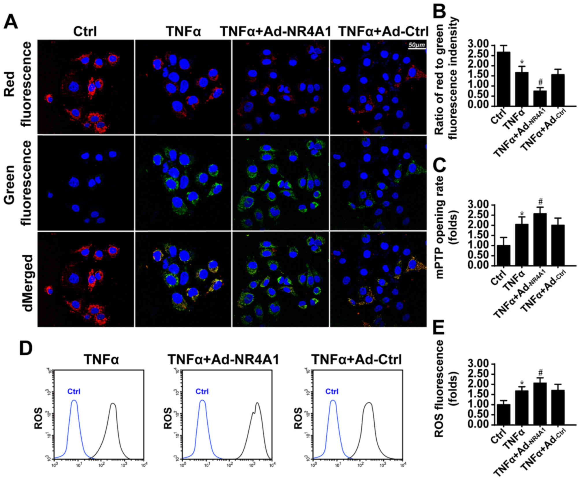
NR4A1 facilitates TNFα-evoked mitochondrial injury.
It has been reported that NR4A1 induces mitochondrial damage
(34). In this study, to determine
whether mitochondrial damage is responsible for the promoting
effects of NR4A1 on TNFα-induced cell death, we detected
mitochondrial function. MMP is the source for the mitochondria to
generate ATP, which fuels a cell's biological function. However, we
observed that NR4A1 further reduced MMP, as revealed by less red
fluorescence and more green fluorescence (Fig. 2A and B). Furthermore, the collapse
of MMP may be a result of the mitochondrial permeability transition
pore (mPTP) opening (35). As
shown in Fig. 2C, compared to the
control group, TNFα induced more mPTP opening. However, regaining
NR4A1 expression further contributed to mPTP opening.
The consequence of mPTP opening and the reduction of
MMP is evidence of mitochondrial metabolism disruption and the
initiation of apoptotic signaling (36). In this study, we first focused on
mitochondrial apoptosis. Mitochondrial apoptosis is characterized
by excessive ROS production and subsequent cellular oxidative
injury, which contributes to the leakage of pro-apoptotic factors
from the mitochondria (29). In
this study, using flow cytometric analysis, we found that TNFα
treatment enhanced the levels of ROS (Fig. 2D and E). Furthermore, the
overexpression of NR4A1 amplified ROS overproduction. In response
to excessive ROS production, the levels of antioxidant factors,
such as GSH, SOD and GPX, were reduced, suggesting an imbalance of
the redox status (Fig. 2F–I). The
enhancement of NR4A1 further consumed antioxidant factors, but
generated greater levels of MDA, an end product of lipid
peroxidation. Finally, we observed greater amounts of Cyt-c
released into the cytoplasm in response to TNFα treatment, which
was similar to the results in the NR4A1 overexpression group
(Fig. 2J). Collectively, these
data suggested that NR4A1 enhanced TNFα-induced mitochondrial
damage.
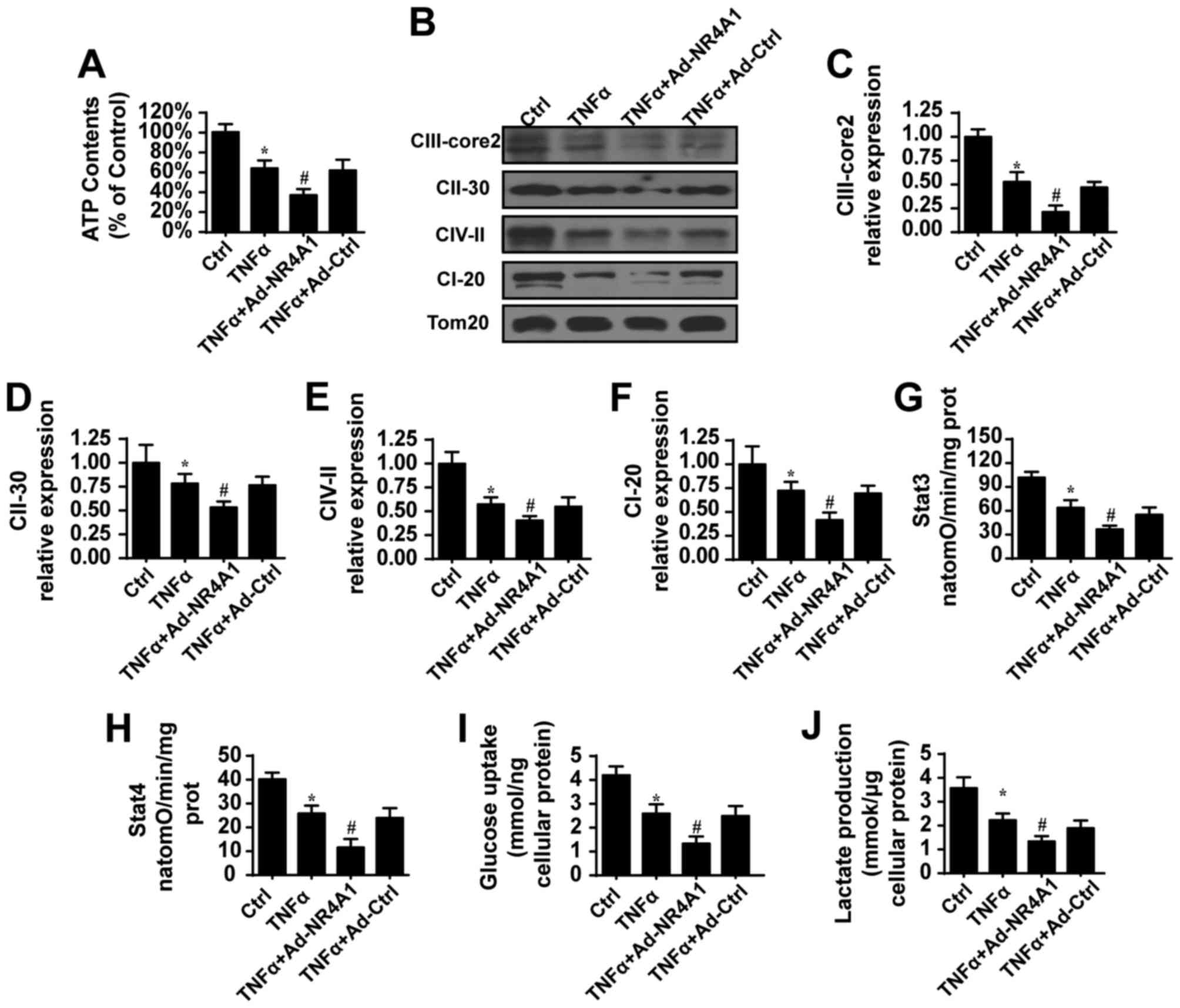
NR4A1 further suppresses mitochondrial
energy metabolism
In addition to cellular apoptosis, mitochondrial
energy production is another determinant for cancer growth and
cellular survival (37).
Considering that NR4A1 promoted mitochondrial apoptosis, we
therefore wished to determine whether NR4A1 hinders mitochondrial
energy production in the presence of TNFα. First, compared to the
control group, TNFα abated the content of ATP, which was further
reduced once NR4A1 was expression was recovered (Fig. 3A), suggesting that NR4A1 has the
ability to influence mitochondrial energy generation. Since ATP is
produced via the mitochondrial respiratory complex, we therefore
examined changes in the mitochondrial respiratory complex. Compared
to the control group, TNFα suppressed the contents of the
mitochondrial respiratory complex (Fig. 3B–F). However, NR4A1 further
suppressed the expression of these factors of the mitochondrial
respiratory complex (Fig. 3B–F).
Correspondingly, mitochondrial Stat3 (Fig. 3G) and Stat4 (Fig. 3H) respiratory functions also
declined when NR4A1 was overexpressed. These data indicated that
NR4A1 impaired mitochondrial respiratory function. The reduced
mitochondrial energy metabolism may be associated with a decrease
in glucose intake and lactate production. Thus, we measured the
content of glucose in medium. As shown in Fig. 3I and J, TNFα indeed reduced glucose
consumption and lactate generation, suggesting the termination of
glycometabolism in GC cells. By contrast, the overexpression of
NR4A1 further limited glucose intake and lactate production. Taken
together, these data indicated that NR4A1 enhanced the suppressive
effects of TNFα on mitochondrial energy metabolism.
NR4A1 inhibits mitophagy to enhance the
sensitivity of GC cells to TNFα
To determine the mechanisms through which NR4A1
enhanced TNFα-induced mitochondrial damage, we focused on the
mitochondria repairing system itself, mitophagy. Damaged
mitochondria activate mitophagy, which facilitates the removal and
clearing of malfunctioning mitochondria via the lysosome, leading
to the preservation of mitochondrial quantity and quality (38). Thus, mitophagy counteracts
TNFα-induced mitochondrial damage (39). Based on this, we first investigated
mitophagy activity. In response to TNFα treatment, the levels of
mito-LC3II, Beclin1, ATG5 and p62 were increased (Fig. 4A–E), suggestive of mitophagy
activation. However, the overexpression of NR4A1 reversed this
tendency, illustrating the inhibitory effect of NR4A1 on mitophagy.
To provide further direct evidence of mitophagy, we used
immunofluorescence to label the mitochondria and lysosomes at the
same time. As shown in Fig. 4F and
G, in the control group, few mitochondria were merged with the
lysosomes. Following treatment with TNFα, most of the mitochondria
were contained by lysosomes, indicative of mitophagy activation.
However, the re-introduction of NR4A1 suppressed the
lysosome-mitochondria interaction, displaying the inhibitory role
of NR4A1 on mitophagy. Subsequently, to determine whether mitophagy
inhibition contributes to the excess mitochondrial damage following
NR4A1 overexpression, we used FCCP to activate mitophagy. After the
activation of mitophagy, caspase-9 activity increased despite the
overexpression of NR4A1 (Fig. 4H).
These data indicated that mitophagy, the protective system of the
mitochondria, was activated by TNFα and contributed to
mitochondrial protection and resistance to apoptosis. The recovery
of NR4A1 expression inhibited mitophagy and enhanced the
sensitivity of GC cells to TNFα-induced cell death.
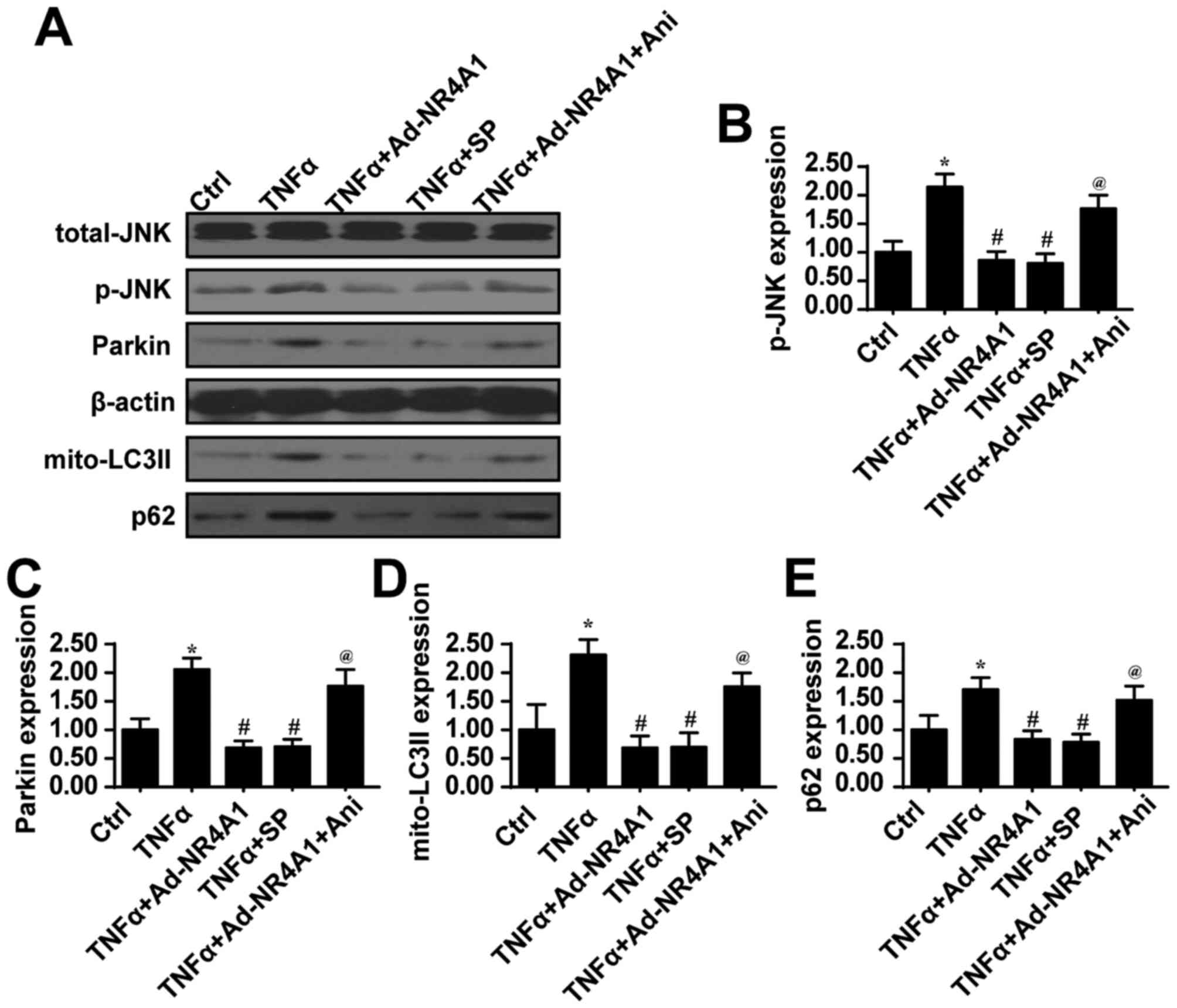
Mitophagy is activated via Parkin through
JNK
Finally, we wished to determine the underlying
signal that modulates mitophagy activity under TNFα treatment
conditions. Parkin is the primary receptor for mitophagy, and it
mediates the damaged mitochondrial removal in several types of
cells (40). Therefore, we first
examined changes in Parkin expression. We found that Parkin
expression was upregulated by TNFα treatment, but was decreased
upon the overexpression of NR4A1 (Fig.
5A–C). This information established the regulatory effects of
TNFα on Parkin. Furthermore, to explain the mechanisms through
which TNFα upregulates Parkin expression, we focused on JNK. JNK is
considered the upstream trigger of Parkin expression (41). To determine whether TNFα regulates
Parkin-required mitophagy via JNK, an inhibitor (SP600125, SP) and
activator (Ani) of JNK were used. As shown in Fig. 5A and B, compared to the control
group, TNFα treatment increased JNK activity as evidenced by
greater levels of phosphorylated JNK. However, NR4A1 overexpression
suppressed TNFα-induced JNK activation. Next, upon the inhibition
of JNK via SP600125 under TNFα treatment, JNK activity was blocked,
and Parkin expression was also inhibited (Fig. 5A–C). By contrast, the activation of
JNK in the cells overexpressing NR4A1 increased Parkin expression
(Fig. 5A–C). In addition, the
results of co-immunofluorescence of p-JNK and Parkin were also in
agreement with the above-mentioned results (Fig. 5F). These data indicated that the
TNFα-induced activation of the JNK pathway was responsible for
Parkin upregulation.
To provide further direct evidence of the role of
JNK in Parkin-mediated mitophagy, we examined markers of mitophagy.
After the blockade of JNK under TNFα treatment, mitophagy was
reduced, as evidenced by less mito-LC3II and p62 expression
(Fig. 5A, D and E), which was
similar to the results observed in the NR4A1 group. By contrast,
the activation of JNK re-evoked the expression of mito-LC3II and
p62 despite the overexpression of NR4A1 (Fig. 5A, D and E).
Finally, to determine whether JNK is also involved
in the resistance of GC cells to TNFα-induced damage, we used PI to
detect cellular damage in cells treated with or without JNK
activator. As shown in Fig. 5G,
the inhibition of JNK increased the number of TNFα-induced
PI-positive cells. However, following NR4A1 overexpression, the
activation of JNK reduced the number of PI-positive cells. Taken
together, these data suggested that JNK/Parkin-dependent mitophagy
was responsible for the therapeutic resistance observed with TNFα
and that NR4A1 enhanced the fatal effects of TNFα via the
inhibition of JNK/Parkin-dependent mitophagy.
Discussion
GC is one of the leading causes of cancer-related
mortality worldwide (42). The
number of patients with GC increases each year, and many of them
are diagnosed at the advanced stages of the disease with overt
metastasis, thus missing the best opportunity for curative surgery
(43). Although patients during
the early stages of GC can be cured by surgery, the majority of
patients with GC are diagnosed at the advanced stages of the
disease and present with extensive invasion, lymphatic metastasis
and other organ metastases (44).
Understanding tumorigenesis may aid in diagnosis the disease stage
and even at earlier stages. GC tumorigenesis is a multistep and
multifactorial process involving diverse genetic alterations,
including the inactivation of tumor suppressor genes, the
activation of oncogenes and the abnormal expression of
cancer-related genes. Thus, it is crucial to investigate the novel
mechanisms that govern the development of GC to elucidate the
molecular mechanisms and develop effective therapeutic
strategies.
Although immunotherapy with IL-2, TNFα and
interferon-α (IFN-α) has been applied to atteunate or intervene
with GC, as the response rate in patients with the disease to such
treatment is only 10–20% (5,45).
In this study, we demonstrated that NR4A1 enhanced the TNFα
therapeutic efficacy by augmenting cellular apoptosis and reducing
drug resistance through mitophagy. To the best of our knowledge,
this is the first study to describe the molecular signal of NR4A1
as the adjuvant to enhance cytokine-based immunotherapy for GC. We
found that the enhancement of NR4A1 further elevated GC cell
apoptosis induced by TNFα. On the one hand, NR4A1 augmented cell
death via strengthening caspase-9-dependent mitochondrial
apoptosis. NR4A1 treatment induced excessive oxidative stress and
evoked extensive mPTP opening. After mPTP opening, the
pro-apoptotic factors in the mitochondria, such as Cyt-c,
were released from the mitochondria into the cytoplasm. On the
other hand, NR4A1 impaired mitochondrial energy production by
suppressing the mitochondria respiratory complex, leading to the
collapse of mitochondria-related energy metabolism. Through the two
above-mentioned mechanisms, NR4A1 enhanced GC cellular damage in
response to TNFα treatment. Considering that NR4A1 was
downregulated following TNFα treatment, according to our findings,
the enhancement of NR4A1 would be useful for tumor treatments in
clinical practice.
In the present study, we also investigated the
mechanisms through which NR4A1 sensitizes GC cells to TNFα. Apart
from the energy production and death signal transmission, the
mitochondria itself have a protection system to reduce excessive
cellular damage, and this is mitophagy (46). Mitophagy neutralizes the damaged
mitochondria via lysosomes, leading to the removal of bad
mitochondria (47). It has been
shown that mitophagy blocks mitochondrial apoptosis and promotes
cellular survival (48). Moreover,
mitochondria also employ mitophagy to provide the energy substrate
to fuel mitochondria energy production (49). Therefore, through mitophagy, the
mitochondria sweep out the defective mitochondria and offer
nutrients to the cell. Given the important role of mitophagy in
cellular protection, we aimed to determine whether mitophagy was
involved in treatment failure or resistance. We found that TNFα
treatment indeed increased the overlap of mitochondria and
lysosomes and the markers of mitophagy, suggesting the activation
of mitophagy. Furthermore, NR4A1 inhibited the mitophagy activity.
By contrast, the activation of mitophagy influenced the
pro-apoptotic effects of NR4A1. These findings indicate that
mitophagy may be the molecular signal for the therapeutic
insensitivity. However, further evidence is required to support
this notion in vivo and in clinical practice.
In this study, we determined that the JNK/Parkin
pathway was the upstream signal that was activated by TNFα to
trigger mitophagy, and NR4A1 suppressed mitophagy via the
inhibition of JNK. In the process of mitophagy, several factors a
Bcl-2 interacting protein 3 (BNIP3), mitofusin 2 (Mfn2) and
dynamin-1-like protein (Drp1) (50–53).
In our study, Parkin was upregulated and led to mitophagy
activation. The underlying mechanism was attributed to the JNK
pathway. TNFα increased JNK activity, which activated Parkin.
Therefore, these findings explain the molecular signal for
mitophagy activation by TNFα treatment, which offers a potential
target to intervene against mitophagy.
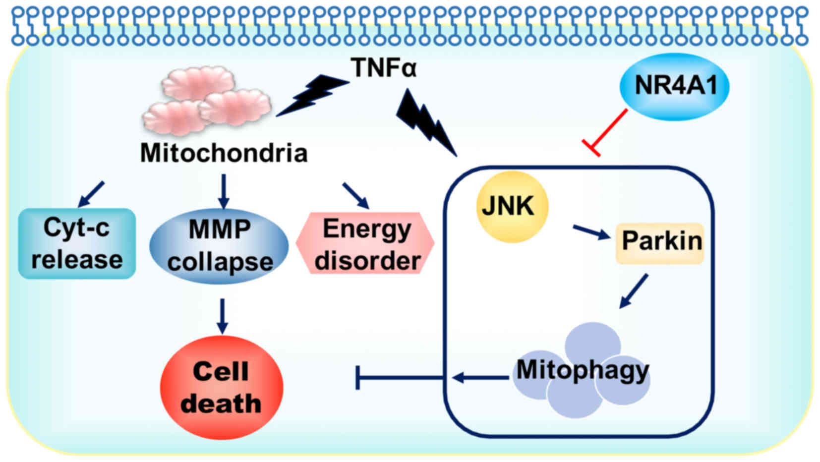
In the present study, we illustrated the important
role of NR4A1 in GC cell apoptosis induced by TNFα (Fig. 6). NR4A1 amplified the therapeutic
sensitivity of GC to TNFα via targeting to the mitochondria. In
this process, mitophagy was the indispensable element in the
protection of the mitochondria and contributed to therapeutic
resistance. NR4A1 abated mitophagy to further augment mitochondrial
apoptosis and energy disruption, finally evoking greater cellular
damage. These findings, on the one hand, explained the mechanisms
underling the drug resistance of GC cells, and on the other hand,
we provided an easy and effective method with which to elevate
TNFα-mediated GC cell death. Thus, the enhancement of NR4A1 may be
a practical and efficient adjuvant for GC treatment. However,
further insight into this combination should be obtained to provide
ample evidence for clinical applications.
Acknowledgments
This study was supported by grants from the project
of Songjiang District Science and Technology Commission (no.
15SJGG29). The funders had no role in the study design, data
collection and analysis, decision to publish, or preparation of the
manuscript.
References
|
1
|
Balakrishnan M, George R, Sharma A and
Graham DY: Changing trends in stomach cancer throughout the world.
Curr Gastroenterol Rep. 19:362017. View Article : Google Scholar : PubMed/NCBI
|
|
2
|
Suzuki H and Mori H: World trends for H.
pylori eradication therapy and gastric cancer prevention strategy
by H. pylori test-and-treat. J Gastroenterol. Nov 14–2017.Epub
ahead of print. View Article : Google Scholar : PubMed/NCBI
|
|
3
|
Lee HS, Kim WH, Kwak Y, Koh J, Bae JM, Kim
KM, Chang MS, Han HS, Kim JM, Kim HW, et al Gastrointestinal
Pathology Study Group of Korean Society of Pathologists; Molecular
Pathology Study Group of Korean Society of Pathologists: Molecular
testing for gastrointestinal cancer. J Pathol Transl Med.
51:103–121. 2017. View Article : Google Scholar : PubMed/NCBI
|
|
4
|
Paoletti X, Oba K, Burzykowski T, Michiels
S, Ohashi Y, Pignon JP, Rougier P, Sakamoto J, Sargent D, Sasako M,
et al GASTRIC (Global Advanced/Adjuvant Stomach Tumor Research
International Collaboration) Group: Benefit of adjuvant
chemotherapy for resectable gastric cancer: A meta-analysis. JAMA.
303:1729–1737. 2010. View Article : Google Scholar : PubMed/NCBI
|
|
5
|
Procaccio L, Schirripa M, Fassan M,
Vecchione L, Bergamo F, Prete AA, Intini R, Manai C, Dadduzio V,
Boscolo A, et al: Immunotherapy in gastrointestinal cancers. BioMed
Res Int. 2017:43465762017. View Article : Google Scholar : PubMed/NCBI
|
|
6
|
Bie Q, Jin C, Zhang B and Dong H: IL-17B:
A new area of study in the IL-17 family. Mol Immunol. 90:50–56.
2017. View Article : Google Scholar : PubMed/NCBI
|
|
7
|
Du LC and Gao R: Role of TNFα -308G/A gene
polymorphism in gastric cancer risk: A case control study and
meta-analysis. Turk J Gastroenterol. 28:272–282. 2017. View Article : Google Scholar : PubMed/NCBI
|
|
8
|
Hong JT, Son DJ, Lee CK, Yoon DY, Lee DH
and Park MH: Interleukin 32, inflammation and cancer. Pharmacol
Ther. 174:127–137. 2017. View Article : Google Scholar : PubMed/NCBI
|
|
9
|
Abozeid M, Rosato A and Sommaggio R:
Immunotherapeutic strategies for gastric carcinoma: A review of
preclinical and clinical recent development. BioMed Res Int.
2017:57912622017. View Article : Google Scholar : PubMed/NCBI
|
|
10
|
Grierson P, Lim KH and Amin M:
Immunotherapy in gastrointestinal cancers. J Gastrointest Oncol.
8:474–484. 2017. View Article : Google Scholar : PubMed/NCBI
|
|
11
|
Procaccio L, Schirripa M, Fassan M,
Vecchione L, Bergamo F, Prete AA, Intini R, Manai C, Dadduzio V,
Boscolo A, et al: Immunotherapy in gastrointestinal cancers. BioMed
Res Int. 2017:43465762017. View Article : Google Scholar : PubMed/NCBI
|
|
12
|
Tsujimoto H, Ono S, Ichikura T, Matsumoto
Y, Yamamoto J and Hase K: Roles of inflammatory cytokines in the
progression of gastric cancer: Friends or foes? Gastric Cancer.
13:212–221. 2010. View Article : Google Scholar : PubMed/NCBI
|
|
13
|
Carrasco-Pozo C, Tan KN, Reyes-Farias M,
De La Jara N, Ngo ST, Garcia-Diaz DF, Llanos P, Cires MJ and Borges
K: The deleterious effect of cholesterol and protection by
quercetin on mitochondrial bioenergetics of pancreatic β-cells,
glycemic control and inflammation: In vitro and in vivo studies.
Redox Biol. 9:229–243. 2016. View Article : Google Scholar : PubMed/NCBI
|
|
14
|
Kakimoto PA and Kowaltowski AJ: Effects of
high fat diets on rodent liver bioenergetics and oxidative
imbalance. Redox Biol. 8:216–225. 2016. View Article : Google Scholar : PubMed/NCBI
|
|
15
|
Kheradpezhouh E, Barritt GJ and Rychkov
GY: Curcumin inhibits activation of TRPM2 channels in rat
hepatocytes. Redox Biol. 7:1–7. 2016. View Article : Google Scholar :
|
|
16
|
Kleszczyński K, Zillikens D and Fischer
TW: Melatonin enhances mitochondrial ATP synthesis, reduces
reactive oxygen species formation, and mediates translocation of
the nuclear erythroid 2-related factor 2 resulting in activation of
phase-2 antioxidant enzymes (γ-GCS, HO-1, NQO1) in ultraviolet
radiation-treated normal human epidermal keratinocytes (NHEK). J
Pineal Res. 61:187–197. 2016. View Article : Google Scholar
|
|
17
|
Ni HM, Williams JA and Ding WX:
Mitochondrial dynamics and mitochondrial quality control. Redox
Biol. 4:6–13. 2015. View Article : Google Scholar :
|
|
18
|
Xu S, Pi H, Zhang L, Zhang N, Li Y, Zhang
H, Tang J, Li H, Feng M, Deng P, et al: Melatonin prevents abnormal
mitochondrial dynamics resulting from the neurotoxicity of cadmium
by blocking calcium-dependent translocation of Drp1 to the
mitochondria. J Pineal Res. 60:291–302. 2016. View Article : Google Scholar : PubMed/NCBI
|
|
19
|
Quijano C, Trujillo M, Castro L and
Trostchansky A: Interplay between oxidant species and energy
metabolism. Redox Biol. 8:28–42. 2016. View Article : Google Scholar : PubMed/NCBI
|
|
20
|
Du K, Ramachandran A and Jaeschke H:
Oxidative stress during acetaminophen hepatotoxicity: Sources,
pathophysiological role and therapeutic potential. Redox Biol.
10:148–156. 2016. View Article : Google Scholar : PubMed/NCBI
|
|
21
|
Pawlak A, Strzadala L and Kalas W:
Non-genomic effects of the NR4A1/Nur77/TR3/NGFIB orphan nuclear
receptor. Steroids. 95:1–6. 2015. View Article : Google Scholar : PubMed/NCBI
|
|
22
|
Wenzl K, Troppan K, Neumeister P and
Deutsch AJ: The nuclear orphan receptor NR4A1 and NR4A3 as tumor
suppressors in hematologic neoplasms. Curr Drug Targets. 16:38–46.
2015. View Article : Google Scholar
|
|
23
|
Wu H, Bi J, Peng Y, Huo L, Yu X, Yang Z,
Zhou Y, Qin L, Xu Y, Liao L, et al: Nuclear receptor NR4A1 is a
tumor suppressor down-regulated in triple-negative breast cancer.
Oncotarget. 8:54364–54377. 2017.PubMed/NCBI
|
|
24
|
Beard JA, Tenga A and Chen T: The
interplay of NR4A receptors and the oncogene-tumor suppressor
networks in cancer. Cell Signal. 27:257–266. 2015. View Article : Google Scholar
|
|
25
|
Alonso-González C, González A,
Martínez-Campa C, Gómez-Arozamena J and Cos S: Melatonin sensitizes
human breast cancer cells to ionizing radiation by downregulating
proteins involved in double-strand DNA break repair. J Pineal Res.
58:189–197. 2015. View Article : Google Scholar : PubMed/NCBI
|
|
26
|
Zhu H, Jin Q, Li Y, Ma Q, Wang J, Li D,
Zhou H and Chen Y: Melatonin protected cardiac microvascular
endothelial cells against oxidative stress injury via suppression
of IP3R-[Ca(2+)] c/VDAC-[Ca(2+)]m axis by activation of MAPK/ERK
signaling pathway. Cell Stress Chaperones. Jul 1–2017.Epub ahead of
print. View Article : Google Scholar
|
|
27
|
Lin C, Chao H, Li Z, Xu X, Liu Y, Hou L,
Liu N and Ji J: Melatonin attenuates traumatic brain injury-induced
inflammation: A possible role for mitophagy. J Pineal Res.
61:177–186. 2016. View Article : Google Scholar : PubMed/NCBI
|
|
28
|
Lin YW, Lee LM, Lee WJ, Chu CY, Tan P,
Yang YC, Chen WY, Yang SF, Hsiao M and Chien MH: Melatonin inhibits
MMP-9 transactivation and renal cell carcinoma metastasis by
suppressing Akt-MAPKs pathway and NF-κB DNA-binding activity. J
Pineal Res. 60:277–290. 2016. View Article : Google Scholar : PubMed/NCBI
|
|
29
|
Zhang Y, Zhou H, Wu W, Shi C, Hu S, Yin T,
Ma Q, Han T, Zhang Y, Tian F, et al: Liraglutide protects cardiac
microvascular endothelial cells against hypoxia/reoxygenation
injury through the suppression of the SR-Ca(2+)-XO-ROS axis via
activation of the GLP-1R/PI3K/Akt/survivin pathways. Free Radic
Biol Med. 95:278–292. 2016. View Article : Google Scholar : PubMed/NCBI
|
|
30
|
de Luxán-Delgado B, Potes Y,
Rubio-González A, Caballero B, Solano JJ, Fernández-Fernández M,
Bermúdez M, Rodrigues Moreira, Guimarães M, Vega-Naredo I, Boga JA,
et al: Melatonin reduces endoplasmic reticulum stress and autophagy
in liver of leptin-deficient mice. J Pineal Res. 61:108–123. 2016.
View Article : Google Scholar : PubMed/NCBI
|
|
31
|
King AL, Mantena SK, Andringa KK,
Millender-Swain T, Dunham-Snary KJ, Oliva CR, Griguer CE and Bailey
SM: The methyl donor S-adenosylmethionine prevents liver hypoxia
and dysregulation of mitochondrial bioenergetic function in a rat
model of alcohol-induced fatty liver disease. Redox Biol.
9:188–197. 2016. View Article : Google Scholar : PubMed/NCBI
|
|
32
|
Mailloux RJ, Craig Ayre D and Christian
SL: Induction of mitochondrial reactive oxygen species production
by GSH mediated S-glutathionylation of 2-oxoglutarate
dehydrogenase. Redox Biol. 8:285–297. 2016. View Article : Google Scholar : PubMed/NCBI
|
|
33
|
Kang JW, Hong JM and Lee SM: Melatonin
enhances mitophagy and mitochondrial biogenesis in rats with carbon
tetrachloride-induced liver fibrosis. J Pineal Res. 60:383–393.
2016. View Article : Google Scholar : PubMed/NCBI
|
|
34
|
Li QX, Ke N, Sundaram R and Wong-Staal F:
NR4A1, 2, 3 - an orphan nuclear hormone receptor family involved in
cell apoptosis and carcinogenesis. Histol Histopathol. 21:533–540.
2006.PubMed/NCBI
|
|
35
|
Dan Dunn J, Alvarez LA, Zhang X and
Soldati T: Reactive oxygen species and mitochondria: A nexus of
cellular homeostasis. Redox Biol. 6:472–485. 2015. View Article : Google Scholar : PubMed/NCBI
|
|
36
|
Zhang J: Teaching the basics of autophagy
and mitophagy to redox biologists–mechanisms and experimental
approaches. Redox Biol. 4:242–259. 2015. View Article : Google Scholar
|
|
37
|
Mukherjee D, Ghosh AK, Dutta M, Mitra E,
Mallick S, Saha B, Reiter RJ and Bandyopadhyay D: Mechanisms of
isoproterenol-induced cardiac mitochondrial damage: Protective
actions of melatonin. J Pineal Res. 58:275–290. 2015. View Article : Google Scholar : PubMed/NCBI
|
|
38
|
Xin Z, Jiang S, Jiang P, Yan X, Fan C, Di
S, Wu G, Yang Y, Reiter RJ and Ji G: Melatonin as a treatment for
gastrointestinal cancer: A review. J Pineal Res. 58:375–387. 2015.
View Article : Google Scholar : PubMed/NCBI
|
|
39
|
Nacarelli T, Azar A and Sell C:
Mitochondrial stress induces cellular senescence in an
mTORC1-dependent manner. Free Radic Biol Med. 95:133–154. 2016.
View Article : Google Scholar : PubMed/NCBI
|
|
40
|
Bernardini JP, Lazarou M and Dewson G:
Parkin and mitophagy in cancer. Oncogene. 36:1315–1327. 2017.
View Article : Google Scholar
|
|
41
|
Jankowski M: The role of JNK pathway in
familial Parkinson's disease. Postepy Biochem. 53:297–303. 2007.In
Polish.
|
|
42
|
Lordick F and Janjigian YY: Clinical
impact of tumour biology in the management of gastroesophageal
cancer. Nat Rev Clin Oncol. 13:348–360. 2016. View Article : Google Scholar : PubMed/NCBI
|
|
43
|
Ishimoto T, Baba H, Izumi D, Sugihara H,
Kurashige J, Iwatsuki M and Tan P: Current perspectives toward the
identification of key players in gastric cancer microRNA
dysregulation. Int J Cancer. 138:1337–1349. 2016. View Article : Google Scholar
|
|
44
|
Scartozzi M, Bittoni A, Pistelli M,
Galizia E, Berardi R, Giampieri R, Faloppi L and Cascinu S: Toward
molecularly selected chemotherapy for advanced gastric cancer:
State of the art and future perspectives. Cancer Treat Rev.
35:451–462. 2009. View Article : Google Scholar : PubMed/NCBI
|
|
45
|
Myint ZW and Goel G: Role of modern
immunotherapy in gastrointestinal malignancies: A review of current
clinical progress. J Hematol Oncol. 10:862017. View Article : Google Scholar : PubMed/NCBI
|
|
46
|
Gao L, Zhao YC, Liang Y, Lin XH, Tan YJ,
Wu DD, Li XZ, Ye BZ, Kong FQ, Sheng JZ, et al: The impaired
myocardial ischemic tolerance in adult offspring of diabetic
pregnancy is restored by maternal melatonin treatment. J Pineal
Res. 61:340–352. 2016. View Article : Google Scholar : PubMed/NCBI
|
|
47
|
Rizzo NR, Hank NC and Zhang J: Detecting
presence of cardiovascular disease through mitochondria respiration
as depicted through biophotonic emission. Redox Biol. 8:11–17.
2016. View Article : Google Scholar : PubMed/NCBI
|
|
48
|
Doskey CM, Buranasudja V, Wagner BA,
Wilkes JG, Du J, Cullen JJ and Buettner GR: Tumor cells have
decreased ability to metabolize H2O2:
Implications for pharmacological ascorbate in cancer therapy. Redox
Biol. 10:274–284. 2016. View Article : Google Scholar : PubMed/NCBI
|
|
49
|
Eirin A, Ebrahimi B, Kwon SH, Fiala JA,
Williams BJ, Woollard JR, He Q, Gupta RC, Sabbah HN, Prakash YS, et
al: Restoration of mitochondrial cardiolipin attenuates cardiac
damage in swine renovascular hypertension. J Am Heart Assoc.
5:e0031182016. View Article : Google Scholar : PubMed/NCBI
|
|
50
|
Zhang W, Ren H, Xu C, Zhu C, Wu H, Liu D,
Wang J, Liu L, Li W, Ma Q, et al: Hypoxic mitophagy regulates
mitochondrial quality and platelet activation and determines
severity of I/R heart injury. eLife. 5:e214072016. View Article : Google Scholar : PubMed/NCBI
|
|
51
|
Zhang W, Siraj S, Zhang R and Chen Q:
Mitophagy receptor FUNDC1 regulates mitochondrial homeostasis and
protects the heart from I/R injury. Autophagy. 13:1080–1081. 2017.
View Article : Google Scholar : PubMed/NCBI
|
|
52
|
Mouton-Liger F, Jacoupy M, Corvol JC and
Corti O: PINK1/Parkin-dependent mitochondrial surveillance: From
pleiotropy to Parkinson's disease. Front Mol Neurosci. 10:1202017.
View Article : Google Scholar : PubMed/NCBI
|
|
53
|
Chourasia AH and Macleod KF: Tumor
suppressor functions of BNIP3 and mitophagy. Autophagy.
11:1937–1938. 2015. View Article : Google Scholar : PubMed/NCBI
|