Introduction
Esophageal cancer is one of the most common types of
cancer in a number of regions worldwide, particularly China, South
America and Western Europe (1).
Esophageal squamous cell carcinoma (ESCC) is the major histological
subtype of esophageal cancer, which predominantly occurs in China
(2). Multimodal treatment for
patients with esophageal tumors has improved the survival rate and
is regarded as the current standard of practice for patients
following surgery (3–5). Neoadjuvant chemoradiotherapy improves
resectability, reduces the extent of disease progression, and
improves local control, disease-free survival and overall survival
rates when compared with surgery alone (6–9). Despite
improvements in surgical techniques, radiotherapy and chemotherapy,
the prognosis for ESCC remains poor, with a 5-year survival rate of
<10% (10). This is frequently due
to extra-esophageal invasion and/or local lymph node metastasis,
which may present in the early stages of ESCC (11). The occurrence and development of
esophageal tumors is caused by the cooperative action of a range of
genes. Investigating the genes involved with the occurrence,
development and metastasis of ESCC may improve the theoretical
foundation of and identify potential targets for the treatment of
this type of tumor.
Beclin 1 was the first mammalian autophagy protein
to be described; it was identified as a homolog of an essential
yeast autophagy gene (Atg6) (12).
Beclin 1 was demonstrated to be a positive regulator of autophagy
(13) and a haplo-insufficient tumor
suppressor that commonly exhibits reduced expression or becomes
monoallelically deleted in certain cancer types (14). However, the expression of Beclin 1 in
ESCC tissues from patients, and its role in the occurrence and
development of ESCC, have yet to be characterized. Furthermore,
data regarding the effect of Beclin 1 expression on ESCC cell
metastasis are not yet available.
In the present study, the expression of Beclin 1
mRNA and protein in ESCC tumor and adjacent normal esophageal
tissue was investigated. It was identified that the expression of
Beclin 1 protein is associated with ESCC progression. The
downregulation of Beclin 1 was also demonstrated to be associated
with the invasion and metastasis of ESCC cells. The results suggest
that Beclin 1 may be a potential therapeutic target in ESCC.
Materials and methods
Patients and tissues
A total of 126 specimens and adjacent normal tissues
(>5 cm from the tumor tissue) obtained from the esophagectomy of
primary ESCC tumors from January 2010 to September 2011 were
collected from the Department of Thoracic Surgery, Ruijin Hospital,
Shanghai Jiao Tong University School of Medicine (Shanghai, China).
Fresh samples were stored immediately in liquid nitrogen. None of
the patients had received preoperative chemotherapy, radiotherapy
or other treatments. The histopathological characteristics of the
126 specimens were assessed.
To determine the disease stage according to the
International Union Against Cancer Tumor, Node, Metastasis (TNM)
classification system (15),
pathology reports and clinical histories from the time of surgery
were reviewed. The patient characteristics, including age, sex,
tumor differentiation and TNM stage (including T, depth of tumor
invasion; N, lymph node metastasis; and M, distant metastasis) were
evaluated. The patients included 92 males and 34 females and ranged
in age from 45 to 80 years (median, 62 years). Approval for the
study was granted by the Ruijin Hospital Ethics Committee. Informed
written consent was obtained from all patients.
Reverse transcription-quantitative
polymerase chain reaction (RT-qPCR)
Changes in Beclin 1 mRNA expression were detected
and quantified relative to GAPDH mRNA expression by qPCR. Total RNA
from ESCC tumor and adjacent tissue was extracted using TRIzol
reagent (Gibco; Thermo Fisher Scientific, Inc., Waltham, MA, USA)
according to the manufacturer's protocol. qPCR testing was
performed using a qPCR system (SYBR® Premix Ex TaqTM II;
Takara Bio, Inc., Japan) according to the manufacturer's protocol.
The primers for Beclin-1 and GAPDH were as follows: Beclin 1
forward, 5′-CTGAGGGATGGAAGGGTC-3′ and reverse
5′-TGGGCTGTGGTAAGTAATG-3′; GAPDH forward, 5′-GAAGGTGAAGGTCGGAGTC-3′
and reverse, 5′-GAAGATGGTGATGGGATTTC-3′. Thermal cycling was
performed with a 20-µl reaction volume in the PRISM 7000 Sequence
Detection system (Applied Biosystems; Thermo Fisher Scientific,
Inc.). The PCR amplification conditions were as follows: 95°C for
10 min, then 40 cycles of 95°C for 5 sec and 60°C for 30 sec. The
expression of Beclin 1 mRNA was calculated and normalized against
GAPDH mRNA expression (16); each
data point was performed in triplicate. The mean value for Beclin 1
expression was used as the cut-off point to define high and low
Beclin 1 expression.
Isolation of proteins and western blot
analysis
A total of 50 µl from each frozen tissue specimen
was ground and homogenized in 1 ml of radioimmunoprecipitation
lysis buffer (RIPA; Santa Cruz Biotechnology, Inc., Dallas, TX,
USA) with complete protease inhibitor cocktail tablets (Roche
Diagnostics, Basel, Switzerland). The homogenate was put into 1.5
ml centrifuge tubes and centrifuged at 16,000 × g for 30 min at
4°C. The concentration of the supernatant was measured with the
bicinchoninic acid protein assay (BCA) method. Total protein (50
µg) was resolved by 10% SDS-PAGE separating gel and transferred
onto 0.22 µm polyvinylidene fluoride (PVDF) membranes. Subsequent
to blocking in 5% skim milk for 2 h at room temperature, the PVDF
membranes were respectively probed with mouse monoclonal goat
anti-human Beclin 1 (cat. no. sc-48341; 1:100; Santa Cruz
Biotechnology, Inc.) and monoclonal mouse anti-GAPDH (cat. no.
sc-47724; 1:5,000; Santa Cruz Biotechnology, Inc.) overnight at
4°C.
Following washing in 1X TBST 3 times, the PVDF
membranes were then incubated with HRP-labeled goat anti-mouse IgG
secondary antibody (cat. no. sc-2039; 1:5,000; Santa Cruz
Biotechnology) at room temperature for 1 h and then were washed
with 0.5% I-Block blocking buffer (Thermo Fisher Scientific, Inc.).
The blots were incubated with chemiluminescence substrate (EMD
Millipore, Billerica, MA, USA) and detected by an enhanced
chemiluminescence assay. Densitometry was performed using Photoshop
software (CS6; Adobe Systems, Inc., San Jose, CA, USA), normalized
to GAPDH from the same blot image. Blot images are representative
of ≥3 independent experiments.
Detection of acidic vesicular
organelles (AVOs) with acridine orange staining
Acridine orange (Sigma-Alrich; Merck KGaA,
Darmstadt, Germany) was added to the culture medium containing the
human esophageal carcinoma EC0706 cell line (Shanghai Institutes
for Biological Science of the Chinese Academy of Sciences,
Shanghai, China) to a final concentration of 1 µg/ml and followed
by incubation in the dark at 20°C for 15 min. Samples were then
examined under a fluorescence microscope. Acridine orange
accumulation in acidic autophagic vacuoles caused the appearance of
a granular distribution of bright red fluorescence in the
cytoplasm, which was indicative of autophagosome formation. The
extent of AVO fluorescence was measured with an excitation
wavelength of 488 nm and an emission wavelength of 515 nm. The
relative amount of AVOs was quantitatively determined according to
the red-to-green fluorescence ratio, which was obtained using
Photoshop software.
Transfection of Beclin 1 siRNA into
EC9706 cells and detection cell invasiveness and metastasis
A Beclin 1-targeting sense sequence and a universal
negative control siRNA (cat. no. 12935-400) were purchased from
Invitrogen (Thermo Fisher Scientic, Inc.). The targeting sequence
for Beclin 1 (5′-GGGTCTAAGACGTCCAACA-3′) and the negative control
siRNA were cloned into the BamHI and EcoRI sites of
the pGSU6-GFP vector (cat. no. GTP600300; Genlantis, Inc., San
Diego, CA, USA). Control and siRNA plasmids, and a recombinant
pcDNA3.1-Beclin 1 plasmid (provided by Professor Qin Ye from
Shanghai Institute of Digestive Surgery, Shanghai Jiao Tong
University School of Medicine) were transfected into the human
esophageal carcinoma EC9706 cells in Dulbecco's modified Eagle's
medium (DMEM; Gibco; Thermo Fisher Scientific, Inc.) with 10% fetal
bovine serum (FBS; Gibco; Thermo Fisher Scientific, Inc.) without
antibiotics at 5% CO2 and 37°C once cells had reached
50% confluence into 6-well plates using Lipofectamine
2000® (cat. no. 11668019; Invitrogen; Thermo Fisher
Scientific, Inc.) according to the manufacturer's protocol. At 48 h
following transfection, cells were washed with PBS three times.
Whole cell lysate was extracted with RIPA lysis buffer, and then a
western blot, as aforementioned, was performed to detect relative
Beclin 1 expression in each group, and to confirm the effectiveness
of Beclin1 RNA interference or upregulation.
Invasion assay
For the invasion assay, the inserted Transwell
chambers (8.0 µm pore size; Corning Incorporated, Corning, NY, USA)
were pretreated with Matrigel® (diluted 1:6 in DMEM; BD
Biosciences, Franklin Lakes, NJ, USA) for 2 h at 37°C. Then, 6×
×104 cells in 200 µl DMEM without serum were added in
the upper chamber, while 600 µl DMEM medium with 10% FBS was placed
in the bottom of wells. Cells were incubated at 37°C with 5%
CO2 for 24 h. Cells that did not move to the lower
chamber were removed from the topside. The cells were then stained
in 2% crystal violet solution for 15 min and immersed in PBS for 10
min. Finally, the number of invading cells were counted under ×200
fields using an inverted microscope (Leica Microsystems, Wetzlar,
Germany). At least five random fields were selected, and the
average number was taken. The data are presented as the mean ±
standard deviation.
Statistical analysis
Statistical analysis was performed using SPSS
software (v.13.0; SPSS, Inc., Chicago, IL, USA). Data are expressed
as the means ± standard deviation of 3 individual experiments.
Comparisons of quantitative data were analyzed using the
χ2 and Student's t-tests between two groups or by
one-way analysis of variance for multiple groups with Fisher's
least significant difference post hoc test. A log-rank test was
performed to compared two survival curves between two groups.
P<0.05 was considered to represent a statistically significant
difference.
Results
Association between Beclin-1 mRNA
expression and clinicopathological features
As summarized in Table
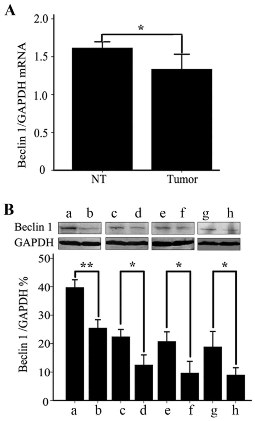
I and Fig. 1A, the expression of
Beclin 1 mRNA in ESCC tissue was significantly lower than in the
adjacent normal esophageal tissue (P<0.05). Tumors that were
less differentiated exhibited lower expression levels of Beclin 1
mRNA (P<0.05). Tumors from early stage (I/II) cancer presented
with a significantly elevated expression of Beclin 1 mRNA compared
with tumors from later stage (III/IV) cancer (P<0.05). The
expression level of Beclin 1 mRNA was significantly lower in tumors
from patients with lymph node metastasis than those without lymph
node involvement (P<0.05). The expression level of Beclin 1 mRNA
was not associated with a patient's age or sex, or the tumor
size.
 | Table I.Beclin 1 mRNA expression in esophageal
squamous cell carcinoma tissue and its association with
clinicopathological factors. |
Table I.
Beclin 1 mRNA expression in esophageal
squamous cell carcinoma tissue and its association with
clinicopathological factors.
|
|
| Beclin 1 mRNA
expression, n |
|
|---|
|
|
|
|
|
|---|
| Pathologic
parameter | Cases, n | High | Low | χ2 |
|---|
| Normal tissue | 126 | 117 | 9 | 31.48a |
| Carcinoma tissue | 126 | 80 | 46 |
|
| Sex |
|
|
|
|
| Male | 92 | 45 | 47 | 0.03 |
|
Female | 34 | 16 | 18 |
|
| Size of tumor,
cm |
|
|
|
|
|
<3 | 72 | 37 | 42 | 0.07 |
| ≥3 | 54 | 24 | 30 |
|
| Age, years |
|
|
|
|
|
<65 | 40 | 18 | 22 | 0.79 |
|
>65 | 86 | 46 | 40 |
|
| Degree of
differentiation |
|
|
| 13.49a |
|
Well/moderately | 70 | 43 | 27 |
|
|
Poorly | 56 | 16 | 40 |
|
| Pathologic stage |
|
|
| 14.12a |
| I/II | 78 | 48 | 30 |
|
|
III/IV | 48 | 13 | 35 |
|
| Lymph node |
|
|
| 32.84a |
|
Negative | 52 | 41 | 11 |
|
|
Positive | 74 | 20 | 54 |
|
Association between Beclin 1 protein
expression and clinicopathological features
As summarized in Table
II and Fig. 1B, the expression of
Beclin 1 protein in ESCC tissue was significantly lower than in the
adjacent normal esophageal tissue (P<0.05). Tumors that were
less differentiated exhibited lower expression levels of Beclin 1
protein (P<0.05). The expression of Beclin 1 protein in the
early stages (I/II) of the cancer was significantly higher than
those in mid-to-late stages (III/IV; P<0.05). The expression of
Beclin 1 protein in tumors from patients with lymph node metastasis
was significantly lower than those without lymph node involvement
(P<0.05). The expression level of Beclin 1 protein was not
associated with a patient's age or sex, or the tumor size.
 | Table II.Beclin 1 protein expressiom in
esophageal squamous cell carcinoma tissue. |
Table II.
Beclin 1 protein expressiom in
esophageal squamous cell carcinoma tissue.
| Pathologic
parameter | Cases, n | Beclin 1
expressiona, % | P-valueb |
|---|
| Type of tissue |
|
| <0.01 |
|
Normal | 126 | 40.60±3.80 |
|
|
Tumor | 126 | 25.32±2.65 |
|
| Degree of
differentiation |
|
| <0.05 |
|
Well/moderately | 70 | 22.27±2.34 |
|
|
Poorly | 56 | 12.35±3.17 |
|
| Clinical stage |
|
| <0.05 |
|
I/II | 78 | 20.63±3.05 |
|
|
III/IV | 48 | 9.52±3.66 |
|
| Lymph node |
|
| <0.05 |
|
Negative | 52 | 18.72±4.81 |
|
|
Positive | 74 | 8.84±2.31 |
|
Confirmation of Beclin 1
overexpression or knockdown
The recombinant pcDNA3.1-Beclin 1 plasmid was
transfected into EC9706 cells to induce overexpression of the
Beclin 1 protein. In another group of EC9706 cells, the level of
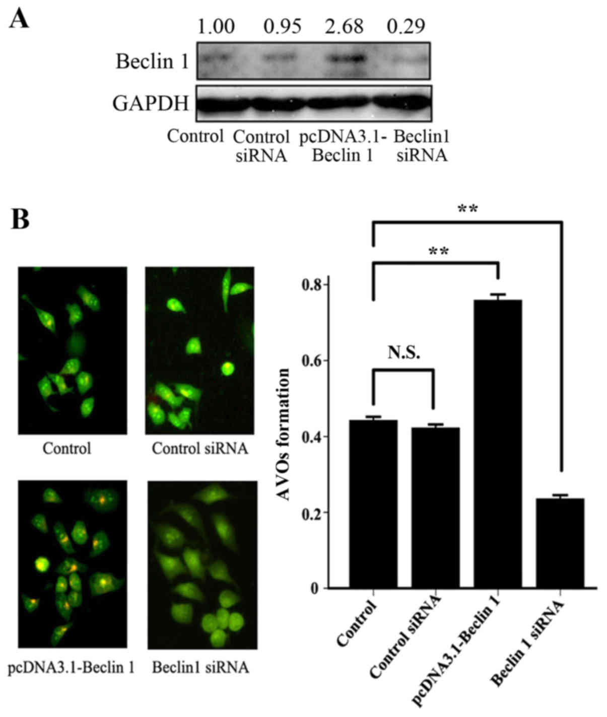
Beclin 1 protein was decreased by RNA interference. Beclin 1
overexpression or knockdown were confirmed using western blotting
and the detection of AVOs with acridine orange. As demonstrated in
in Fig. 2, the expression levels of
Beclin 1 protein were significantly reduced in EC9706 cells
transfected with Beclin 1 siRNA (P<0.05), whereas the level was
significantly increased in EC9706 cells transfected with the
pcDNA3.1-Beclin 1 plasmid (P<0.01).
Role of Beclin 1 expression level on
the invasiveness of the EC9706 ESCC cell line
As included in Table
III, the EC9706 cells transfected with pcDNA3.1-Beclin 1
exhibited a significant decrease in the number of invasive cells
crossing the polycarbonate membrane of the Transwell invasion
chamber (P<0.01), whereas the EC9706 cells transfected with
Beclin 1 siRNA cells exhibited a significant increase in the number
of invasive cells (P<0.01), compared with the untransfected
control group. The expression of Beclin 1 protein and cell
invasiveness did not differ significantly between the untransfected
and negative control siRNA control groups. These data indicated
that an elevated expression level of Beclin 1 protein may decrease
the invasiveness of the EC9706 cells, while a lowered expression
level of Beclin 1 protein may promote the invasion of EC9706
cells.
 | Table III.The association between the
invasiveness of EC9706 esophageal squamous cell carcinoma cells
with the relative expression of Beclin 1. |
Table III.
The association between the
invasiveness of EC9706 esophageal squamous cell carcinoma cells
with the relative expression of Beclin 1.
| Groups | Beclin 1
expressiona, % | Number of invasive
cells |
|---|
| Untransfected
control group | 20.35±3.02 | 243.32±38.23 |
| Control
siRNA-transfected group | 21.30±2.18 | 225.36±28.16 |
| pcDNA3.1-Beclin
1-transfected group | 38.34±5.25 |
40.65±15.24b |
| Beclin 1
siRNA-transfected group | 5.65±1.27 |
382.36±45.73b |
Kaplan-Meier cumulative survival
curve
The median follow-up period was 33.59 months, with a
range of 8–69 months. The 1-, 2- and 3-year survival rates for the
entire patient group were 93.65, 58.73 and 34.92%, respectively.
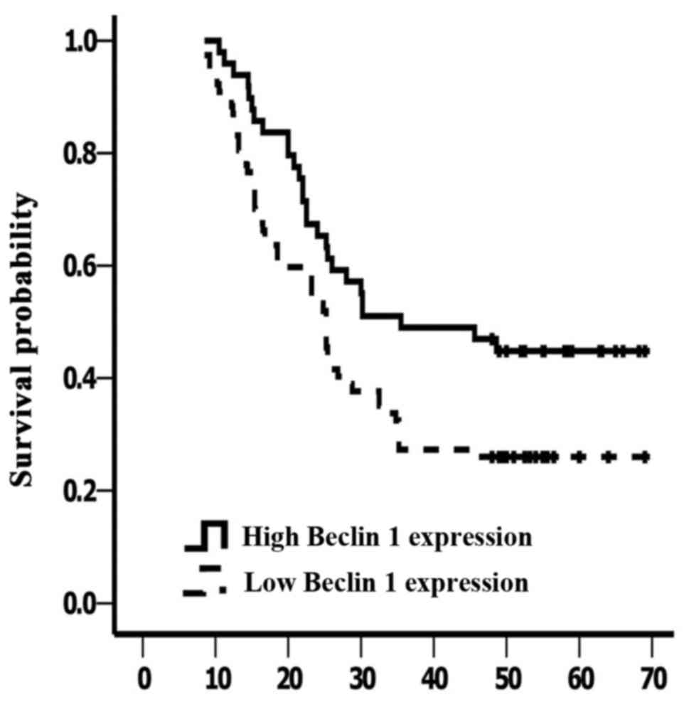
The survival rate of the Beclin 1-high group was significantly
improved compared with the Beclin 1-low group (P<0.01; Fig. 3).
Discussion
The reported pattern of Beclin 1 expression in
gastrointestinal cancer is not consistent. Previous studies have
reported that Beclin 1 and autophagy were suppressed in
hepatocellular carcinoma and pancreatic cancer (17), but that they were elevated in colon
and gastric cancer (18), compared
with adjacent normal tissue. Ding et al (19) confirmed that the expression of Beclin
1 in liver cancer tissue was significantly decreased compared with
adjacent tissue, and that Beclin 1 expression was associated with
the occurrence and development of liver cancer. In the present
study, it was determined that the level of mRNA Beclin 1 expression
relative to GAPDH in ESCC tissue and adjacent normal tissues in
patients was 63.49 and 92.85%, respectively. The expression of
Beclin 1 mRNA and protein in ESCC were significantly downregulated
compared with adjacent normal esophageal tissue (P<0.05). The
more highly undifferentiated esophageal cells demonstrated lower
expression levels of Beclin 1 mRNA and protein (P<0.05). The
expression levels of Beclin 1 mRNA and protein were significantly
higher in patients with early stage tumors (I/II; P<0.05). The
expression levels of Beclin 1 mRNA and protein in tumors from
patients with lymph node metastasis were significantly decreased
vs. tumors from patients without lymph node metastasis
(P<0.05).
Furthermore, the association between Beclin 1
expression and clinicopathological factors was assessed in 126
patients with ESCC. A high expression of Beclin 1, as determined by
the mean expression level, was observed in 63.5% of ESCC tumors.
Lymph node metastasis, the degree of tumor differentiation and the
clinical stage were significantly associated with Beclin-1
expression; however, there was no significant difference in the
expression of Beclin 1 associated with other features (age, sex,
and size of primary carcinoma; Tables
I and II). Previous study has
revealed that autophagy serves an important role in many
physiological functions; defects in this process have been linked
to many types of cancer (20,21). Beclin 1, an autophagy execution
protein, is considered a tumor suppressor protein in tumor biology
(14,22,23). The
reduction of Beclin 1 expression in tumor tissue observed in the
present study may be associated with the function of basal
autophagy in maintaining homeostasis in normal tissue (20,21).
Studies in transgenic mouse models have shown that the monoallelic
deletion of Beclin 1 promotes tumor development (24). The growth of colorectal cancer cells
that overexpress Beclin 1 was observed to be reduced compared with
mock-transfected cells (25). Beclin
1 deficiency is also associated with increased angiogenesis
(26,27).
In the present study, EC9706 cells were transfected
with the pcDNA3.1-Beclin 1 plasmid or a Beclin 1 siRNA. Changes in
cell invasiveness in these cells were then observed; EC9706 cells
transfected with the pcDNA3.1-Beclin 1 plasmid were confirmed to
overexpress Beclin 1 protein (P<0.01) and a significantly
reduced number of cells crossed the Transwell chamber compared with
the empty vector-transfected and untransfected control groups
(P<0.01). EC9706 cells transfected with Beclin 1 siRNA exhibited
significantly reduced Beclin 1 protein expression (P<0.01) and a
significantly increased number of cells crossing the Transwell
chamber compared with the control siRNA-transfected and the
untransfected control groups (P<0.01). These results indicated
that downregulating Beclin 1 may promote cell invasion, while
upregulating Beclin 1 may reduce the invasiveness. We hypothesize
that Beclin 1 is a microtubule-associated protein, and that it may
regulate cell motility through modulating the stability of
microtubules.
Surgical resection followed by multimodal therapy is
considered as the standard treatment for patients with localized
ESCC (3–5). However, the problem of how to identify
the patients likely to benefit from surgery is unresolved. In the
present study, the elevated expression of Beclin 1 was identified
as a favorable prognostic factor for ESCC patients.
In conclusion, it was identified that the expression
of Beclin 1 is associated with the progression of ESCC, and that
Beclin 1 upregulation is associated with a reduction in the
invasive capabilities of ESCC cells. Examining the expression of
Beclin 1 may contribute to improving the understanding of the
prognosis of ESCC, and Beclin 1 may be a potential therapeutic
target for treating ESCC.
Acknowledgements
The present study was supported by the Shanghai
Ruijin North Hospital Fund (2014ZY05) and Shanghai Charity
Foundation for Cancer Research. The authors would like to thank Dr
Zhiyong Du, Dr Songling Yu, Dr Guiyang Zhang and Dr Guangtao Sun
from the Department of Thoracic Surgery, Ruijin Hospital, Shanghai
Jiao Tong University School of Medicine (Shanghai, China) for the
collection of ESCC specimens and discussion during research.
References
|
1
|
Pickens A and Orringer MB: Geographical
distribution and racial disparity in esophageal cancer. Ann Thorac
Surg. 76:S1367–S1369. 2003. View Article : Google Scholar : PubMed/NCBI
|
|
2
|
Zhang J, Jiang Y, Wu C, Cai S, Wang R,
Zhen Y, Chen S, Zhao K, Huang Y, Luketich J and Chen H: Comparison
of clinicopathologic features and survival between eastern and
western population with esophageal squamous cell carcinoma. J
Thorac Dis. 7:1780–1786. 2015.PubMed/NCBI
|
|
3
|
Cui Z, Tian Y, He B, Li H, Li D, Liu J,
Cai H, Lou J, Jiang H, Shen X and Peng K: Associated factors of
radiation pneumonitis induced by precise radiotherapy in 186
elderly patients with esophageal cancer. Int J Clin Exp Med.
8:16646–16651. 2015.PubMed/NCBI
|
|
4
|
Nasr JY and Schoen RE: Prevalence of
adenocarcinoma at esophagectomy for Barrett's esophagus with high
grade dysplasia. J Gastrointest Oncol. 2:34–38. 2011.PubMed/NCBI
|
|
5
|
Wagner TD, Khushalani N and Yang GY:
Clinical T2N0M0 carcinoma of thoracic esophagus. J Thorac Dis.
2:36–42. 2010.PubMed/NCBI
|
|
6
|
Jabbour SK and Thomas CR Jr: Radiation
therapy in the postoperative management of esophageal cancer. J
Gastrointest Oncol. 1:102–111. 2010.PubMed/NCBI
|
|
7
|
Das P: Esophageal cancer: Is preoperative
chemoradiation the new standard? J Gastrointest Oncol. 1:68–69.
2010.PubMed/NCBI
|
|
8
|
Kleinberg L: Does postoperative radiation
therapy benefit patients with esophageal cancer? J Gastrointest
Oncol. 1:70–71. 2010.PubMed/NCBI
|
|
9
|
Prasanna PG, Stone HB, Wong RS, Capala J,
Bernhard EJ, Vikram B and Coleman CN: Normal tissue protection for
improving radiotherapy: Where are the gaps? Transl Cancer Res.
1:35–48. 2012.PubMed/NCBI
|
|
10
|
Sun GP, Wan X, Xu SP, Wang H, Liu SH and
Wang ZG: Antiproliferation and apoptosis induction of paeonol in
human esophageal cancer cell lines. Dis Esophagus. 21:723–729.
2008. View Article : Google Scholar : PubMed/NCBI
|
|
11
|
Yu C, Fan S, Sun Y and Pickwell-Macpherson
E: The potential of terahertz imaging for cancer diagnosis: A
review of investigations to date. Quant Imaging Med Surg. 2:33–45.
2012.PubMed/NCBI
|
|
12
|
Yue Z, Jin S, Yang C, Levine AJ and Heintz
N: Beclin 1, an autophagy gene essential for early embryonic
development, is a haploinsufficient tumor suppressor. Proc Natl
Acad Sci USA. 100:pp. 15077–15082. 2003; View Article : Google Scholar : PubMed/NCBI
|
|
13
|
Huang W, Choi W, Hu W, Mi N, Guo Q, Ma M,
Liu M, Tian Y, Lu P, Wang FL, et al: Crystal structure and
biochemical analyses reveal Beclin 1 as a novel membrane binding
protein. Cell Res. 22:473–489. 2012. View Article : Google Scholar : PubMed/NCBI
|
|
14
|
Yang MC, Wang HC, Hou YC, Tung HL, Chiu TJ
and Shan YS: Blockade of autophagy reduces pancreatic cancer stem
cell activity and potentiates the tumoricidal effect of
gemcitabine. Mol Cancer. 14:1792015. View Article : Google Scholar : PubMed/NCBI
|
|
15
|
Talsma K, van Hagen P, Grotenhuis BA,
Steyerberg EW, Tilanus HW, van Lanschot JJ and Wijnhoven BP:
Comparison of the 6th and 7th editions of the UICC-AJCC TNM
classification for esophageal cancer. Ann Surg Oncol. 19:2142–2148.
2012. View Article : Google Scholar : PubMed/NCBI
|
|
16
|
Du H, Yang W, Chen L, Shen B, Peng C, Li
H, Ann DK, Yen Y and Qiu W: Emerging role of autophagy during
ischemia-hypoxia and reperfusion in hepatocellular carcinoma. Int J
Oncol. 40:2049–2057. 2012.PubMed/NCBI
|
|
17
|
Fiorini C, Cordani M, Gotte G, Picone D
and Donadelli M: Onconase induces autophagy sensitizing pancreatic
cancer cells to gemcitabine and activates Akt/mTOR pathway in a
ROS-dependent manner. Biochim Biophys Acta. 1853:549–560. 2015.
View Article : Google Scholar : PubMed/NCBI
|
|
18
|
Li BX, Li CY, Peng RQ, Wu XJ, Wang HY, Wan
DS, Zhu XF and Zhang XS: The expression of beclin 1 is associated
with favorable prognosis in stage IIIB colon cancers. Autophagy.
5:303–306. 2009. View Article : Google Scholar : PubMed/NCBI
|
|
19
|
Ding ZB, Shi YH, Zhou J, Qiu SJ, Xu Y, Dai
Z, Shi GM, Wang XY, Ke AW, Wu B and Fan J: Association of autophagy
defect with a malignant phenotype and poor prognosis of
hepatocellular carcinoma. Cancer Res. 68:9167–9175. 2008.
View Article : Google Scholar : PubMed/NCBI
|
|
20
|
Levine B and Kroemer G: Autophagy in the
pathogenesis of disease. Cell. 132:27–42. 2008. View Article : Google Scholar : PubMed/NCBI
|
|
21
|
Mathew R, Karantza-Wadsworth V and White
E: Role of autophagy in cancer. Nat Rev Cancer. 7:961–967. 2007.
View Article : Google Scholar : PubMed/NCBI
|
|
22
|
Koneri K, Goi T, Hirono Y, Katayama K and
Yamaguchi A: Beclin 1 gene inhibits tumor growth in colon cancer
cell lines. Anticancer Res. 27:1453–1457. 2007.PubMed/NCBI
|
|
23
|
Toton E, Lisiak N, Sawicka P and
Rybczynska M: Beclin-1 and its role as a target for anticancer
therapy. J Physiol Pharmacol. 65:459–467. 2014.PubMed/NCBI
|
|
24
|
Qu X, Yu J, Bhagat G, Furuya N, Hibshoosh
H, Troxel A, Rosen J, Eskelinen EL, Mizushima N, Ohsumi Y, et al:
Promotion of tumorigenesis by heterozygous disruption of the beclin
1 autophagy gene. J Clin Invest. 112:1809–1820. 2003. View Article : Google Scholar : PubMed/NCBI
|
|
25
|
Burada F, Nicoli ER, Ciurea ME, Uscatu DC,
Ioana M and Gheonea DI: Autophagy in colorectal cancer: An
important switch from physiology to pathology. World J Gastrointest
Oncol. 7:271–284. 2015. View Article : Google Scholar : PubMed/NCBI
|
|
26
|
Teng RJ, Du J, Welak S, Guan T, Eis A, Shi
Y and Konduri GG: Cross talk between NADPH oxidase and autophagy in
pulmonary artery endothelial cells with intrauterine persistent
pulmonary hypertension. Am J Physiol Lung Cell Mol Physiol.
302:L651–L663. 2012. View Article : Google Scholar : PubMed/NCBI
|
|
27
|
Lee SJ, Kim HP, Jin Y, Choi AM and Ryter
SW: Beclin 1 deficiency is associated with increased
hypoxia-induced angiogenesis. Autophagy. 7:829–839. 2011.
View Article : Google Scholar : PubMed/NCBI
|


