Introduction
Angiogenesis is the formation of new capillaries
from preexisting blood vessels and it is an important mechanism
involved in various pathological processes, including inflammation
and tumor growth. Antiangiogenic therapy is being investigated as a
potentially powerful novel therapy for cancer and other
angiogenesis-dependent diseases.
Endostatin is one of the most potent inhibitors of
angiogenesis and may induce tumor regression in mice (1,2).
Clinical trials on the antitumor effects of endostatin are
currently ongoing (3). Originally,
endostatin was purified from a conditioned medium of murine
hemangioendothelioma cells as a proteolytically cleaved fragment of
type XVIII collagen. The generation of endostatin may be achieved
by cleavage of collagen by cathepsin L (4), matrilysin (5) or elastase (6). Endostatin activated by proteolytic
processing (7) may inhibit
endothelial cell proliferation, migration/invasion and tube
formation. The inhibitory action of endostatin has been attributed
to its binding to the α5β1 integrin receptor (8) and possibly to its low-affinity binding
to glypican-1 and -4 or its high-affinity binding to an
unidentified molecule on endothelial cells (9). Blockage of VEGF/VEGFR
signaling (10,11), inhibition of metalloproteinases,
e.g., MMP-2 (12), and
downregulation of c-MYC and cyclin D1(13,14),
are examples of the mechanisms through which endostatin signaling
may lead to reduced endothelial cell survival, motility and
invasion. A number of physiological functions of endostatin have
been identified. The endostatin levels are elevated in certain
types of cancer and chronic inflammatory diseases, e.g., rheumatoid
arthritis (15) and diabetic
retinopathy (16). Platelets were
shown to sequester endostatin (17)
for later release, e.g., to modulate wound healing. Endostatin also
suppresses vascular permeability (18).
The mechanism of action of angiogenesis inhibitors
on endothelial cells and their receptors has not been elucidated.
Studies with platelet factor 4 (19) and thrombospondin (20) indicated that their heparin-binding
domains were involved, by competing with the angiogenic basic
fibroblast growth factor for binding to proteoglycan receptors. A
strong affinity for heparin was also demonstrated for angiostatin
and endostatin (21,22). However, other studies reported an
inhibitory action of non-heparin-binding thrombospondin fragments
(23,24), which may bind to the CD36 receptor
(25), suggesting a complex array
of biologically active sites. A similar complexity apparently
exists for angiostatin, in which four individual kringle domains
each exhibit antiproliferative activity, although to different
extents (26). There was apparently
no correlation with lysine affinity; however, whether there is a
correlation with heparin affinity remains to be established. The
diversity of angiogenesis inhibitors suggests that each component
may require its own precise molecular analysis and it is possible
that no common interaction mechanism exists.
The specific functional site of endostatin has not
yet been determined. Certain investigators (27) have constructed two mutants of
endostatin, EM1 (9 amino residues of C-terminal were deleted) and
EM2 (17 amino residues of C-terminal were deleted). EM1 and EM2
were administered to a renal cell carcinoma tumor xenograft model.
EM1 retained the natural biological activity of endostatin, whereas
EM2 exhibited loss of function. The results obtained indicated that
C-terminal conservation may be crucial for the biological activity
of endostatin.
To determine the function of endostatin in ascites
hepatoma cells, we constructed a mutant of endostatin designated as
EM13, by deleting 13 amino residues (LCIENSFMTSFSK) from its
C-terminal. Plasmids that encode EM13 and wild-type endostatin,
were then transfected into H22 ascites hepatoma cells. Transfected
cells were implanted into nude mice and the resulting tumors were
measured and examined. Based on those results we aimed to determine
the function of endostatin and whether the 13 amino residues of its
C-terminal are indispensable to its biological function.
Materials and methods
Plasmids and materials
Plasmid pEGFP-N2 was a kind gift from Professor
Jianing Zhang, Dalian Medical University. pMD-18-T vector was
purchased from Takara Biotechnology Co., Ltd. (Dalian, China). The
RNA LA PCR™ kit (AMV) Ver. 2.1, the PCR Agarose Gel DNA
Purification kit, the MiniBEST Plasmid Purification kit Ver. 2.0,
XhoI and SacII restriction enzymes, T4 DNA ligase,
DL2000 DNA Marker, λ-HindIII, X-gal and IPTG were also
purchased from Takara Biotechnology Co., Ltd. DEPC, RPMI-1640, G418
and Lipofectamine 2000 Transfection reagent were purchased from
Invitrogen (Carlsbad, CA, USA).
Construction of pEGFP-N2-endostatin and
pEGFP- N2-EM13
The cDNA of endostatin was amplified from plasmid
pBV220-endostatin by F01 and R02 primers. The cDNA of EM13 (amino
acids 1-171), was amplified from mouse liver tissue by RT-PCR with
the primers 5′-CTGCTCGAGATGCATACTCATCAGGACTT-3′ and
5′-TAACCGCGGGACGATGTAGCTGTTGTGGC-3′. The product was incorporated
into the pMD-18-T vector. After the plasmid was sequenced, the
result revealed that the EM13 sequence contained in pMD18-T-EM13
was identical to the endostatin sequence (1-513) in the GenBank
database.
To construct pEGFP-N2-endostatin (full-length), the
PCR product of endostatin was excised (XhoI and
SacII). It was then inserted into an
XhoI/SacII site of the pEGFP-N2 vector. Following
construction of pEGFP-N2-EM13 (amino acids 1-171), the cDNA of EM13
was excised from pMD-18-T-EM13 (XhoI and SacII) and
inserted into the pEGFP-N2 vector (XhoI/SacII).
Cell culture, transfection and creation
of stable cell lines
H22 cells were cultured in RPMI-1640 medium
containing 10% fetal bovine serum, 100 U/ml penicillin, 100 μg/ml
streptomycin and 50 μg/ml L-glutamine in a humidified incubator at
37°C with 5% CO2. H22 cells were seeded at a density of
1×10⁶ cells/60-mm dish and transfected with
pEGFP-N2-endostatin, pEGFP-N2-EM13 and pEGFP-N2 vector with
Lipofectamine 2000 (Invitrogen), according to the manufacturer’s
instructions. After being transfected for 24 h, H22 cells were
determined by fluorescence microscopy and PCR to grow into stable
cell lines and the transfected cells were screened in a selection
medium containing 600 μg/ml G418. The screened cells were then
cultured with G418 at a sustained concentration of 200 μg/ml.
Antitumor effect in vivo
Nude BALB/c mice, weighing 18–25 g, were used for
the studies of endostatin on tumor growth in vivo. Six mice
were injected subcutaneously with H22 cells stably transfected with
pEGFP-N2-endostatin, pEGFP-N2-EM13 or vector control. After 16
days, the tumor weight was quantified. The density of blood vessels
in each tumor tissue was examined under a light microscope
following hematoxylin and eosin (H&E) staining. The blood
vessel densities of each group were measured.
Results
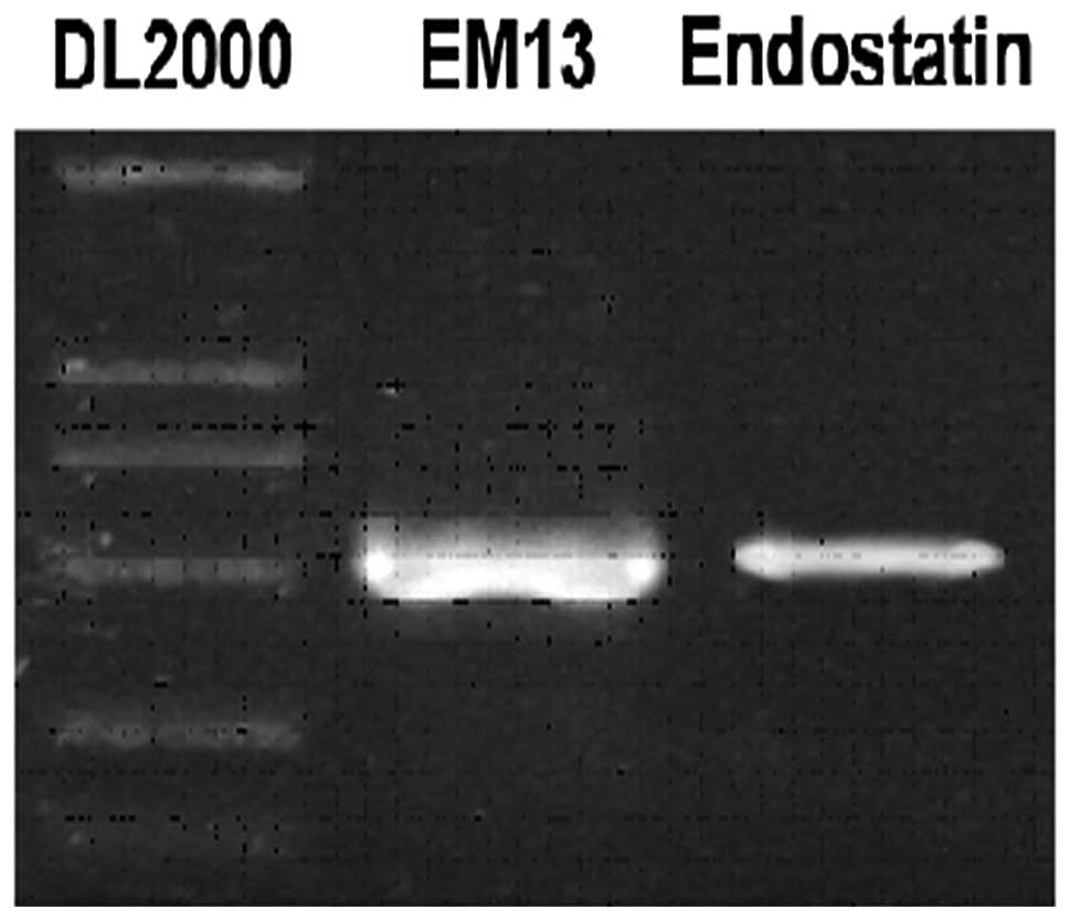
Extraction of total RNA and amplification
of EM13
Total RNA was extracted by TRIzol reagent from the
mouse liver. Following extraction, the RNA was resolved into 30 μl
ddH2O. According to the endostatin sequence in GenBank,
two primers were designed to amplify the full sequence of
endostatin, except for the 13 aminos in the C-terminal. The
recognition sites of XhoI and SacII were introduced
into the upstream (R01: 5′-CTGCTCGAGATGCATACTCATCAGGACTT-3′) and
downstream (F01: 5′-TAACCGCGGGACGATGTAGCTGTTGTGGC-3′) primers,
respectively. The product of RT-PCR with these primers was
designated as EM13. The cDNA of endostatin was also amplified by
PCR with primers F01 and R02 (Fig.
1), with pBV220-endostatin plasmid as the template.
Construction of expressing plasmid
The PCR products were incorporated into vector
pMD18-T following purification. Clones with plasmid pMD18-T-EM13
were verified by PCR with primers F01 and R01 (Fig. 2A). One clone was selected and
cultured and plasmid pMD18-T-EM13 was harvested (Fig. 2B). The plasmid was sequenced and the
results demonstrated that the endostatin sequence contained in
pMD18-T-EM13 was identical to the endostatin sequence in
GenBank.
 | Figure 2(A) Construction of expressing
plasmid. Screening for endostatin 13-T-positive clones by
polymerase chain reaction (PCR): Lane 1, DL2000 DNA marker; Lanes
2–6, PCR products no. 1–5. (B) Screening for endostatin
13-T-positive clones by plasmid extracting: Lane 1, DL2000 DNA
marker; lane 2, λ-HindIII DNA marker; and lane 3, plasmid of
positive clone pMD18-T-EM13. (C) Digestion of endostatin 13-T,
endostatin, pEGFP-N2 by SacII/XhoI: Lane 1, vector
pEGFP-N2 digested by SacII/XhoI; lane 2, endostatin
digested by SacII/XhoI; lane 3, pMD18-T-EM13 digested
by SacII/XhoI; lane 4, DL2000 DNA marker; lane 5,
λ-HindIII DNA marker; lane 6, purification of pMD18-T-EM13
digestion product; lane 7, purification of endostatin digestion
product; and lane 8, purification of pEGFP-N2 digestion product.
(D) Screening for positive clones by PCR: Lane 1, DL2000 DNA
marker; lane 2–5, PCR products no. 11–14; lanes 6 and 11, negative
control of respective PCR; lanes 8–10, PCR products no. 21–23. (E)
Plasmid pEGFP-N2-EM13 and pEGFP-N2-endostatin. Lane 1, DL2000 DNA
marker; lane 2, pEGFP-N2-EM13; lane 3, pEGFP-N2-endostatin; and
lane 4, λ-HindIII DNA marker. (F) Plasmid pEGFP-N2-EM13 and
pEGFP-N2-endostatin were digested with XhoI and
SacII. Lane 1, DL2000 DNA marker; lane 2, pEGFP-N2-EM13 was
digested with XhoI and SacII; lane 3,
pEGFP-N2-endostatin was digested with XhoI and SacII;
and lane 4, λ-HindIII DNA marker. |
The plasmid pMD18-T-EM13 and the fragment of
endostatin were digested with XhoI and SacII and the
fragment of endostatin and EM13 was inserted into the plasmid
pEGFP-N2, which was also digested by XhoI and SacII
(Fig. 2C). Through PCR screening, 4
clones possibly carrying pEGFP-N2-endostatin and 3 clones possibly
carrying pEGFP-N2-EM13 were selected (Fig. 2D). One clone from each group was
selected for harvesting plasmids (Fig.
2E). The two plasmids were also verified by digestion with
XhoI and SacII (Fig.
2F).
Endostatin inhibits tumor growth and
blood vessel formation
Plasmids pEGFP-N2-endostatin and pEGFP-N2-EM13 were
transfected into H22 cells. After 36 h the efficiency of
transfection was examined under a fluorescent microscope (the
transfected cells exhibited green fluorescence). To obtain stable
cell lines, transfected cells were screened in a selection medium
containing 600 μg/ml G418 and the screened cells were then cultured
with G418 at a sustained concentration of 200 μg/ml.
To determine whether endostatin is able to suppress
tumorigenesis and the formation of capillaries, H22 cells and H22
cells constantly transfected with plasmids pEGFP-N2-endostatin or
pEGFP-N2-EM13 were implanted into BALB/c mice (six mice/group).
Sixteen days later, the mice were sacrificed, the tumors were
weighed and the density of blood vessels was also measured by
H&E staining. Compared to the untransfected group, the tumor
growth of the group transfected with endostatin was relatively slow
and exhibited a prolonged incubation period. However, the result
did not differ significantly between the untransfected group and
the group transfected with endostatin. The tumor weight of the
group transfected with EM13, compared to the untransfected group,
exhibited no statistically significant difference (Table I). Following H&E staining, the
histological examination revealed a relatively low density of tumor
capillaries in the group transfected with endostatin compared to
the untransfected group, while the group transfected with EM13
exhibited no significant difference compared to the untransfected
group (Fig. 3).
 | Table IInhibitory effect of endostatin on
tumor growth. |
Table I
Inhibitory effect of endostatin on
tumor growth.
| Groups | Tumor weight (g) | P-value |
|---|
| Untransfected | 1.2489±0.4166 | - |
| PEGFP-N2-vector | 1.0973±0.3472 | 0.510 |
|
PEGFP-N2-endostatin | 0.8914±0.1027 | 0.091 |
| PEGFP-N2-EM13 | 1.2005±0.3736 | 0.837 |
Our results have demonstrated that endostatin may
play an important role in inhibiting tumor growth and the formation
of blood vessels and that the 13 amino acids at the C-terminal may
be a region indispensable to the biological activity of
endostatin.
Discussion
Angiogenesis is the sprouting of capillaries from
preexisting blood vessels by the proliferation, differentiation and
migration of endothelial cells. This physiological process is
closely regulated by a delicate balance between pro- and
antiangiogenic factors. An imbalance of the angiogenic process
contributes to the development of a number of disorders. It is well
established that angiogenesis is vital for the development,
progression and metastasis of a number of human solid tumors.
The growth and progression of solid tumors beyond 2
mm3 is dependent on the recruitment of angiogenic
vessels and an expansion of the tumor vasculature. Investigations
have been mainly focused on the inhibitors of angiogenesis that are
required for tumor growth and metastases. Previous studies
(10–14) have clearly defined some of the
angiogenic factors that contribute to tumor growth. In this study,
the potential antitumorigenic activity of endostatin and EM13 in
the H22 mouse hepatocellular carcinoma model was investigated.
The plasmids expressing endostatin and EM13 gene
were transfected into hepatoma H22 cells with the cationic
liposome-mediated method. Following G418 screening, the cells were
subcutaneously inoculated in BALB/c inbred mice. Our data have
demonstrated that there was no significant difference in tumor
weight between the control and transfected groups. The H&E
staining demonstrated that the group transfected with endostatin
exhibited a relatively low density of tumor capillaries compared to
the control group; however, the group transfected with EM13
exhibited the same result as the untransfected group.
The results suggested that the group transfected
with endostatin exhibited tumor growth inhibition to a certain
extent. The tumor cell types determine the difference of the
sensitivity of endostatin. The relatively low quantity of blood
vessels in the ascitic hepatoma may have no effect on tumor weight
between the untransfected and endostatin groups (Table I).
Cancer cells produce various vascular growth factors
which may induce the host blood vessels to grow into the tumor and
ensure nutrition supply. The growth velocity and biological
characteristics of the tumor are associated with angiogenesis and
different types of tumors exhibit different intensities of
angiogenic activity. Tumor cells that arise from ascites hepatoma
and HL60 cells (a leukemia cell line) do not possess angiogenic
activity. However, following inoculation of Ehrlich ascites cells
into experimental animals and the formation of a solid tumor, the
angiogenic activity reappears, indicating that angiogenic activity
is closely associated with tumor type and living environment. A
possible mechanism explaining this phenomenon is that it may be
easy for the floating cells to uptake nutrients from a liquid
environment. Due to the ascites tumor having a relatively low
density of blood vessels, endostatin exerts almost no effect.
In a previous study by Peroulis et
al(28), C6 glioma cells
transfected with endostatin by the cationic liposome-mediated
method were subcutaneously inoculated into rats. The expression of
endostatin detected by RT-PCR and western blot analysis was low and
the tumor inhibitory effect was not significant. Although a number
of studies reported that endostatin treatment reduced tumor growth
rates and induced the regression of established tumors (2,29–31),
complete tumor inhibition was not readily achieved.
The assessment of the endostatin and EM13 treatment
models demonstrated that endostatin therapy alone may not be
sufficient for the complete regression of all types of tumors. The
persistent tumor growth and the variations in the extent of
regression suggest that the targeting of tumor angiogenesis alone
may not effectively treat all tumors. Therefore, endostatin
treatment administered in combination with chemo- or immunotherapy
may lead to tumor growth arrest and a significantly reduced tumor
growth rate.
Acknowledgements
We would like to thank Ping Yan for their help with
the immunofluorescence assay. This study was supported by grants
from NSFC (30870550), the Chinese State Key Program in Basic
Research (2009CB521804) and the Liaoning Education Department
(2008T034).
References
|
1
|
Boehm T, Folkman J, Browder T and O’Reilly
MS: Antiangiogenic therapy of experimental cancer does not induce
acquired drug resistance. Nature. 390:404–407. 1997. View Article : Google Scholar
|
|
2
|
Li J, Dong X, Xu Z, Jiang X, Jiang H,
Krissansen GW and Sun X: Endostatin gene therapy enhances the
efficacy of paclitaxel to suppress breast cancers and metastases in
mice. J Biomed Sci. 15:99–109. 2008. View Article : Google Scholar : PubMed/NCBI
|
|
3
|
Herbst RS, Hess KR, Tran HT, Tseng JE,
Mullani NA, Charnsangavej C, Madden T, Davis DW, McConkey DJ,
O’Reilly MS, Ellis LM, Pluda J, Hong WK and Abbruzzese JL: Phase I
study of recombinant human endostatin in patients with advanced
solid tumors. J Clin Oncol. 20:3792–3803. 2002. View Article : Google Scholar : PubMed/NCBI
|
|
4
|
Felbor U, Dreier L, Bryant RA, Ploegh HL,
Olsen BR and Mothes W: Secreted cathepsin L generates endostatin
from collagen XVIII. EMBO J. 19:1187–1194. 2000. View Article : Google Scholar : PubMed/NCBI
|
|
5
|
Lin HC, Chang JH, Jain S, Gabison EE, Kure
T, Kato T, Fukai N and Azar DT: Matrilysin cleavage of corneal
collagen type XVIII NC1 domain and generation of a 28-kDa fragment.
Invest Ophthalmol Vis Sci. 42:2517–2524. 2001.PubMed/NCBI
|
|
6
|
Wen W, Moses MA, Wiederschain D, Arbiser
JL and Folkman J: The generation of endostatin is mediated by
elastase. Cancer Res. 59:6052–6056. 1999.PubMed/NCBI
|
|
7
|
Ferreras M, Felbor U, Lenhard T, Olsen BR
and Delaisse J: Generation and degradation of human endostatin
proteins by various proteinases. FEBS Lett. 486:247–251. 2000.
View Article : Google Scholar : PubMed/NCBI
|
|
8
|
Sudhakar A, Sugimoto H, Yang C, Lively J,
Zeisberg M and Kalluri R: Human tumstatin and human endostatin
exhibit distinct antiangiogenic activities mediated by αvβ3 and
α5β1 integrins. Proc Natl Acad Sci USA. 100:4766–4771.
2003.PubMed/NCBI
|
|
9
|
Karumanchi SA, Jha V, Ramchandran R,
Karihaloo A, Tsiokas L, Chan B, Dhanabal M, Hanai JI, Venkataraman
G, Shriver Z, Keiser N, Kalluri R, Zeng H, Mukhopadhyay D, Chen RL,
Lander AD, Hagihara K, Yamaguchi Y, Sasisekharan R, Cantley L and
Sukhatme VP: Cell surface glypicans are low-affinity endostatin
receptors. Mol Cell. 7:811–822. 2001. View Article : Google Scholar : PubMed/NCBI
|
|
10
|
Ling Y, Yang Y, Lu N, You QD, Wang S, Gao
Y, Chen Y and Guo QL: Endostar, a novel recombinant human
endostatin, exerts antiangiogenic effect via blocking VEGF-induced
tyrosine phosphorylation of KDR/Flk-1 of endothelial cells. Biochem
Biophys Res Commun. 361:79–84. 2007. View Article : Google Scholar
|
|
11
|
Kim YM, Hwang S, Kim YM, Pyun BJ, Kim TY,
Lee ST, Gho YS and Kwon YG: Endostatin blocks vascular endothelial
growth factor-mediated signaling via direct interaction with
KDR/Flk-1. J Biol Chem. 277:27872–27879. 2002. View Article : Google Scholar : PubMed/NCBI
|
|
12
|
Kim YM, Jang JW, Lee OH, Yeon J, Choi EY,
Kim KW, Lee ST and Kwon YG: Endostatin inhibits endothelial and
tumor cellular invasion by blocking the activation and catalytic
activity of matrix metalloproteinase. Cancer Res. 60:5410–5413.
2000.PubMed/NCBI
|
|
13
|
Hanai J, Dhanabal M, Karumanchi SA,
Albanese C, Waterman M, Chan B, Ramchandran R, Pestell R and
Sukhatme VP: Endostatin causes G1 arrest of endothelial cells
through inhibition of cyclin D1. J Biol Chem. 277:16464–16469.
2002. View Article : Google Scholar : PubMed/NCBI
|
|
14
|
Shichiri M and Hirata Y: Antiangiogenesis
signals by endostatin. FASEB J. 15:1044–1053. 2001. View Article : Google Scholar : PubMed/NCBI
|
|
15
|
Hebbar M, Peyrat JP, Hornez L, Hatron PY,
Hachulla E and Devulder B: Increased concentrations of the
circulating angiogenesis inhibitor endostatin in patients with
systemic sclerosis. Arthritis Rheum. 43:889–893. 2000. View Article : Google Scholar : PubMed/NCBI
|
|
16
|
Funatsu H, Yamashita H, Noma H, Shimizu E,
Yamashita T and Hori S: Stimulation and inhibition of angiogenesis
in diabetic retinopathy. Jpn J Ophthalmol. 45:577–584. 2001.
View Article : Google Scholar : PubMed/NCBI
|
|
17
|
Ma L, del Soldato P and Wallace JL:
Divergent effects of new cyclooxygenase inhibitors on gastric ulcer
healing: Shifting the angiogenic balance. Proc Natl Acad Sci USA.
99:13243–13247. 2002. View Article : Google Scholar : PubMed/NCBI
|
|
18
|
Hajitou A, Grignet C, Devy L, Berndt S,
Blacher S, Deroanne CF, Bajou K, Fong T, Chiang Y, Foidart JM and
Noël A: The antitumoral effect of endostatin and angiostatin is
associated with a down-regulation of vascular endothelial growth
factor expression in tumor cells. FASEB J. 16:1802–1804.
2002.PubMed/NCBI
|
|
19
|
Maione TE, Gray GS, Petro J, Hunt AJ,
Donner AL, Bauer SI, Carson HF and Sharpe RJ: Inhibition of
angiogenesis by recombinant human platelet factor-4 and related
peptides. Science. 247:77–79. 1990. View Article : Google Scholar : PubMed/NCBI
|
|
20
|
Vogel T, Guo N, Krutzsch HC, Blake DA,
Hartmann J, Mendelovitz S, Panet A and Roberts DD: Modulation of
endothelial cell proliferation, adhesion and motility by
recombinant heparin-binding domain and synthetic peptides from the
type I repeats of thrombospondin. J Cell Biochem. 53:74–84. 1993.
View Article : Google Scholar : PubMed/NCBI
|
|
21
|
O’Reilly MS, Holmgren L, Shing Y, Chen C,
Rosenthal RA, Moses M, Lane WS, Chao Y, Sage EH and Folkman J:
Angiostatin: a novel angiogenesis inhibitor that mediates the
suppression of metastases by a Lewis lung carcinoma. Cell.
79:315–328. 1994.PubMed/NCBI
|
|
22
|
O’Reilly MS, Boehm T, Shing Y, Fukai N,
Vasois G, Lane WS, Flynn E, Birkhead JR, Olsen BR and Folkman J:
Endostatin: an endogenous inhibitor of angiogenesis and tumor
growth. Cell. 88:277–285. 1997.
|
|
23
|
Good DJ, Polverini PJ, Rastinejad F, Le
Beau MM, Lemons RS, Frazier WA and Bouck NP: A tumor
suppressor-dependent inhibitor of angiogenesis is immunologically
and functionally indistinguishable from a fragment of
thrombospondin. Proc Natl Acad Sci USA. 87:6624–6628. 1990.
View Article : Google Scholar : PubMed/NCBI
|
|
24
|
Tolsma SS, Volpert OV, Good DJ, Frazier
WA, Polverini PJ and Bouck N: Peptides derived from two separate
domains of the matrix protein thrombospondin-1 have anti-angiogenic
activity. J Cell Biol. 122:497–511. 1993. View Article : Google Scholar : PubMed/NCBI
|
|
25
|
Dawson DW, Pearce SF, Zhong R, Silverstein
RL, Frazier WA and Bouck NP: CD36 mediates the in vitro inhibitory
effects of thrombospondin-1 on endothelial cells. J Cell Biol.
138:707–717. 1997. View Article : Google Scholar : PubMed/NCBI
|
|
26
|
Cao Y, Ji RW, Davidson D, Schaller J,
Marti D, Söhndel S, McCance SG, O’ Reilly M, Llinás M and Folkman
J: Kringle domains of human angiostatin. Characterization of the
anti-proliferative activity on endothelial cells. J Biol Chem.
271:29461–29467. 1996. View Article : Google Scholar : PubMed/NCBI
|
|
27
|
Dhanabal M, Ramchandran R, Volk R, et al:
Endostatin: yeast production, mutants, and antitumor effect in
renal cell carcinoma. Cancer Res. 59:189–197. 1999.PubMed/NCBI
|
|
28
|
Peroulis I, Jonas N and Saleh M:
Antiangiogenic activity of endostatin inhibits C6 glioma growth.
Int J Cancer. 97:839–845. 2002. View Article : Google Scholar : PubMed/NCBI
|
|
29
|
Yokoyama Y, Sedgewich G and Ramakrishnan
S: Endostatin binding to ovarian cancer cells inhibits peritoneal
attachment and dissemination. Cancer Res. 67:10813–10822. 2007.
View Article : Google Scholar : PubMed/NCBI
|
|
30
|
Liu F, Tan G, Li J, et al: Gene transfer
of endostatin enhances the efficacy of doxorubicin to suppress
human hepatocellular carcinomas in mice. Cancer Sci. 98:1381–1387.
2007. View Article : Google Scholar : PubMed/NCBI
|
|
31
|
Hoffmann S, Wunderlich A, Celik I, et al:
Paneling human thyroid cancer cell lines for candidate proteins for
targeted antiangiogenic therapy. J Cell Biochem. 98:954–965. 2006.
View Article : Google Scholar : PubMed/NCBI
|