Motor fiber organization in the extratemporal trunk of the facial nerve in rats: A retrograde Fluoro-Gold study
- Authors:
- Published online on: September 7, 2012 https://doi.org/10.3892/etm.2012.701
- Pages: 844-848
Metrics: Total
Views: 0 (Spandidos Publications: | PMC Statistics: )
Total PDF Downloads: 0 (Spandidos Publications: | PMC Statistics: )
Abstract
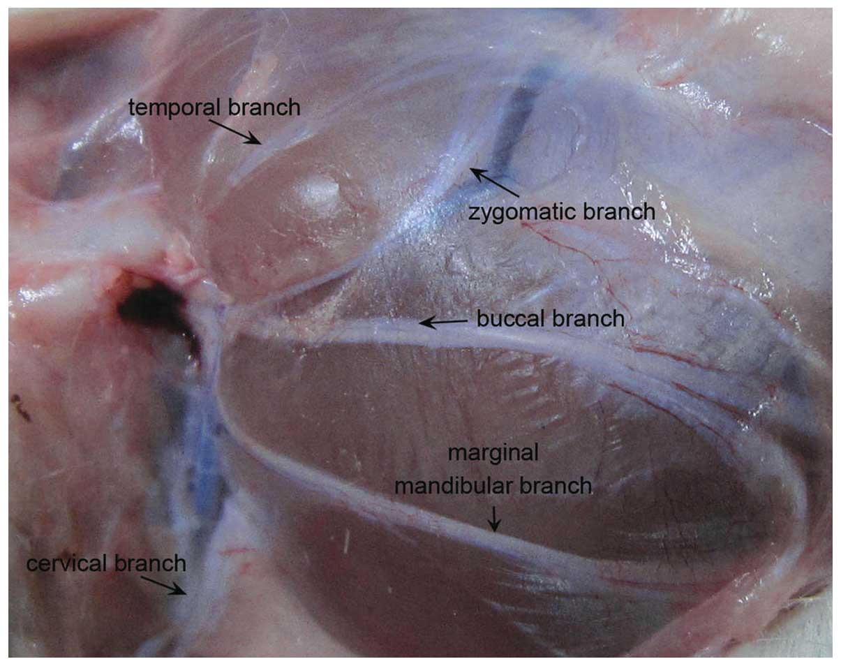
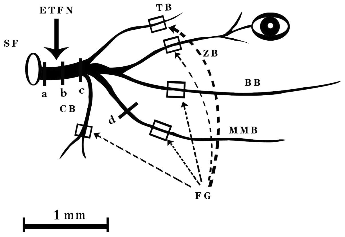
Understanding the microanatomy of the facial nerve is vital to functional restoration of facial nerve injury. This study aimed to locate the spatial orientation of five branches in the extratemporal trunk of the rat facial nerve (ETFN). Fifteen adult Sprague-Dawley albino rats were divided randomly into five groups corresponding to the five facial nerves. Fluoro-Gold™ (FG) was applied to one branch in all three rats in each group. The trunk of the facial nerve was cut at three points for fluorescence detection. Staining results showed that each branch of the facial motor nerve had a topographical orientation in the distal part of the ETFN. The temporal branch was located in the medial and acroscopic quadrant of the nerve trunk. The zygomatic branch was located in the lateral and acroscopic quadrant. The buccal branch occupied the upper half of the nerve trunk, whereas the mandibular branch occupied the lower half. The cervical branch presented a square-shaped distribution in the lateral nerve trunk. In the middle part of the ETFN, the topographical orientation remained clear, but the FG-labeled zone was extended to some extent. In the stylomastoid foramen region, all branches diffused, thereby blurring the orientation. In conclusion, each branch of the facial motor nerve had a topographical orientation and distribution in the crotch and middle part of the ETFN, but the branches diffused near the stylomastoid foramen.