Transection method for shortening the rat spine and spinal cord
- Authors:
- Published online on: November 29, 2012 https://doi.org/10.3892/etm.2012.841
- Pages: 384-388
Metrics: Total
Views: 0 (Spandidos Publications: | PMC Statistics: )
Total PDF Downloads: 0 (Spandidos Publications: | PMC Statistics: )
Abstract
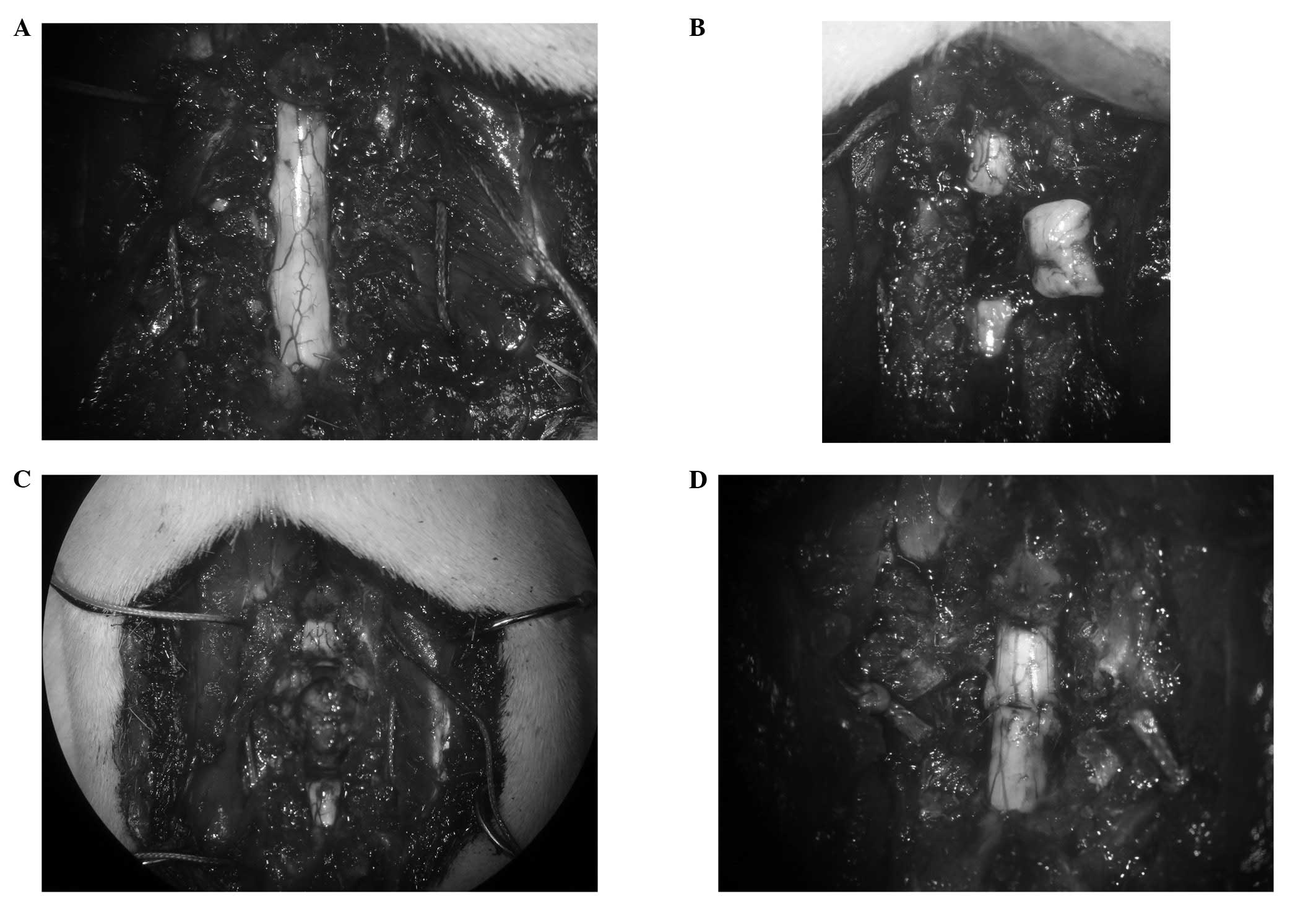
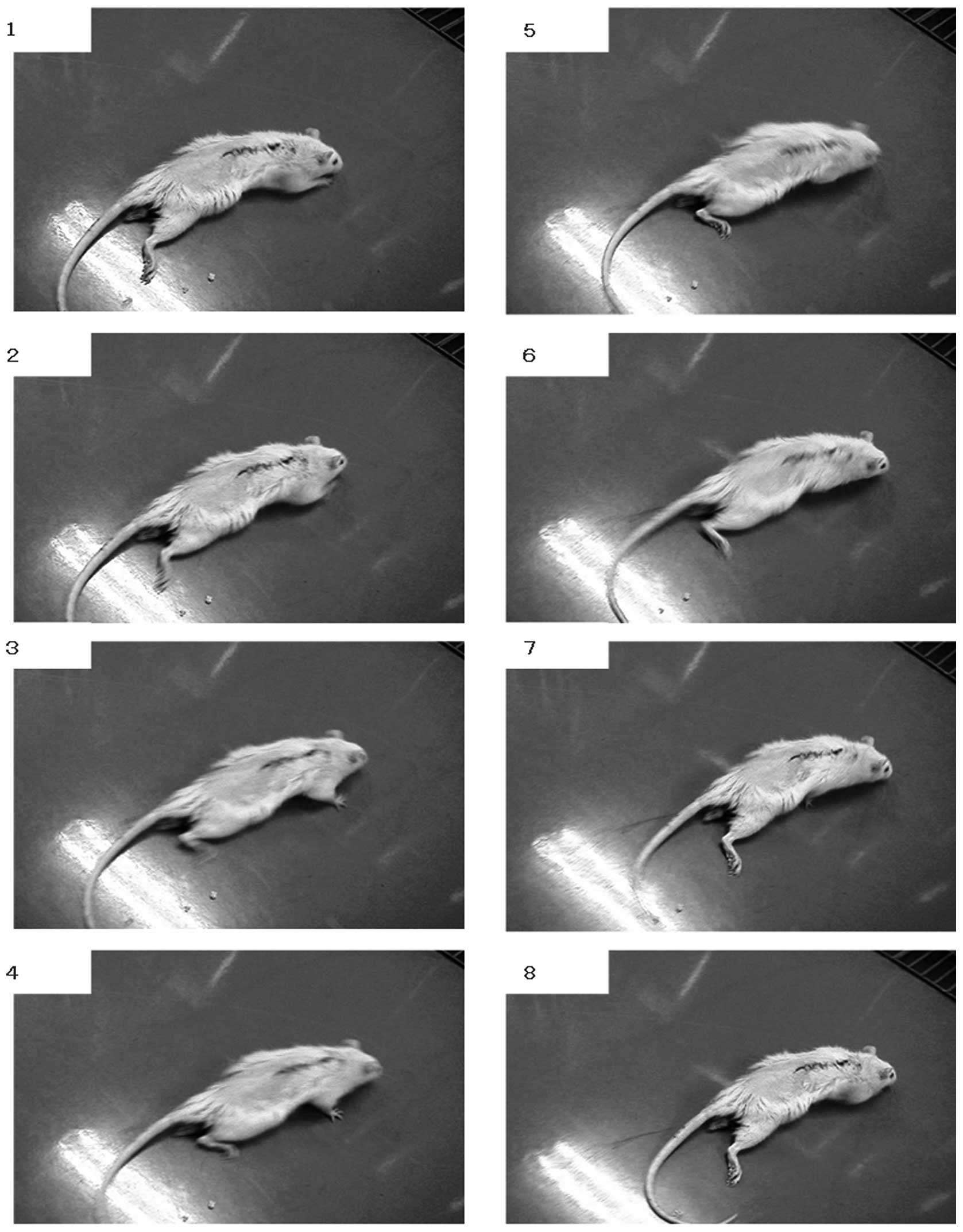
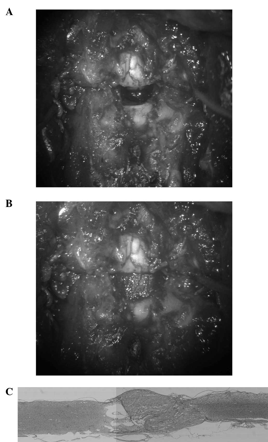
Previous studies have presented evidence which indicates that the regeneration of axons in the spinal cord occurs following spinal cord transection in young rats. However, in a transection‑regeneration model, the completeness of the transection is often a matter of dispute. We established a method for shortening the rat spine and spinal cord to provide a spinal cord injury (SCI) model in which there was no doubt about whether the axonal transection was complete. In the future, this model may be applied to the chronic period of complete paralysis following SCI. Adult, female Wistar rats (220‑250g) were used in the study. The spinal cord was exposed and a 4‑mm‑long segment of the spinal cord was removed at Th8. Subsequently, the Th7/8 and Th8/9 discs were cut between the stumps of the spinal cord to remove the Th8 vertebra. The stitches which had been passed through the 7th and 9th ribs bilaterally were tied gradually to bring together the stumps of the spinal cord. Almost all the rats survived until the end of the experiment. Uncoordinated movements of the hind limbs in locomotion were observed at 4 weeks after surgery. However coordinated movements of the hind limbs in locomotion were not observed until the end of the experiment. After 12 weeks, an intracardiac perfusion was performed to remove the thoracic spine and the spinal cord. There were no signs of infection. The bone fusion of the Th7 and Th9 vertebrae was observed to be complete in all specimens and the alignment of the thoracic spine was maintained. The spinal canal was also correctly reconstituted. The stumps of the spinal cord were connected. Light microscopy of the cord showed that scar tissue intervened at the connection site. Cavitation inhibiting the axonal regeneration was also observed. This model was also made on the assumption that glial scar tissue inhibits axonal regeneration in chronic SCI. Axonal regeneration was not observed across the transected spinal cord in this model. Attempts should be made to minimize the damage to the spinal cord and the surgery time for successful axonal regeneration to occur. The model developed in this study may be useful in the study of axonal regeneration in SCI.