Application of 256-slice computed tomography with low radiation doses in neonates with hypoxic-ischemic encephalopathy
- Authors:
View Affiliations
Affiliations: Radiology Department, Qilu Hospital, Shandong University, Jinan, Shandong 250012, P.R. China
- Published online on: September 30, 2013 https://doi.org/10.3892/etm.2013.1322
-
Pages:
1414-1416
Metrics: Total
Views: 0 (Spandidos Publications: | PMC Statistics: )
Metrics: Total PDF Downloads: 0 (Spandidos Publications: | PMC Statistics: )
This article is mentioned in:
Abstract
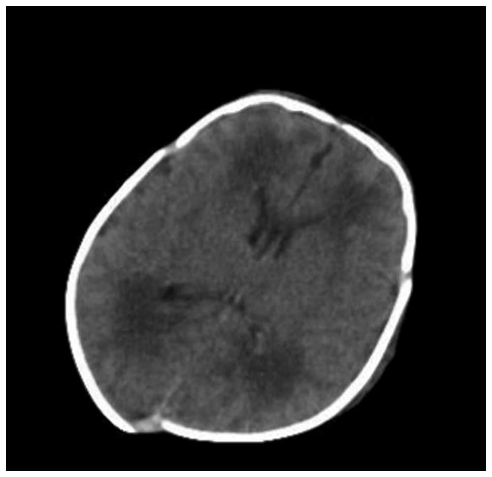
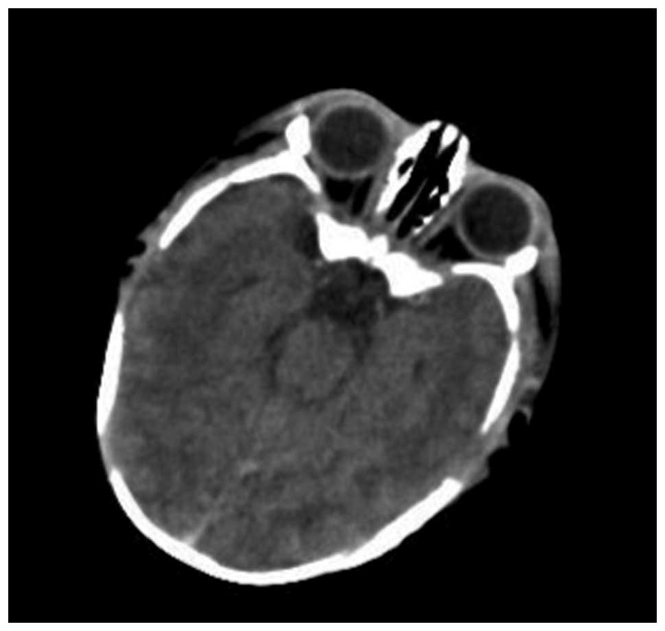
Hypoxic-ischemic encephalopathy (HIE), an injury or disease with lack of oxygen in the brain, may occur at any stage in childhood but the exact mechanisms that cause HIE remain unknown. In this study, 150 newborns suspected of having neonatal HIE and scheduled for a brain CT scan were randomly assigned to three equally sized groups as follows: standard dose group (120 kV, 250 mAsec), low dose group 1 (120 kV, 150 mAsec) and low dose group 2 (120 kV, 50 mAsec). All other acquisition parameters were the same in all groups. The CT dose index (CTDI), dose length product (DLP) and the image noise were compared among the three groups. The image quality was evaluated by blinded readers. The DLP of low dose group 2 was 19.3% of that of the standard dose group without a significant difference (P>0.05). The image noise of the low dose group 1 was greater than that of the standard dose group with a significant difference (P<0.01). Low dose scanning is feasible in the screening of HIE in neonates and is beneficial in protecting newborns against unnecessary radiation damage.
View References
1
|
McKinney AM, Teksam M, Felice R, et al:
Diffusion-weighted imaging in the setting of diffuse cortical
laminar necrosis and hypoxic-ischemic encephalopathy. AJNR Am J
Neuroradiol. 25:1659–1665. 2004.PubMed/NCBI
|
2
|
Fee SC, Malee K, Deddish R, et al: Severe
acidosis and subsequent neurologic status. Am J Obstet Gynecol.
162:802–806. 1990. View Article : Google Scholar : PubMed/NCBI
|
3
|
Ferrari F, Todeschini A, Guidotti I, et
al: General movements in full-term infants with perinatal asphyxia
are related to Basal Ganglia and thalamic lesions. J Pediatr.
158:904–911. 2011. View Article : Google Scholar : PubMed/NCBI
|
4
|
Berrington de González A, Mahesh M, Kim
KP, et al: Projected cancer risks from computed tomographic scans
performed in the United States in 2007. Arch Int Med.
169:2071–2077. 2009.PubMed/NCBI
|
5
|
Mori S, Endo M, Nishizawa K, et al:
Comparison of patient doses in 256-slice CT and 16-slice CT
scanners. Br J Radiol. 79:56–61. 2006. View Article : Google Scholar : PubMed/NCBI
|
6
|
Endo M, Mori S, Tsunoo T and Miyazaki H:
Magnitude and effects of x-ray scatter in a 256-slice CT scanner.
Med Phys. 33:3359–3368. 2006. View Article : Google Scholar : PubMed/NCBI
|
7
|
Pearce MS, Salotti JA, Little MP, et al:
Radiation exposure from CT scans in childhood and subsequent risk
of leukaemia and brain tumours: a retrospective cohort study.
Lancet. 380:499–505. 2012. View Article : Google Scholar : PubMed/NCBI
|
8
|
Strauss KJ, Goske MJ, Kaste SC, et al:
Image gently: ten steps you can take to optimize image quality and
lower CT dose for pediatric patients. AJR Am J Roentgenol.
94:868–873. 2010. View Article : Google Scholar : PubMed/NCBI
|