Construction of functional tissue‑engineered bone using cell sheet technology in a canine model
- Authors:
- Tao Chen
- Yanhui Wang
- Lingxue Bu
- Ningyi Li
View Affiliations
Affiliations: Department of Oral and Maxillofacial Surgery, Affiliated Hospital of Qingdao University Medical College, Qingdao, Shandong 266003, P.R. China
- Published online on: January 29, 2014 https://doi.org/10.3892/etm.2014.1514
-
Pages:
958-962
Metrics: Total
Views: 0 (Spandidos Publications: | PMC Statistics: )
Metrics: Total PDF Downloads: 0 (Spandidos Publications: | PMC Statistics: )
This article is mentioned in:
Abstract
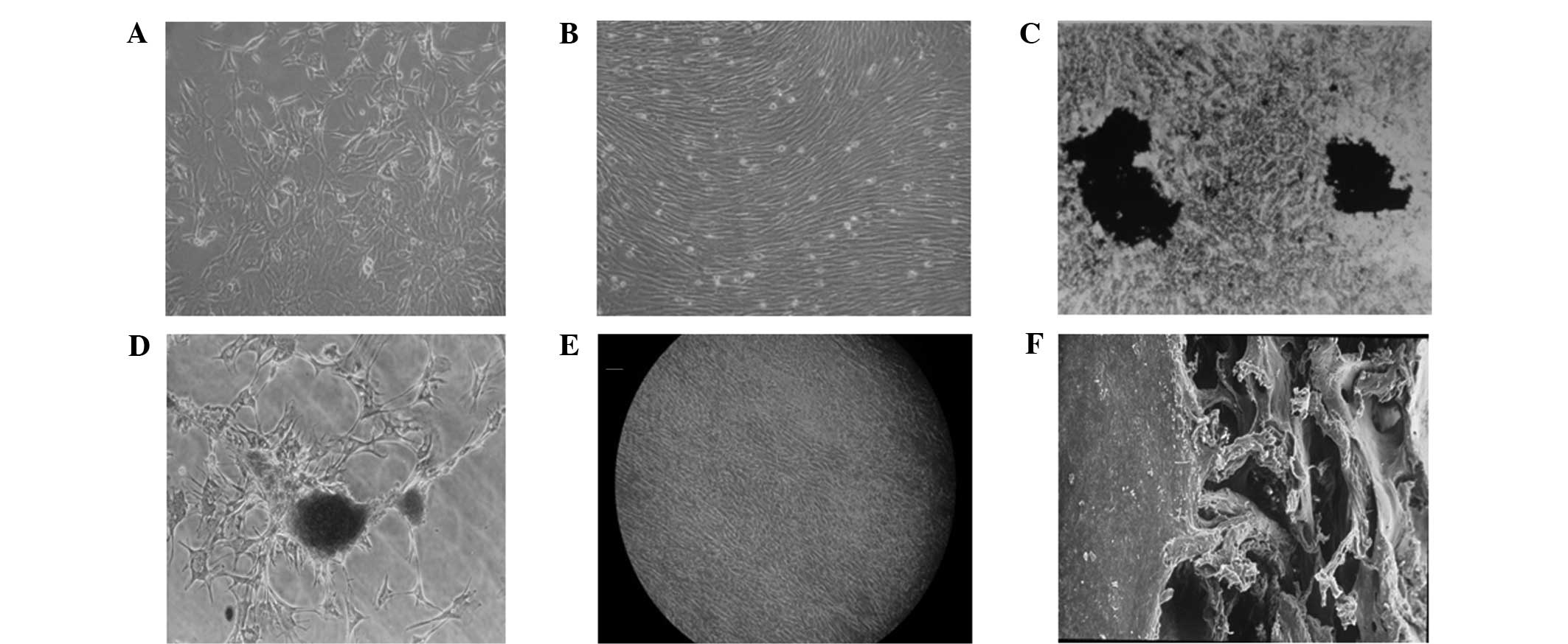
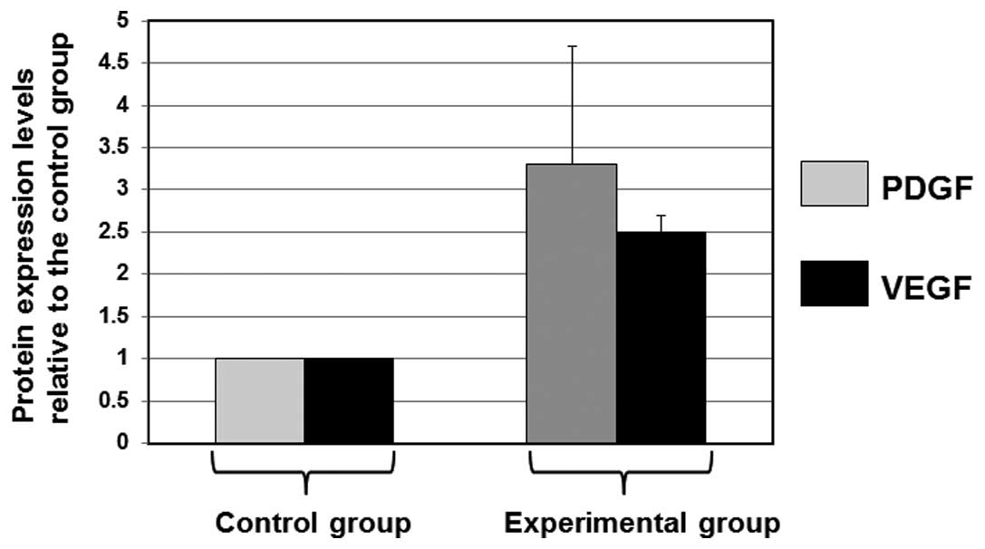
The aim of the present study was to construct functional tissue‑engineered bone with cell sheet technology and compare the efficacy of this method with that of traditional bone tissue engineering techniques. Canine bone mesenchymal stem cells (BMSCs) were isolated using density gradient centrifugation and then cultured. The BMSCs were induced to differentiate into osteoblasts and cultured in temperature‑responsive culture dishes. The BMSCs detached automatically from the temperature‑responsive culture dishes when the temperature was reduced to 20˚C, forming an intact cell sheet. Demineralized bone matrix (DBM) and platelet‑rich plasma (PRP) were prepared and used to construct a DBM/PRP/BMSC cell sheet/BMSC complex, which was implanted under the left latissimus dorsi muscle in a dog model. A DBM/PRP/BMSC complex was used as a control and implanted under the right latissimus dorsi muscle in the dog model. Immunoblot assays were performed to detect the levels of growth factors. Osteogenesis was observed to be induced significantly more effectively in the DBM/PRP/BMSC cell sheet/BMSC implants than in the DBM/PRP/BMSC implants. Immunoblot assay results indicated that the levels of the growth factors platelet-derived growth factor (PDGF) and vascular endothelial growth factor (VEGF) in the experimental group were 3.2‑ and 2.5‑fold higher compared with those in the control group, respectively. These results indicated that the BMSC cell sheets were functional and more effective than the control cell complex. Therefore, cell sheet technology may be used for the effective construction of functional tissue‑engineered bone with ideal properties.
View References
1
|
Yan H and Tsujii K: Thermo-responsive
poly(N-isopropylacrylamide) gel containing polymeric surfactant
poly(2-(methacryloyloxyl)decylphosphate): correlation between rapid
collapsing characters and micelles of polymeric surfactant. J Oleo
Sci. 57:401–405. 2008. View Article : Google Scholar
|
2
|
Yamada N, Okano T, Sakai H, et al:
Thermo-responsive polymeric surfaces; control of attachment and
detachment of cultured cells. Makromol Chem Rapid Commun.
11:571–576. 1990. View Article : Google Scholar
|
3
|
Shimizu T, Yamato M, Kikuchi A and Okano
T: Cell sheet engineering for myocardial tissue reconstruction.
Biomaterials. 24:2309–2316. 2003. View Article : Google Scholar : PubMed/NCBI
|
4
|
Jing H, Li NY, Tan S, et al: In vitro
culture and cell sheet preparation of bone marrow mesenchymal
cells. Guo Ji Kou Qiang Yi Xue Za Zhi. 37:272–274. 2010.(In
Chinese).
|
5
|
Nishida K, Yamato M, Hayashida Y, et al:
Functional bioengineered corneal epithelial sheet grafts from
corneal stem cells expanded ex vivo on a temperature-responsive
cell culture surface. Transplantation. 77:379–385. 2004. View Article : Google Scholar
|
6
|
Gao Z, Chen F, Zhang J, et al:
Vitalisation of tubular coral scaffolds with cell sheets for
regeneration of long bones: a preliminary study in nude mice. Br J
Oral Maxillofac Surg. 47:116–122. 2009. View Article : Google Scholar : PubMed/NCBI
|
7
|
Yang J, Yamato M, Nishida K, et al: Cell
delivery in regenerative medicine: the cell sheet engineering
approach. J Control Release. 116:193–203. 2006. View Article : Google Scholar : PubMed/NCBI
|
8
|
Weibrich G, Kleis WK and Hafner G: Growth
factor levels in the platelet rich plasma produced by 2 different
methods: curasan-type PRP kit versus PCCS PRP system. Int J Oral
Maxillofac Implants. 17:184–190. 2002.PubMed/NCBI
|
9
|
Schmitz JP and Hollinger JO: The biology
of platelet-rich plasma. J Oral Maxillofac Surg. 59:1119–1121.
2001. View Article : Google Scholar : PubMed/NCBI
|
10
|
Weibrich G, Kleis WK, Hafner G and Hitzler
WE: Growth factor levels in platelet-rich plasma and correlations
with donor age, sex, and platelet count. J Craniomaxillofac Surg.
30:97–102. 2002. View Article : Google Scholar : PubMed/NCBI
|
11
|
Eppley BL, Woodell JE and Higgins J:
Platelet quantification and growth factor analysis from
platelet-rich plasma: implications for wound healing. Plast
Reconstr Surg. 114:1502–1508. 2004. View Article : Google Scholar : PubMed/NCBI
|
12
|
Li NY, Yuan RT, Chen T, et al: Effect of
platelet-rich plasma and latissimus dorsi muscle flap on
osteogenesis and vascularization of tissue-engineered bone in dogs.
J Oral Maxillofac Surg. 67:1850–1858. 2009. View Article : Google Scholar : PubMed/NCBI
|