A novel animal model of osteonecrosis of the femoral head induced using a magnetic resonance imaging‑guided argon‑helium cryotherapy system
- Authors:
- Published online on: March 14, 2014 https://doi.org/10.3892/etm.2014.1625
- Pages: 1525-1528
Metrics: Total
Views: 0 (Spandidos Publications: | PMC Statistics: )
Total PDF Downloads: 0 (Spandidos Publications: | PMC Statistics: )
Abstract
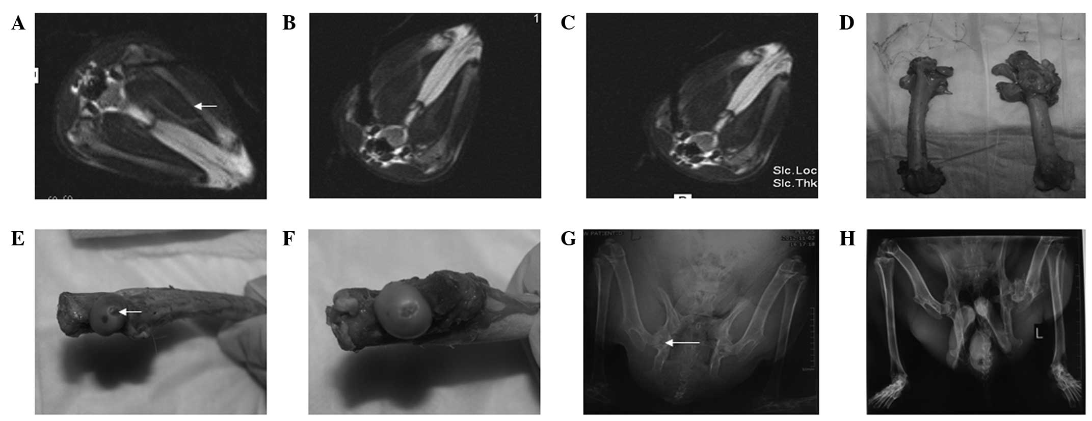
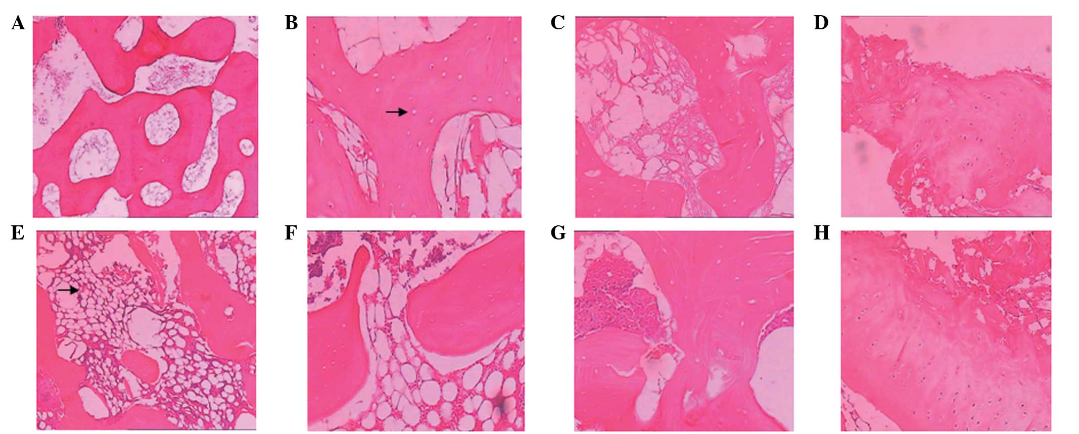
The aim of the present study was to establish a novel animal model of osteonecrosis of the femoral head (ONFH) using a magnetic resonance imaging (MRI)‑guided argon‑helium cryotherapy system. A total of 48 rabbits were used to generate the ONFH models. In group I, the left femoral head of the rabbits received two cycles of argon‑helium freezing‑thawing under MRI guidance, while in group II, the right femoral head of each rabbit received only one cycle of argon‑helium freezing‑thawing. X‑ray and histological examinations were performed. The percentages of lacunae in the femoral heads of group I at weeks 4, 8 and 12 following surgery (49.75±3.17, 62.06±4.12 and 48.25±2.76%, respectively) were higher than those in group II (39.13±4.48, 50.69±3.84 and 37.50±3.86%, respectively). In addition, the percentage of empty lacunae in group I was 62.06% at week 8 following surgery. Therefore, an animal model of ONFH was successfully established using an argon‑helium cryotherapy system. The percentage of empty lacunae in group I was higher than that in group II at weeks 4, 8 and 12 after surgery.