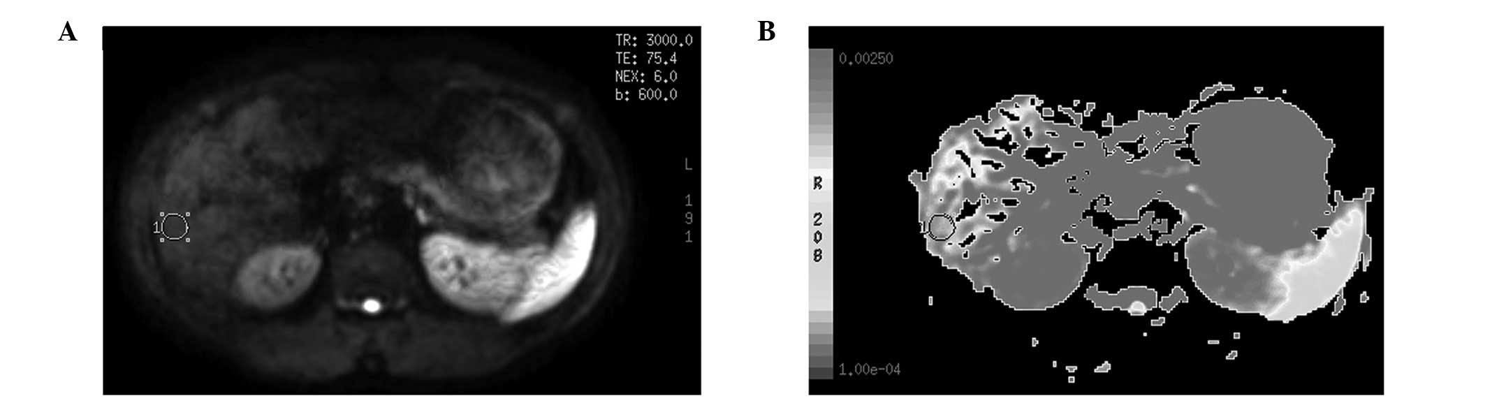
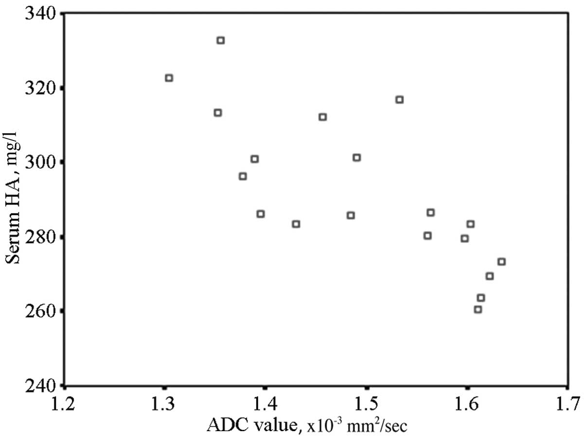
|
1
|
Bakan AA, Inci E, Bakan S, Gokturk S and
Cimilli T: Utility of diffusion-weighted imaging in the evaluation
of liver fibrosis. Eur Radiol. 22:682–687. 2012. View Article : Google Scholar : PubMed/NCBI
|
|
2
|
Yin M, Talwalkar JA, Glaser KJ, et al:
Dynamic postprandial hepatic stiffness augmentation assessed with
MR elastography in patients with chronic liver disease. AJR Am J
Roentgenol. 197:64–70. 2011. View Article : Google Scholar : PubMed/NCBI
|
|
3
|
Do RK, Chandarana H, Felker E, et al:
Diagnosis of liver fibrosis and cirrhosis with diffusion-weighted
imaging: value of normalized apparent diffusion coefficient using
the spleen as reference organ. AJR Am J Roentgenol. 195:671–676.
2010. View Article : Google Scholar
|
|
4
|
Papanikolaou N, Gourtsoyianni S,
Yarmenitis S, Maris T and Gourtsoyiannis N: Comparison between
two-point and four-point methods for quantification of apparent
diffusion coefficient of normal liver parenchyma and focal lesions.
Value of normalization with spleen. Eur J Radiol. 73:305–309. 2010.
View Article : Google Scholar
|
|
5
|
Sandrasegaran K, Akisik FM, Lin C, et al:
Value of diffusion-weighted MRI for assessing liver fibrosis and
cirrhosis. AJR Am J Roentgenol. 193:1556–1560. 2009. View Article : Google Scholar : PubMed/NCBI
|
|
6
|
Gourtsoyianni S, Papanikolaou N,
Yarmenitis S, Maris T, Karantanas A and Gourtsoyiannis N:
Respiratory gated diffusion-weighted imaging of the liver: value of
apparent diffusion coefficient measurements in the differentiation
between most commonly encountered benign and malignant focal liver
lesions. Eur Radiol. 18:486–492. 2008. View Article : Google Scholar
|
|
7
|
Taouli B, Tolia AJ, Losada M, et al:
Diffusion-weighted MRI for quantification of liver fibrosis:
preliminary experience. AJR Am J Roentgenol. 189:799–806. 2007.
View Article : Google Scholar : PubMed/NCBI
|
|
8
|
Ismail MH and Pinzani M: Reversal of liver
fibrosis. Saudi J Gastroenterol. 15:72–79. 2009. View Article : Google Scholar
|
|
9
|
Faria SC, Ganesan K, Mwangi I, et al: MR
imaging of liver fibrosis: current state of the art. Radiographics.
29:1615–1635. 2009. View Article : Google Scholar : PubMed/NCBI
|
|
10
|
Eccles CL, Haider EA, Haider MA, Fung S,
Lockwood G and Dawson LA: Change in diffusion weighted MRI during
liver cancer radiotherapy: preliminary observations. Acta Oncol.
48:1034–1043. 2009. View Article : Google Scholar : PubMed/NCBI
|
|
11
|
Lewin M, Poujol-Robert A, Boëlle PY, et
al: Diffusion-weighted magnetic resonance imaging for the
assessment of fibrosis in chronic hepatitis C. Hepatology.
46:658–665. 2007. View Article : Google Scholar : PubMed/NCBI
|
|
12
|
Wang Y, Ganger DR, Levitsky J, et al:
Assessment of chronic hepatitis and fibrosis: comparison of MR
elastography and diffusion-weighted imaging. AJR Am J Roentgenol.
196:553–561. 2011. View Article : Google Scholar : PubMed/NCBI
|
|
13
|
Sun J, Shi YX, Zhang ZY, Zhang ZQ, Wu JT
and Wan HY: Combination of MRI ADC value and biochemical indicators
for the assessment of fibrosis in chronic hepatitis: Preliminary
results. Lin Chuang Fang She Xue Za Zhi. 43:616–619. 2010.(In
Chinese).
|
|
14
|
Binkovitz LA, El-Youssef M, Glaser KJ, Yin
M, Binkovitz AK and Ehman RL: Pediatric MR elastography of hepatic
fibrosis: principles, technique and early clinical experience.
Pediatr Radiol. 42:402–409. 2012. View Article : Google Scholar : PubMed/NCBI
|
|
15
|
Kamel IR, Bluemke DA, Ramsey D, et al:
Role of diffusion weighted imaging in estimating tumor necrosis
after chemoembolization of hepatocellular carcinoma. AJR Am J
Roentgenol. 181:708–710. 2003. View Article : Google Scholar : PubMed/NCBI
|
|
16
|
Kamada K, Nakanishi T, Kitamoto M, et al:
Long-term prognosis of patients undergoing transcatheter arterial
chemoembolization for unresectable hepatocellular carcinoma:
comparison of cisplatin lipiodol suspension and doxorubicin
hydrochloride emulsion. J Vasc Interv Radiol. 12:847–854. 2001.
View Article : Google Scholar
|
|
17
|
Aguirre DA, Behling CA, Alpert E,
Hassanein TI and Sirlin CB: Liver fibrosis: noninvasive diagnosis
with double contrast material-enhanced MR imaging. Radiology.
239:425–437. 2006. View Article : Google Scholar : PubMed/NCBI
|
|
18
|
Aubé C, Racineux PX, Lebigot J, et al:
Diagnosis and quantification of hepatic fibrosis with diffusion
weighted MR imaging: preliminary results. J Radiol. 85:301–306.
2004.(In French).
|
|
19
|
Koinuma M, Ohashi I, Hanafusa K and
Shibuya H: Apparent diffusion coefficient measurements with
diffusion-weighted magnetic resonance imaging for evaluation of
hepatic fibrosis. J Magn Reson Imaging. 22:80–85. 2005. View Article : Google Scholar
|
|
20
|
Taouli B and Koh DM: Diffusion-weighted MR
imaging of the liver. Radiology. 254:47–66. 2010. View Article : Google Scholar : PubMed/NCBI
|