Diagnosis and treatment of a dural arteriovenous fistula presenting with progressive parkinsonism and dementia: A case report and literature review
- Authors:
- Chen Ma
- Qiaoli Lu
- Wanchao Shi
- Zhiguo Su
- Yujun Zhao
- Chen Li
- Zhenlin Liu
View Affiliations
Affiliations: Department of Neurology, The Fifth Central Hospital of Tianjin, Tianjin 300450, P.R. China, Department of Neurosurgery, The Fifth Central Hospital of Tianjin, Tianjin 300450, P.R. China
- Published online on: December 9, 2014 https://doi.org/10.3892/etm.2014.2122
-
Pages:
523-526
Metrics: Total
Views: 0 (Spandidos Publications: | PMC Statistics: )
Metrics: Total PDF Downloads: 0 (Spandidos Publications: | PMC Statistics: )
This article is mentioned in:
Abstract
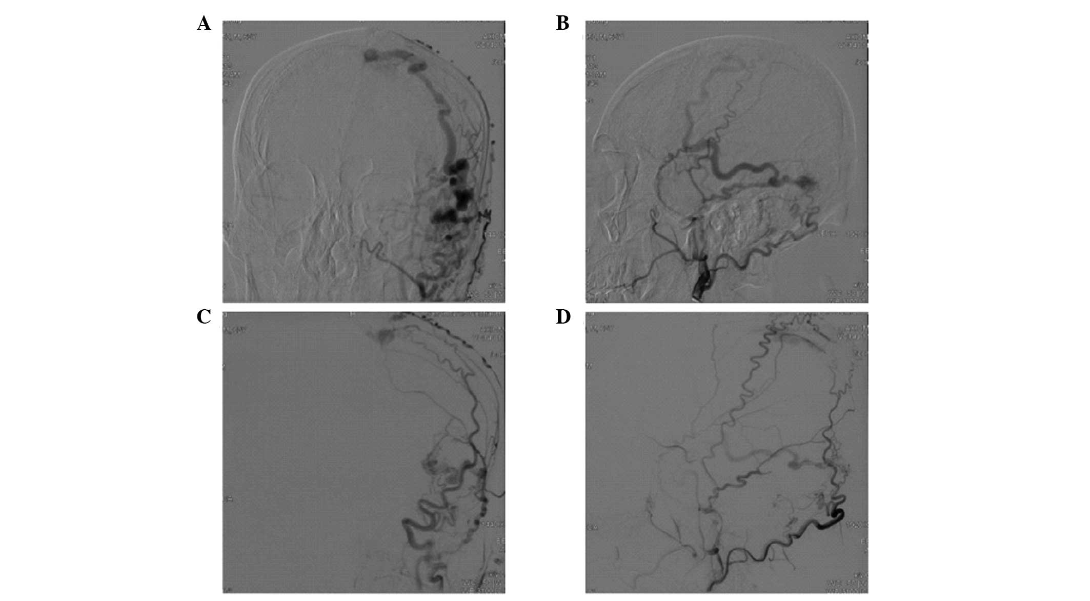
A dural arteriovenous fistula (DAVF) presenting with parkinsonism and dementia is rare; thus, is easily misdiagnosed. The present study reports the case of a 62‑year‑old male with mobility disabilities and a cognitive disorder. The initial symptoms were progressive symmetrical limb stiffness and weakness without significant limb tremor, and subsequently the appearance of progressive memory loss, behavioral abnormalities and a decline in the activities of daily living. Cranial magnetic resonance imaging (MRI) revealed an enlarged vascular shadow at the meninges of the left temporal lobe. In addition, digital subtraction angiography (DSA) revealed a DAVF in the left temporal region, fed by the bilateral middle meningeal arteries and meningeal branches of the vertebral artery, which were enlarged abnormally, with poor venous reflux to the superior sagittal sinus. The patient was treated with transarterial embolization therapy. Intraoperative angiography showed almost complete embolization of the DAVF. At day 3 following the surgery, the muscle tension of the bilateral limbs decreased significantly. After two weeks, the memory ability of the patient had recovered to the level prior to the onset, and the gait was stable. At one month post‑surgery, the patient was able to take care of himself completely, and after three months, a stereotactic treatment was conducted for the residual fistula. At the one year follow‑up, neurological examination revealed that the patient had recovered normally. In conclusion, progressive parkinsonism and dementia with an abnormal flow void shadow on cranial MRI films should be considered as a possible diagnosis of a DAVF. In these cases, DSA and endovascular treatment are recommended as soon as possible.
View References
1
|
Jahan R, Gobin YP, Glenn B, Duckwiler GR
and Viñuela F: Transvenous embolization of a dural arteriovenous
fistula of the cavernous sinus through the contralateral pterygoid
plexus. Neuroradiology. 40:189–193. 1998. View Article : Google Scholar : PubMed/NCBI
|
2
|
Cha KC, Yeon JY, Kim GH, Jeon P, Kim JS
and Hong SC: Clinical and angiographic results of patients with
dural arteriovenous fistula. J Clin Neurosci. 20:536–542. 2013.
View Article : Google Scholar : PubMed/NCBI
|
3
|
Kuwayama N, Kubo M, Tsumura K, Yamamoto H
and Endo S: Hemodynamic status and treatment of aggressive dural
arteriovenous fistulas. Acta Neurochir Suppl. 94:123–126. 2005.
View Article : Google Scholar : PubMed/NCBI
|
4
|
Kai Y, Hamada J, Morioka M, et al:
Arteriovenous fistulas at the cervicomedullary junction presenting
with subarachoid hemorrhage: six case reports with special
Reference to the angiographic pattern of venous drainage. AJNR Am J
Neuroradiol. 26:1949–1954. 2005.PubMed/NCBI
|
5
|
He HW, Jiang CH, Wu ZX, Li YX and Wang ZC:
Transvenous embolization of cavernous dural arteriovenous fistula:
report of 28 cases. Clin Med J (Engl). 120:2229–2232. 2007.
|
6
|
Chen L, Mao Y and Zhou LF: Local chronic
hypoperfusion secondary to sinus high pressure seems to be mainly
responsible for the formation of intracranial dural arteriovenous
fistula. Neurosurgery. 64:973–983. 2009. View Article : Google Scholar : PubMed/NCBI
|
7
|
Tsai LK, Jeng JS, Liu HM, Wang HJ and Yip
PK: Intracranial dural arteriovenous fistulas with or without
cerebral sinus thrombosis: analysis of 69 patients. J Neurol
Neurosurg Psychiatry. 75:1639–1641. 2004. View Article : Google Scholar : PubMed/NCBI
|
8
|
Xavier J, Cruz R, Stocker A, et al: Dural
fistulas of the anterior cranial fossa. Acta Med Port. 14:71–75.
2001.(In Portuguese). PubMed/NCBI
|
9
|
Kajitani M, Yagura H, Kawahara M, et al:
Treatable fluctuating Parkinsonism and dementia in a patient with a
dural arteriovenous fistula. Mov Disord. 22:437–439. 2007.
View Article : Google Scholar : PubMed/NCBI
|
10
|
Matsumura A, Oda M, Hozuki T, Imai T and
Shimohama S: Dural arteriovenous fistula in a case of dementia with
bithalamic MR lesions. Neurology. 7l:l5532008.
|
11
|
Yamanouchi H and Nagura H: Neurological
signs and frontal white matter lesions in vascular Parkinsonism. A
clinicopathological study. Stroke. 28:965–969. 1997. View Article : Google Scholar : PubMed/NCBI
|
12
|
Gandhi D, Chen J, Pearl M, et al:
Intracranial dural arteriovenous fistulas: classification, imaging
findings, and treatment. AJNR Am J Neuroradiol. 33:1007–1013. 2012.
View Article : Google Scholar : PubMed/NCBI
|
13
|
Ming Y, Zhong L, Ze Y, et al: Comparison
among CT, MRI and DSA features of dural ateriovenous fistula.
Zhongguo Linchuang Shenjing Waike Zazhi. 11:201–203. 2006.(In
Chinese).
|
14
|
Pelz DM, Lownie SP, Fox AJ and Rosso D:
Intracranial dural arteriovenous fistulae with pial venous
drainage: combined endovascular-neurosurgical therapy. Can J Neurol
Sci. 24:210–218. 1997.PubMed/NCBI
|
15
|
Jiang C, Lv X, Li Y, Zhang J and Wu Z:
Endovascular treatment of high-risk tentorial dural arteriovenous
fistulas: clinical outcomes. Neuroradiology. 51:103–111. 2009.
View Article : Google Scholar
|