Clinical analysis of a patient with a benign lesion of the pharynx misdiagnosed as functional thyroid cancer metastasis
- Authors:
- Renfei Wang
- Jian Tan
- Guizhi Zhang
- Qiang Jia
View Affiliations
Affiliations: Department of Nuclear Medicine, Tianjin Medical University General Hospital, Tianjin 300052, P.R. China
- Published online on: March 9, 2015 https://doi.org/10.3892/etm.2015.2335
-
Pages:
1871-1874
Metrics: Total
Views: 0 (Spandidos Publications: | PMC Statistics: )
Metrics: Total PDF Downloads: 0 (Spandidos Publications: | PMC Statistics: )
This article is mentioned in:
Abstract
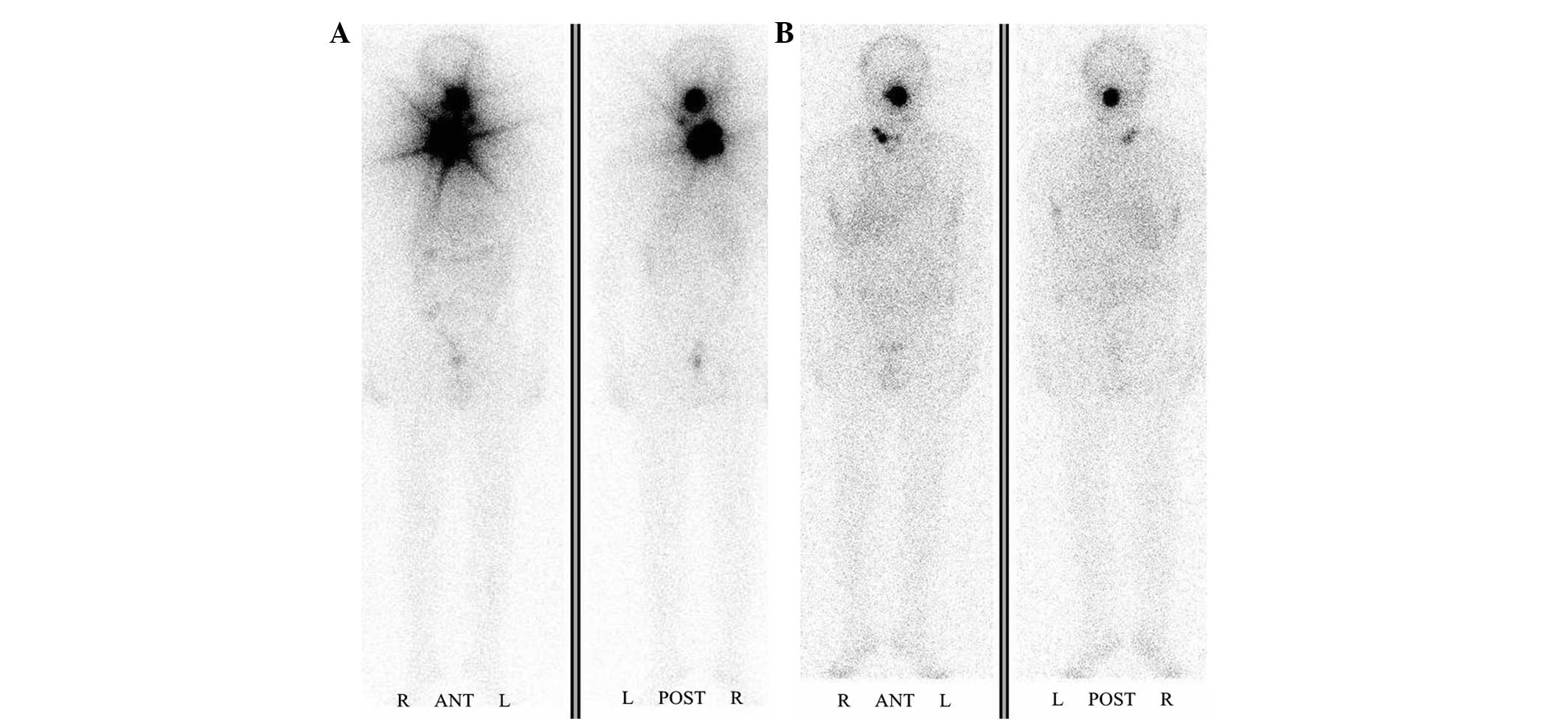
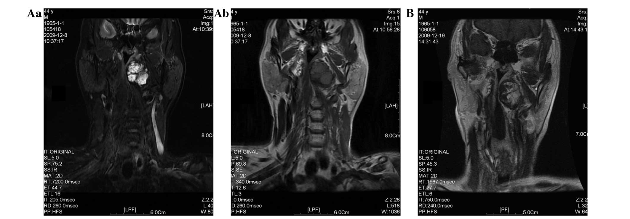
Papillary thyroid carcinoma (PTC) usually has a lower malignancy rate and better prognosis than other types of thyroid cancer. It tends to spread through the lymphatic system, and the percentage of lymph node metastasis to the neck is 50‑70% in patients at the time of diagnosis. Iodine-131 (131I) whole‑body scanning is very valuable in the diagnosis and positioning of functional metastatic lesions. However, a variety of comprehensive test results are frequently required to make an appropriate judgment. The present case report describes the case of a patient with PTC where a benign lesion of the pharynx was misdiagnosed as functional metastasis. Some remnant thyroid tissue and an abnormal accumulation of 131I in the oropharynx and at the right side of the neck were observed by 131I whole‑body scanning one week after the first therapeutic dose. Following the second therapeutic dose, quasi‑circular trapping of 131I by the pharynx persisted. Magnetic resonance imaging enhanced scanning of the oropharynx showed markedly uneven enhancement inside the lesion, and it was considered as a metastatic lesion. However, the serological test results of the patient revealed that there was no increase in the thyroglobulin level and did not support the diagnosis of progressive disease. Therefore, a biopsy on the lesion was conducted under local anesthesia. The histopathological examination exhibited salivary gland tissue and chronic inflammation. Thus, it was necessary to analyze the imaging results, as well as serologic testing outcomes and pathological findings comprehensively, in order to make the correct diagnosis.
View References
1
|
Cobin RH, Gharib H, Bergman DA, et al:
Thyroid Carcinoma Task Force: AACE/AAES medical/surgical guidelines
for clinical practice: management of thyroid carcinoma. American
Association of Clinical Endocrinologists. American College of
Endocrinology. Endocr Prac. 7:202–220. 2001.
|
2
|
Al-Humadi H, Zarrros A, Al-Saigh R and
Liapi C: Genetic basis and gene therapy trials for thyroid cancer.
Cancer Genomics Proteomics. 7:31–49. 2010.PubMed/NCBI
|
3
|
Hay ID: Papillary thyroid carcinoma.
Endocrinol Metab Clin North Am. 19:545–576. 1990.PubMed/NCBI
|
4
|
Sarkar SD, Kalapparambath TP and Palestro
CJ: Comparison of 123I and 131I for whole-body imaging in thyroid
cancer. J Nucl Med. 5:632–634. 2002.
|
5
|
Grewal RK, Tuttle RM, Fox J, et al: The
effect of posttherapy 131I SPECT/CT on risk classification and
management of patients with differentiated thyroid cancer. J Nucl
Med. 9:1361–1367. 2010. View Article : Google Scholar
|
6
|
Chinese Society of Endocrinology,
Endocrine Group of Surgery Branch of Chinese Medical Association,
Committee for Head and Neck Oncology of Chinese Anti-cancer
Association, Society of Nuclear Medicine of Chinese Medical
Association, . Clinical guidelines for the diagnosis and management
of thyroid nodules and differentiated thyroid cancer. Zhonghua He
Yi Xue Yu Fen Zi Ying Xiang Za Zhi. 33:96–115. 2013.[In
Chinese].
|
7
|
Leger AF, Pellan M, Dagousset F, et al: A
case of stunning of lung and bone metastases of papillary thyroid
cancer after a therapeutic dose (3.7 GBq) of 131I and review of the
literature: implications for sequential treatments. Br J Radiol.
78:428–432. 2005. View Article : Google Scholar : PubMed/NCBI
|
8
|
Lind P and Kohlfürst S: Respective roles
of thyroglobulin, radioiodine imaging and positron emission
tomography in the assessment of thyroid cancer. Semin Nucl Med.
36:194–205. 2006. View Article : Google Scholar : PubMed/NCBI
|
9
|
Spanu A, Solinas ME, Chessa F, et al: 131I
SPECT/CT in the follow-up of differentiated thyroid carcinoma:
incremental value versus planar imaging. J Nucl Med. 50:184–190.
2009. View Article : Google Scholar : PubMed/NCBI
|
10
|
Mitchell G, Pratt BE, Vini L, et al: False
positive 131I whole body scans in thyroid cancer. Br J Radiol.
73:627–635. 2000. View Article : Google Scholar : PubMed/NCBI
|
11
|
Ma C, Kuang A, Xie JW and Ma TK: Possible
explanations for patients with discordant findings of serum
thyroglobulin and 131I whole-body scanning. J Nucl Med.
46:1473–1480. 2005.PubMed/NCBI
|
12
|
Mandel L and Liu F: Salivary gland injury
resulting from exposure to radioactive iodine: case reports. J Am
Dent Assoc. 138:1582–1587. 2007. View Article : Google Scholar : PubMed/NCBI
|