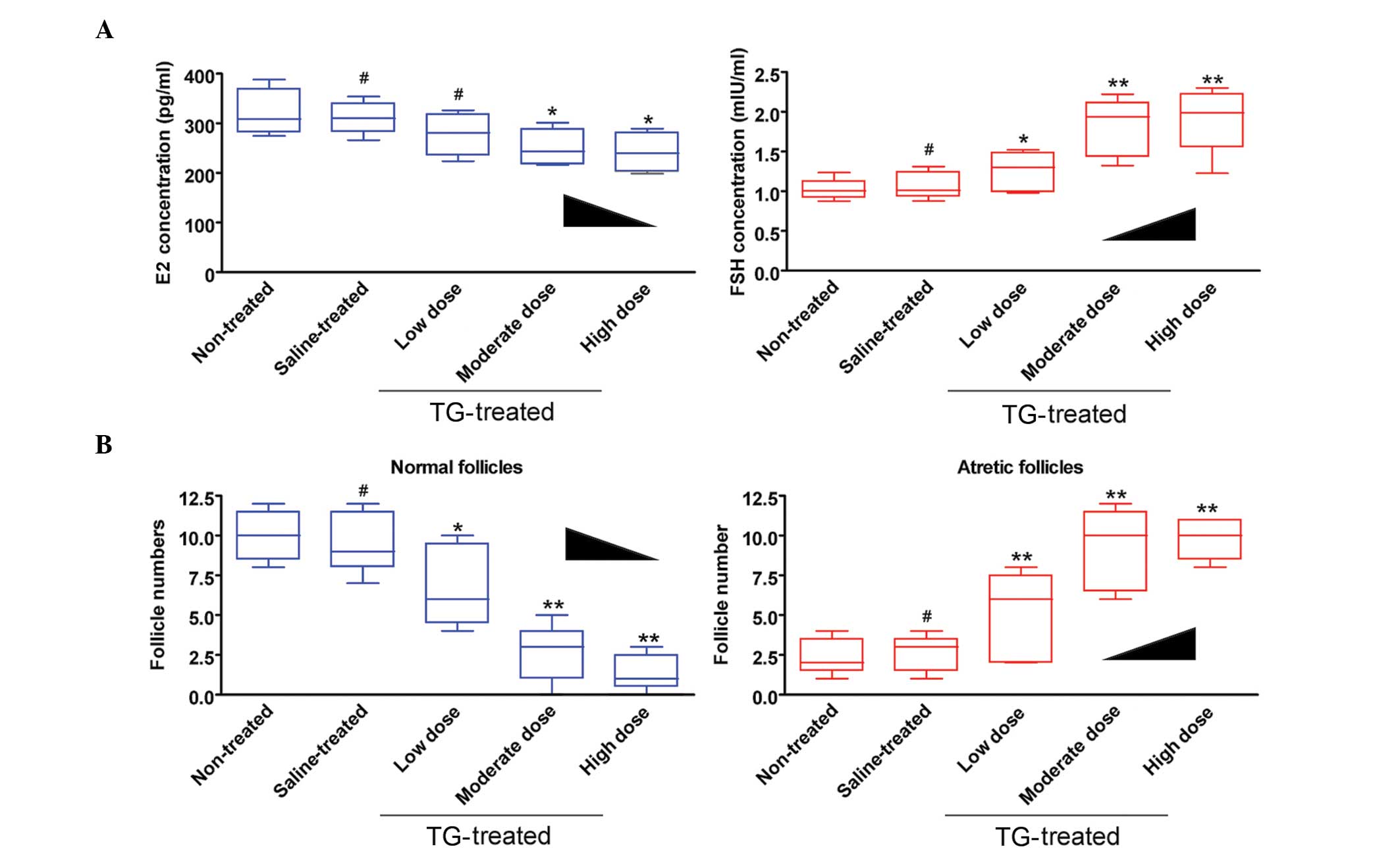
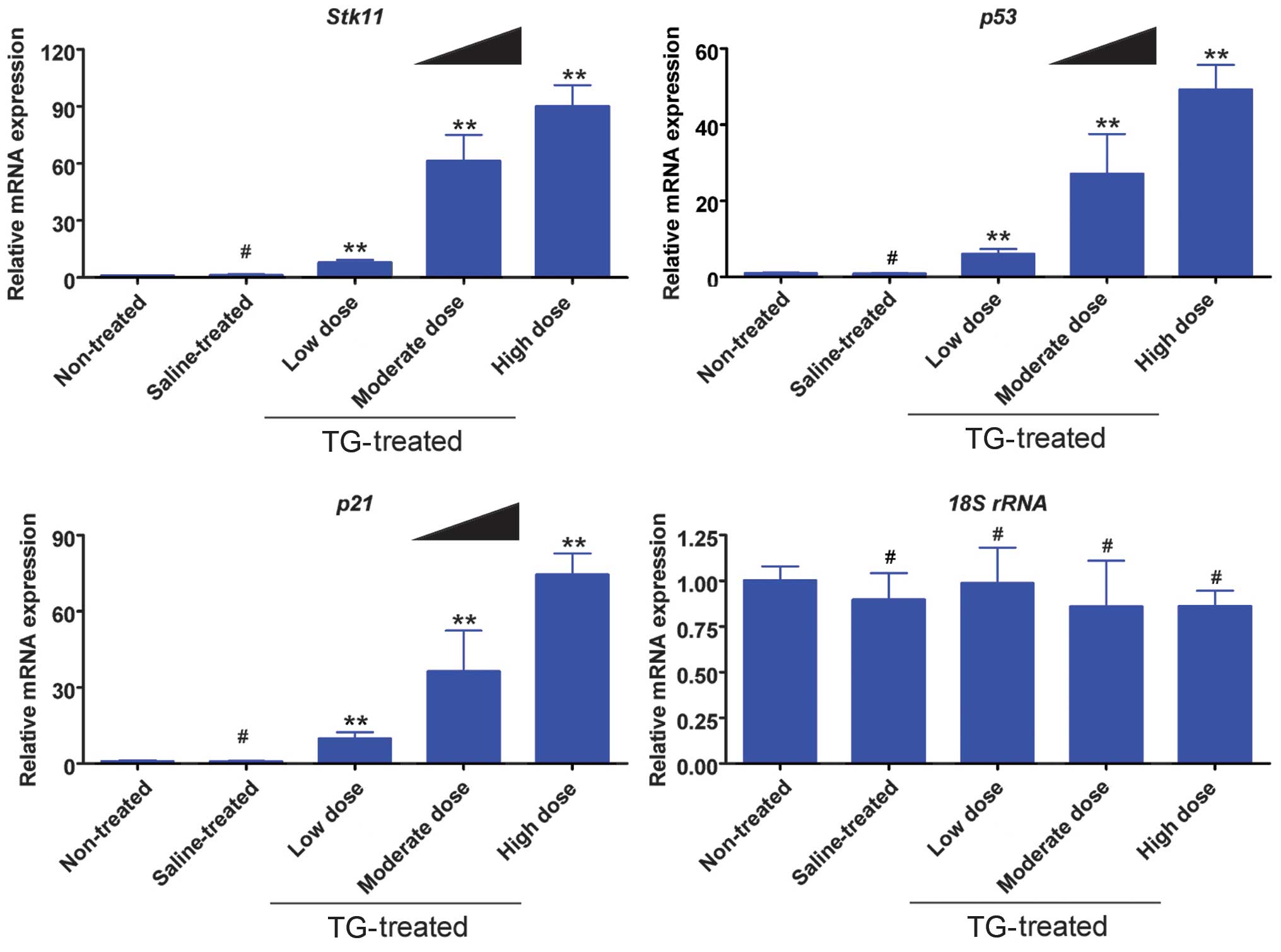
Tripterygium glycosides induce premature ovarian failure in rats by promoting p53 phosphorylation and activating the serine/threonine kinase 11-p53-p21 signaling pathway
- Authors:
- Te Liu
- Lina Zhang
- Suwei Wang
- Chuan Chen
- Jin Zheng
View Affiliations
Affiliations: Department of Gynecology, Longhua Hospital, Shanghai University of Traditional Chinese Medicine, Shanghai 200031, P.R. China, Shanghai Geriatric Institute of Chinese Medicine, Longhua Hospital, Shanghai University of Traditional Chinese Medicine, Shanghai 200031, P.R. China
- Published online on: May 18, 2015 https://doi.org/10.3892/etm.2015.2498
-
Pages:
12-18
-
Copyright: © Liu
et al. This is an open access article distributed under the
terms of Creative
Commons Attribution License.
Metrics: Total
Views: 0 (Spandidos Publications: | PMC Statistics: )
Metrics: Total PDF Downloads: 0 (Spandidos Publications: | PMC Statistics: )
This article is mentioned in:
Abstract
Premature ovarian failure (POF) is a typical pathological disease of the reproductive system in aging females. Infection, inflammation, immune abnormalities, genetic mutation, radiotherapy and chemotherapy can cause POF. Tripterygium glycosides (TGs) are a component extracted from the Chinese herb Tripterygium wilfordii Hook. f., also known as Huangteng. Although TGs have been used to treat various diseases, drug resistance and toxicity can affect patients. The aim of the present study was to investigate the mechanism of TG‑induced POF in rats. The rats were treated with different concentrations of TG, and pathology assays showed that the TG‑induced POF was predominantly composed of interstitial cells in a fibrous matrix with a reduced number of follicles at each stage and an increased number of collapsed oocytes. Furthermore, reverse transcription‑quantitative polymerase chain reaction (PCR) and immunohistochemistry assays indicated that the expression levels of serine/threonine kinase 11 (Stk11), p53 p21 and activated caspase‑3 were elevated significantly in the TG‑treated groups. Serine 15 phosphorylation of p53 was also enhanced significantly in the TG‑treated groups. In addition, a chromatin immunoprecipitation‑PCR assay revealed that the TGs induced p53 activation and enhanced the transcription of p21. In conclusion, TGs induce apoptosis and necrosis in rat ovarian tissues, as well as POF, via p53 phosphorylation and activation of the Stk11-p53-p21 signaling pathway.
View References
1
|
Beck-Peccoz P and Persani L: Premature
ovarian failure. Orphanet J Rare Dis. 1:92006. View Article : Google Scholar : PubMed/NCBI
|
2
|
Vujović S, Kanazir S, Ivović M, et al:
Collagen type I alpha 1 gene polymorphism in premature ovarian
failure. Srp Arh Celok Lek. 141:344–348. 2013. View Article : Google Scholar : PubMed/NCBI
|
3
|
Liu T, Huang Y, Guo L, Cheng W and Zou G:
CD44+/CD105+ human amniotic fluid mesenchymal stem cells survive
and proliferate in the ovary long-term in a mouse model of
chemotherapy-induced premature ovarian failure. Int J Med Sci.
9:592–602. 2012. View Article : Google Scholar : PubMed/NCBI
|
4
|
Liu T, Qin W, Huang Y, Zhao Y and Wang J:
Induction of estrogen-sensitive epithelial cells derived from
human-induced pluripotent stem cells to repair ovarian function in
a chemotherapy-induced mouse model of premature ovarian failure.
DNA Cell Biol. 32:685–698. 2013. View Article : Google Scholar : PubMed/NCBI
|
5
|
Liu T, Huang Y, Zhang J, et al:
Transplantation of human menstrual blood stem cells to treat
premature ovarian failure in mouse model. Stem Cells Dev.
23:1548–1557. 2014. View Article : Google Scholar : PubMed/NCBI
|
6
|
Li J, Shen F, Guan C, et al: Activation of
Nrf2 protects against triptolide-induced hepatotoxicity. PLoS One.
9:e1006852014. View Article : Google Scholar : PubMed/NCBI
|
7
|
Li XJ, Jiang ZZ and Zhang LY: Triptolide:
progress on research in pharmacodynamics and toxicology. J
Ethnopharmacol. 155:67–79. 2014. View Article : Google Scholar : PubMed/NCBI
|
8
|
Zhou J, Xi C, Wang W, et al:
Triptolide-induced oxidative stress involved with Nrf2 contribute
to cardiomyocyte apoptosis through mitochondrial dependent
pathways. Toxicol Lett. 230:454–466. 2014. View Article : Google Scholar : PubMed/NCBI
|
9
|
Zeng PY and Berger SL: LKB1 is recruited
to the p21/WAF1 promoter by p53 to mediate transcriptional
activation. Cancer Res. 66:10701–10708. 2006. View Article : Google Scholar : PubMed/NCBI
|
10
|
Vaahtomeri K and Mäkelä TP: Molecular
mechanisms of tumor suppression by LKB1. FEBS Lett. 585:944–951.
2011. View Article : Google Scholar : PubMed/NCBI
|
11
|
Ollila S and Mäkelä TP: The tumor
suppressor kinase LKB1: lessons from mouse models. J Mol Cell Biol.
3:330–340. 2011. View Article : Google Scholar : PubMed/NCBI
|
12
|
Shen DZ, Xin SL, Chen C and Liu T: Effect
of atorvastatin on expression of TLR4 and NF-κB p65 in
atherosclerotic rabbits. Asian Pac J Trop Med. 6:493–496. 2013.
View Article : Google Scholar : PubMed/NCBI
|