Introduction
Essential hypertension is a common chronic disease
that has no identifiable cause. Essential hypertension may increase
the risk of coronary heart disease, stroke, kidney failure and
heart failure, all of which may be seriously harmful to human
health (1). In recent years, due to
progressively aging populations, improved living standards and
stress, the incidence of essential hypertension has increased
significantly (2). The prevalence of
essential hypertension in young patients is increasing, and there
is thus an urgent requirement for methods to prevent and treat
essential hypertension.
Cardiac hypertrophy is a common pathological
manifestation in patients with essential hypertension, which is
primarily caused by prolonged cardiac stress. The myocardium
thickens in order to compensate for the increase in blood pressure,
eventually leading to heart failure (3). A previous study demonstrated that gene
expression profiles in cardiac mast cells differ significantly
between patients with cardiac hypertrophy and healthy donors, and
cardiac hypertrophy is reversible in the early stages (4). Therefore, elucidation of the molecular
mechanisms of essential hypertension accompanied by cardiac
hypertrophy is of clinical significance in the diagnosis and
treatment of hypertrophic cardiomyopathy.
microRNA (miRNA) is a type of small endogenic
non-coding RNA molecule (18–22 nucleotides) that regulates gene
expression by binding to 3′-untranslated regions of target mRNA.
miRNA molecules are widely present in the tissues and blood, with
good stability. The expression levels of various miRNA in cardiac
mast cells exhibit significant alterations in cardiac hypertrophy,
which suggests that miRNAs serve key functions in the progression
of cardiac hypertrophy (5,6). miRNA-208 is highly expressed in the
serum of patients with myocardial infarction, which indicates that
miR-208 may be a suitable biomarker for the diagnosis of myocardial
damage (7). Oliveira-Carvalho et
al reported a strong association of miR-208 expression in
cardiac mast cells with hypertrophic cardiomyopathy, suggesting
that miR-208 may be involved in the progression of the condition,
although the mechanism remains unclear (8). miR-208 promotes the proliferation of
esophageal squamous cell carcinoma cells by regulating the gene
sex-determining region Y-box 6 (SOX6) (9). The SOX gene family encodes a group of
transcription factors defined by the conserved high mobility group
DNA-binding domain, which is closely associated with a variety of
types of cell differentiation and embryonic development (10,11).
However, the role of SOX6 in hypertrophic cardiomyopathy remains
unknown. In the present study, the expression levels of miR-208 and
SOX6 in the peripheral blood of patients with essential
hypertension accompanied by left ventricular hypertrophy were
determined by western blot analysis and quantitative polymerase
chain reaction (qPCR) assay. Furthermore, the mechanism underlying
the affects of miR-208 in the progression of cardiac hypertrophy
were further studied.
Materials and methods
Reagents
TRIzol was purchased from Invitrogen Life
Technologies (Carlsbad, CA, USA). Takara PrimeScript RT Reagent kit
and SYBR PrimeScript RT-PCR kit II (Perfect Real Time) were
purchased from Takara Bio, Inc. (Otsu, Japan). Rabbit anti-rat SOX6
polyclonal antibodies (sc-20092), rabbit anti-rat α-sarcomeric
actin (α-SA) monoclonal antibodies (sc-135072) and DAPI were
purchased from Santa Cruz Biotechnology, Inc. (Santa Cruz, CA,
USA). Rabbit anti-rat glyceraldehyde 3-phosphate dehydrogenase
(GAPDH; MB001H) monoclonal antibodies and horseradish peroxidase
(HRP)-conjugated goat anti-rabbit antibodies were purchased from
Bioworld Technology, Inc. (BS1327; St. Louis Park, MN, USA).
High-glucose Dulbecco's modified Eagle's medium (H-DMEM) and fetal
bovine serum (FBS) were purchased from Gibco Life Technologies
(Carlsbad, CA, USA). Phenylephrine (PE) was from Sigma-Aldrich (St.
Louis, MO, USA) and antagomiR-208 was from Guangzhou RiboBio Co.,
Ltd. (Guangzhou, China).
Clinical data
A total of consecutive 50 patients with essential
hypertension were enrolled in this study. All patients had left
ventricular hypertrophy diagnosed by ultrasound. The patient
population with essential hypertension included 31 men and 19
women, aged between 17 and 66 years, with an average age of 63.4
years. Duration of illness in these patients was >5 years. A
total of 30 healthy individuals were enrolled in this study
consecutively as a control group. Peripheral blood was collected
from the patients and healthy individuals. Prior written and
informed consent were obtained from each patient. The study was
approved by the ethics review board of Zhumadian Central Hospital
(Zhumadia, China).
Cell culture and transfection
Primary cultures of neonatal rat cardiomyocytes were
established according to a procedure published previously (12). Briefly, hearts excised from neonatal
Sprague-Dawley rats (Sibeifu Beijing Laboratory Animal Science and
Technology Co., Ltd., Beijing, China) were cut into 1.0-mm pieces
and digested in phosphate-buffered saline (PBS) with 0.5%
collagenase II and trypsin (Gibco Life Technologies) for 5 min at
37°C. The supernatant of the heart tissue digests was transferred
to a new tube, and the digestion was repeated twice. The heart
tissue digests were suspended in H-DMEM supplemented with 10% FBS.
The cardiomyocytes in suspension were collected and seeded in a
25-ml culture flask containing H-DMEM supplemented with 10% FBS.
Cells were incubated at 37°C in 5% CO2. In order to
establish an in vitro cellular model of cardiac hypertrophy,
60% confluent primary cardiomyocytes were treated with PE at a
final concentration of 100 µM for 72 h in serum-free H-DMEM. After
a 48-h culture, the establishment of a cellular model of cardiac
hypertrophy was confirmed by bicinchoninic acid assay (Biyuntian,
Beijing, China) and expression of hypertrophy associated genes,
including β-myosin heavy chain (β-MHC), α-SA and atrial natriuretic
peptide (ANP).
Transfection with antagomiR-208 was performed in 70%
confluent cardiomyocytes. Prior to transfection, cardiomyocyte in
the logarithmic phase were transferred to a 24-well plate and
incubated in serum-free H-DMEM at 37°C in 5% CO2 for 24
h. The medium was replaced with 200 µl serum-free RPMI-1640 medium
(Gibco Life Technologies) mixed with 4 µl antagomiR-208 (40
pmol/µl). Analyses were performed at 72 h after transfection. An
miR-208 independent sequence was used as negative control.
Reverse transcription (RT)-qPCR
assay
Total RNA was extracted from peripheral blood
samples and cardiomyocytes using TRIzol reagent. RNA was quantified
using electrophoresis and spectrophotometric determination of the
260/280 nm optical density ratio. All RNA was reverse transcribed
into cDNA with PrimeScript RT Reagent kits. qPCR was conducted
using SYBR PrimeScript RT-PCR kit II (Perfect Real Time) in
accordance with the manufacturer's instructions. U6 was used as an
internal control against which miR-208, SOX6, β-MHC, α-SA and ANP
were compared. The oligonucleotide primers used (Table I) were synthesized by Shanghai
Invitrogen Biotechnology Co., Ltd. (Shanghai, China). For each
sample, the qPCR assay was repeated at least three times.
 | Table I.Primer sequences. |
Table I.
Primer sequences.
| Gene | Sequences
(5′-3′) |
|---|
| miR-208 | F:
5′-CTTTTGGCCCGGGTTATAC-3′ |
|
| R:
5′-CTGACATCCTCTAGGCTGG-3′ |
| SOX6 | F:
5′-GTCATCCAGAGCACTTAT-3′ |
|
| R:
5′-TCTAAAGACTGGAAGGAG-3′ |
| U6 | F:
5′-CTCGCTTCGGCAGCACA-3′ |
|
| R:
5′-AACGCTTCACGAATTTGCGT-3′ |
| β-MHC | F:
5′-GACAGATGAAGACGATGAC-3′ |
|
| R:
5′-TGATGCTCCAAGGTAACT-3′ |
| α-SA | F:
5′-GTAACAACAGCATAAGAA-3′ |
|
| R:
5′-TTACAGGTATTACAGAGAA-3′ |
| ANP | F:
5′-TACGCCTTCTTCAACATT-3′ |
|
| F:
5′-ACGAGTATGCTTGCTTAG-3′ |
Immunofluorescence staining
Cardiomyocytes cultured on coverslips were fixed
with acetone for 1 h and washed with deionized H2O for 5
min at room temperature, which was repeated three times. In order
to inactivate endogenous peroxidase, 3% fresh prepared hydrogen
peroxide was added and the cardiomyocytes were incubated at room
temperature for 20 min. After washing with PBS, the coverslips were
blocked in 5% bovine serum albumin for 1 h. The primary anti-α-SA
antibodies (1:200) were added and the cardiomyocytes were incubated
in the dark overnight at 4°C. After rinsing, secondary fluorescein
isothiocyanate-conjugated rabbit anti-rat IgG antibodies (1:200;
Abcam, Cambridge, MA, USA) were added and the cardiomyocytes were
incubated for 30 min at 37°C. After staining the nucleus with DAPI
for 5 min, the coverslips were visualized under an IX83 inverted
fluorescence microscope (Olympus Corporation, Tokyo, Japan) at high
magnification (x200).
Western blot analysis
Cardiomyocytes were homogenized in
radioimmunoprecipitation assay buffer. Total proteins were
separated on 12% SDS-PAGE gels and then analyzed by immunoblot
analysis. GAPDH was used as an internal control. The primary
polyclonal rabbit anti-rat SOX6 (1:1,000) and anti-rat GAPDH
antibodies (1:5,000) were used. The secondary antibodies used were
HRP-conjugated goat anti-rabbit antibodies (1:1,000) and goat
anti-rat antibodies (1:3,000). The western blots were visualized
using BeyoECL Plus reagent (Beyotime Institute of Biotechnology,
Haimen, China). Image quantification was performed using Quantity
One software (Bio-Rad Laboratories, Inc., Hercules, CA, USA). The
experiments were repeated at least three times.
Statistical analysis
All results are expressed as the mean ± standard
deviation. All statistical analyses were performed using SPSS
software for Windows, version 11.0 (SPSS Inc., Chicago, IL, USA).
Paired t-test was used to analyze comparisons between groups and
analysis of paired data. P<0.05 was considered to indicate a
statistically significantly difference.
Results
Expression levels of miR-208 are
increased in the peripheral blood of patients with left ventricular
hypertrophy
In order to detect the expression levels of miR-208
in the peripheral blood of patients with left ventricular
hypertrophy and healthy individuals, qPCR assays were performed. As
shown in Fig. 1A, the expression
levels of miR-208 in the peripheral blood of patients with left
ventricular hypertrophy were ~3.21±0.1-fold higher compared with
those of healthy individuals (P<0.05). Therefore, the qPCR
results indicate that expression levels of miR-208 are increased in
the peripheral blood of patients with left ventricular
hypertrophy.
PE-stimulated cardiomyocytes are
significantly larger than normal cardiomyocytes
In order to determine the morphological differences
between the cellular model of cardiac hypertrophy and normal
cardiomyocytes, immunofluorescence staining was performed. As shown
in Fig. 1B, primary neonatal rat
cardiomyocytes exhibited a typical polygon-shape and rod-shape.
However, following stimulation with 100 µM PE for 72 h (Fig. 1C), cardiomyocytes was significantly
larger compared with the normal cardiomyocytes that did not receive
PE treatment. These results show that the PE-stimulated
cardiomyocytes in the cellular model of cardiac hypertrophy were
significantly larger than normal cardiomyocytes.
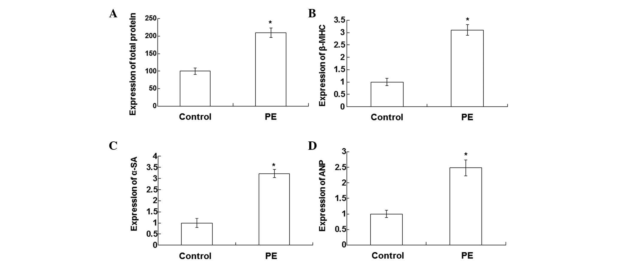
Expression levels of
hypertrophy-associated genes are increased in the PE-stimulated
cardiomyocytes
In order to determine the expression levels of
hypertrophy-associated genes in a cellular model of cardiac
hypertrophy, qPCR assays were performed. Total proteins were
detected in the PE-stimulated and normal cardiomyocytes using a
bicinchoninic acid assay. As shown in Fig. 2A, the total protein levels in the
PE-stimulated cardiomyocytes were significantly increased compared
with those in normal cardiomyocytes (P<0.05). The qPCR results
showed that the expression levels of β-MHC (Fig. 2B), α-SA (Fig. 2C) and ANP (Fig. 2D) in the PE-stimulated cardiomyocytes
were significantly increased compared with those in the normal
cardiomyocytes (P<0.05). These results indicate that the
expression levels of hypertrophy-associated genes are increased in
the PE-stimulated cardiomyocytes.
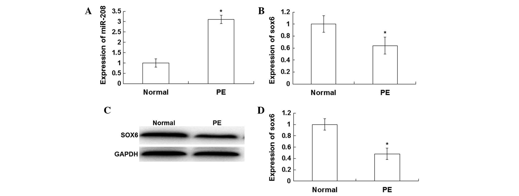
miR-208 expression levels are
increased, while SOX6 expression levels are decreased, in the
PE-stimulated cardiomyocytes
In order to investigate the expression levels of
miR-208 and SOX6 in the PE-stimulated cardiomyocytes and normal
cardiomyocytes, qPCR and western blot assays were performed. The
qPCR results showed that miR-208 expression levels were
significantly increased (4.23±0.21-fold) in the PE-stimulated
cardiomyocytes compared with those in the normal cardiomyocytes
(P<0.05; Fig. 3A). However, SOX6
expression levels were significantly decreased (0.68±0.17-fold) in
the PE-stimulated cardiomyocytes compared with those in the normal
cardiomyocytes (P<0.05; Fig. 3B).
As shown by the western blotting results in Fig. 3C, the expression levels of SOX6 were
significantly reduced in the PE-stimulated cardiomyocytes compared
with those in the normal cardiomyocytes. Relative values indicated
that the quantity of SOX6 expression levels in the PE-stimulated
cardiomyocytes were ~0.48±0.11-fold lower compared with those in
the normal cardiomyocytes (P<0.05; Fig. 3D). These results suggest that miR-208
expression levels are increased, whereas SOX6 expression levels are
decreased in the PE-stimulated cardiomyocytes.
miR-208 is associated with cardiac
hypertrophy by negative regulation of SOX6
In order to investigate the association between the
expression levels of miR-208 and SOX6, transfection of the
PE-stimulated cardiomyocytes with antagomiR-208 was performed. The
mRNA and protein expression levels of miR-208 and SOX6 were
detected using qPCR and western blot analysis, respectively. As
shown in Fig. 4A, following
transfection with antagomiR-208 in the PE-stimulated
cardiomyocytes, SOX6 expression levels were significantly increased
compared with those in untransfected PE-stimulated cardiomyocytes
and negative control cells. The results of the quantitative western
blot analysis are presented in Fig.
4B. The expression levels of SOX6, detected by qPCR assay, were
significantly increased in the antagomiR-208-transfected
cardiomyocytes compared with those of the untransfected
PE-stimulated and negative control cardiomyocytes (Fig. 4C). Following transfection of the
PE-stimulated cardiomyocytes with antagomiR-208, the total proteins
were significantly decreased (data not shown). The qPCR results
showed that the expression levels of β-MHC (Fig. 5A), α-SA (Fig. 5B) and ANP (Fig. 5C) in the PE-stimulated cardiomyocytes
after transfection with antagomiR-208 were significantly decreased
compared with those without transfection (P<0.05).
These results indicate that the inhibition of
miR-208 resulted in increased expression levels of SOX6. Therefore,
miR-208 may be associated with cardiac hypertrophy, mediated by the
negative regulation of SOX6.
Discussion
Cardiac hypertrophy is associated with the
expression of various genes. Expression of the transcription factor
myocardin may be inhibited by nuclear factor κ-light-chain-enhancer
of activated B cells, which modifies the gene expression profile in
cardiomyocytes, thereby inhibiting cardiac hypertrophy (13). Endothelin-1 is able to promote
cardiac hypertrophy via the upregulation of ANP (14). The stimulation of transient receptor
potential cation channel subfamily V member 1 can activate the
PPAR-σ signaling pathway, which plays an important role in
inhibiting cardiac hypertrophy. Therefore, identification of new
hypertrophy-associated genes and their underlying molecular
mechanisms has clinical significance for the diagnosis and
treatment of patients with essential hypertension accompanied with
cardiac hypertrophy.
In cardiac hypertrophy, the expression of various
miRNAs in cardiac mast cells exhibit significant differences from
those in healthy individuals, which involve a range of signal
pathways. Increased expression of miR-1 significantly inhibits
cardiac hypertrophy via the regulation of connexin 43 (15). In addition, adrenaline and
endothelin-induced cardiac hypertrophy can be inhibited by miR-133
in vitro. The expression of miR-133 is significantly
decreased in cardiac mast cells, which suggests that miR-133 is
closely associated with the development of cardiac hypertrophy.
Recent studies have indicated that miR-423, miR-34, miR-190 and
other miRNA molecules may be involved in the development of cardiac
hypertrophy (16,17).
In the present study, immunofluorescence staining,
western blot analysis and qPCR assays were performed to evaluate
the expression levels of miR-208 and SOX6 and their effects on the
progression of cardiac hypertrophy. The association between the
expression levels of miR-208 and SOX6 was investigated in a
cellular model of cardiac hypertrophy, and by transfecting a
proportion of the PE-treated rat cardiomyocytes with antagomiR-208.
The regulatory mechanism of miR-208 was further studied. The
present results showed that the expression levels of miR-208 were
significantly increased in the peripheral blood of patients with
left ventricular hypertrophy compared with those of healthy
individuals (P<0.05). The PE-stimulated cardiomyocytes were
significantly larger than normal cardiomyocytes. The expression
levels of hypertrophy related genes, including β-MHC, α-SA and ANP,
were increased in the PE-stimulated cardiomyocytes (P<0.05). The
qPCR results showed that the miR-208 expression levels were
significantly increased in the PE-stimulated cardiomyocytes
compared with normal cardiomyocytes (P<0.05). However, SOX6
expression levels were significantly decreased in the PE-stimulated
cardiomyocytes compared with the normal cardiomyocytes (P<0.05).
In the PE-stimulated cardiomyocytes that were transfected with
antagomiR-208, the SOX6 expression levels were significantly
increased. In addition, the total proteins and the mRNA expression
levels of hypertrophy-associated genes were significantly
decreased, which indicated that cardiac hypertrophy may be reduced
by inhibiting miR-208 by transfection with antagomiR-208.
In conclusion, miR-208 expression levels are
increased in the peripheral blood of patients with cardiac
hypertrophy. The expression levels of miR-208 are associated with
cardiac hypertrophy by negative regulation of SOX6. These results
suggest that miR-208 may be useful as a novel therapeutic and
diagnostic marker for cardiac hypertrophy.
Acknowledgements
The authors thanks Professor Xuemin Pang at the
Department of Cardiology, Zhumadian Central Hospital (Zhumadian,
China) for support.
References
|
1
|
Milouk FZ, El Bakkali M, Coghlan L,
Lachhab A, Aboudrar S and Benjelloun H: Kinetics of orthostatic
blood pressure in primary hypertension. Int Cardiovasc Res J.
8:83–88. 2014.PubMed/NCBI
|
|
2
|
Prkačin I, Balenovic D, Cavrić G, Bartolek
D and Bulum T: Importance of standardized stepwise screening in
patients with resistant hypertension. Acta Med Croatica.
68:111–115. 2014.PubMed/NCBI
|
|
3
|
Shah AS, Chin CW, Vassiliou V, Cowell SJ,
Doris M, Kwok TC, Semple S, Zamvar V, White AC, McKillop G, et al:
Left ventricular hypertrophy with strain and aortic stenosis.
Circulation. 130:1607–1616. 2014. View Article : Google Scholar : PubMed/NCBI
|
|
4
|
Kuster DW, Mulders J, Ten Cate FJ, Michels
M, Dos Remedios CG, da Costa Martins PA, van der Velden J and
Oudejans CB: MicroRNA transcriptome profiling in cardiac tissue of
hypertrophic cardiomyopathy patients with MYBPC3 mutations. J Mol
Cell Cardiol. 65:59–66. 2013. View Article : Google Scholar : PubMed/NCBI
|
|
5
|
Bernardo BC, Gao XM, Tham YK, Kiriazis H,
Winbanks CE, Ooi JY, Boey EJ, Obad S, Kauppinen S, Gregorevic P, et
al: Silencing of miR-34a attenuates cardiac dysfunction in a
setting of moderate, but not severe, hypertrophic cardiomyopathy.
PLoS One. 9:e903372014. View Article : Google Scholar : PubMed/NCBI
|
|
6
|
Hernandez-Torres F, Aranega AE and Franco
D: Identification of regulatory elements directing
miR-23a-miR-27a-miR-24-2 transcriptional regulation in response to
muscle hypertrophic stimuli. Biochim Biophys Acta. 1839:885–897.
2014. View Article : Google Scholar : PubMed/NCBI
|
|
7
|
Wang GK, Zhu JQ, Zhang JT, Li Q, Li Y, He
J, Qin YW and Jing Q: Circulating microRNA: A novel potential
biomarker for early diagnosis of acute myocardial infarction in
humans. Eur Heart J. 31:659–666. 2010. View Article : Google Scholar : PubMed/NCBI
|
|
8
|
Oliveira-Carvalho V, Carvalho VO, Silva
MM, Guimarães GV and Bocchi EA: MicroRNAs: A new paradigm in the
treatment and diagnosis of heart failure? Arq Bras Cardiol.
98:362–369. 2012. View Article : Google Scholar : PubMed/NCBI
|
|
9
|
Li H, Zheng D, Zhang B, Liu L, Ou J, Chen
W, Xiong S, Gu Y and Yang J: miR-208 promotes cell proliferation by
repressing SOX6 expression in human esophageal squamous cell
carcinoma. J Transl Med. 12:1962014. View Article : Google Scholar : PubMed/NCBI
|
|
10
|
Lee KE, Seo J, Shin J, Ji EH, Roh J, Kim
JY, Sun W, Muhr J, Lee S and Kim J: Positive feedback loop between
Sox2 and Sox6 inhibits neuronal differentiation in the developing
central nervous system. Proc Natl Acad Sci USA. 111:2794–2799.
2014. View Article : Google Scholar : PubMed/NCBI
|
|
11
|
Scott O, Pugh J, Kiddoo D, Sonnenberg LK,
Bamforth S and Goez HR: Global developmental delay, progressive
relapsing-remitting parkinsonism and spinal syrinx in a child with
SOX6 mutation. J Child Neurol. 29:NP164–NP167. 2014. View Article : Google Scholar : PubMed/NCBI
|
|
12
|
Prasad V, Lorenz JN, Lasko VM, et al:
Ablation of plasma membrane Ca2+-ATPase isoform 4
prevents development of hypertrophy in a model of hypertrophic
cardiomyopathy. J Mol Cell Cardiol. 77:53–63. 2014. View Article : Google Scholar : PubMed/NCBI
|
|
13
|
Liao XH, Wang N, Zhao DW, Zheng DL, Zheng
L, Xing WJ, Zhou H, Cao DS and Zhang TC: NF-κB (p65) negatively
regulates myocardin-induced cardiomyocyte hypertrophy through
multiple mechanisms. Cell Signal. 26:2738–2748. 2014. View Article : Google Scholar : PubMed/NCBI
|
|
14
|
You HW, Chen X, You HJ, Zhang YQ, Cai Y
and Liu GZ: Role of endothelin-1 and its receptors on hypertrophy
or proliferation of cultured cardial cells. Zhongguo Yi Xue Ke Xue
Yuan Xue Bao. 28:520–523. 2006.PubMed/NCBI
|
|
15
|
Wu Y, Ma XJ, Wang HJ, Li WC, Chen L, Ma D
and Huang GY: Expression of Cx43-related microRNAs in patients with
tetralogy of Fallot. World J Pediatr. 10:138–144. 2014. View Article : Google Scholar : PubMed/NCBI
|
|
16
|
Feng HJ, Ouyang W, Liu JH, Sun YG, Hu R,
Huang LH, Xian JL, Jing CF and Zhou MJ: Global microRNA profiles
and signaling pathways in the development of cardiac hypertrophy.
Braz J Med Biol Res. 47:361–368. 2014. View Article : Google Scholar : PubMed/NCBI
|
|
17
|
Huang J, Sun W, Huang H, Ye JI, Pan W,
Zhong Y, Cheng C, You X, Liu B, Xiong L and Liu S: miR-34a
modulates angiotensin II-induced myocardial hypertrophy by direct
inhibition of ATG9A expression and autophagic activity. PLoS One.
9:e943822014. View Article : Google Scholar : PubMed/NCBI
|