Relationship between deep medullary veins in susceptibility‑weighted imaging and ipsilateral cerebrovascular reactivity of middle cerebral artery in patients with ischemic stroke
- Authors:
- Published online on: March 28, 2016 https://doi.org/10.3892/etm.2016.3198
- Pages: 2217-2220
-
Copyright: © Han et al. This is an open access article distributed under the terms of Creative Commons Attribution License.
Metrics: Total
Views: 0 (Spandidos Publications: | PMC Statistics: )
Total PDF Downloads: 0 (Spandidos Publications: | PMC Statistics: )
Abstract
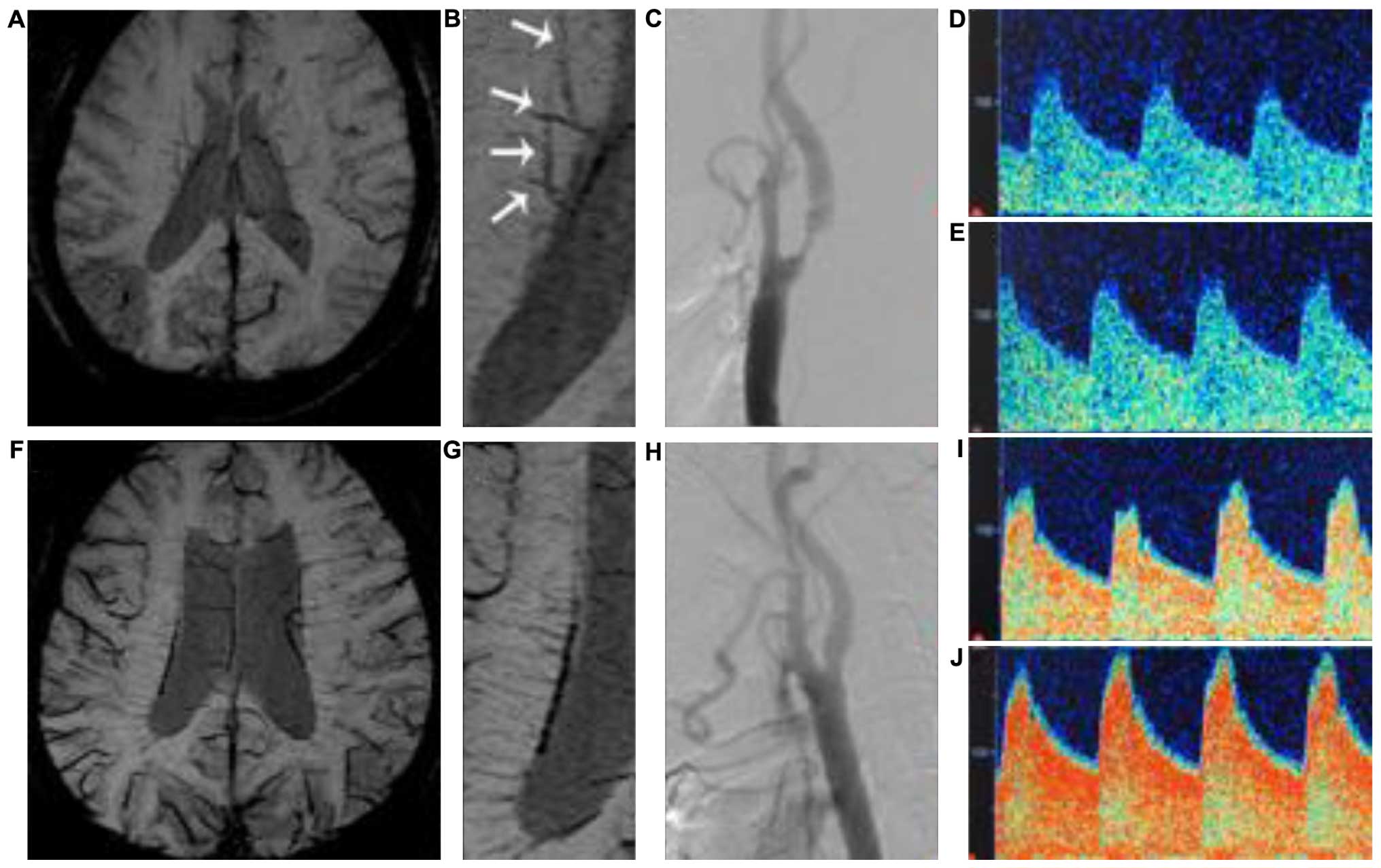
Deep cerebral veins have been recently associated with the severity of hemodynamic impairment in moyamoya disease. The aim of the current study was to determine the correlation of deep medullary veins (DMVs) in susceptibility‑weighted imaging (SWI) with ipsilateral cerebrovascular reactivity (CVR) of and anterior cecebrocervical artery stenosis in patients with ischemic stroke. Patients with unilateral TIA or infarction who underwent 3.0 T magnetic resonance imaging SWI, digital subtraction angiography and transcranial Doppler with CO2 stimulation within the first 7 days of hospitalization were retrospectively selected. CVR and stenosis of anterior cerebrocervical arteries were compared between different DMVs stages in symptomatic hemispheres (SHs) and asymptomatic hemispheres (AHs). A total of 61 patients were subsequently included in the present study. A univariate analysis was conducted and results for age (PAHs=0.004, PSHs=0.006), hypertension (PAHs=0.008, PSHs=0.020), current smoking (PAHs=0.006, PSHs=0.021), CVR (PAHs=0.000, PSHs=0.000), and artery stenosis (PAHs=0.000, PSHs=0.000) were obtained. The results suggested statistically significant differences between DMVs grades in SHs and AHs. A subsequent multivariate analysis revealed that CVR (ORAHs=0.925, 95% CIAHs: 0.873‑0.981; ORSHs=0.945, 95% CISHs: 0.896‑0.996), and artery stenosis (ORAH=3.147, 95% CIAH: 1.010‑9.806; ORSHs=2.882, 95% CISHs: 1.017‑8.166) were independent risk factors of DMVs. In conclusion, 3.0 T SWI was useful in detecting the DMVs around the lateral ventricle in patients with atherosclerotic ischemic stroke. CVR and stenosis of anterior cerebrocervical arteries were independent risk factors for ipsilateral DMVs in SHs and AHs.