Proteomic analysis of cerebrospinal fluid in amyotrophic lateral sclerosis
- Authors:
- Published online on: March 31, 2016 https://doi.org/10.3892/etm.2016.3210
- Pages: 2095-2106
-
Copyright: © Chen et al. This is an open access article distributed under the terms of Creative Commons Attribution License.
Metrics: Total
Views: 0 (Spandidos Publications: | PMC Statistics: )
Total PDF Downloads: 0 (Spandidos Publications: | PMC Statistics: )
Abstract
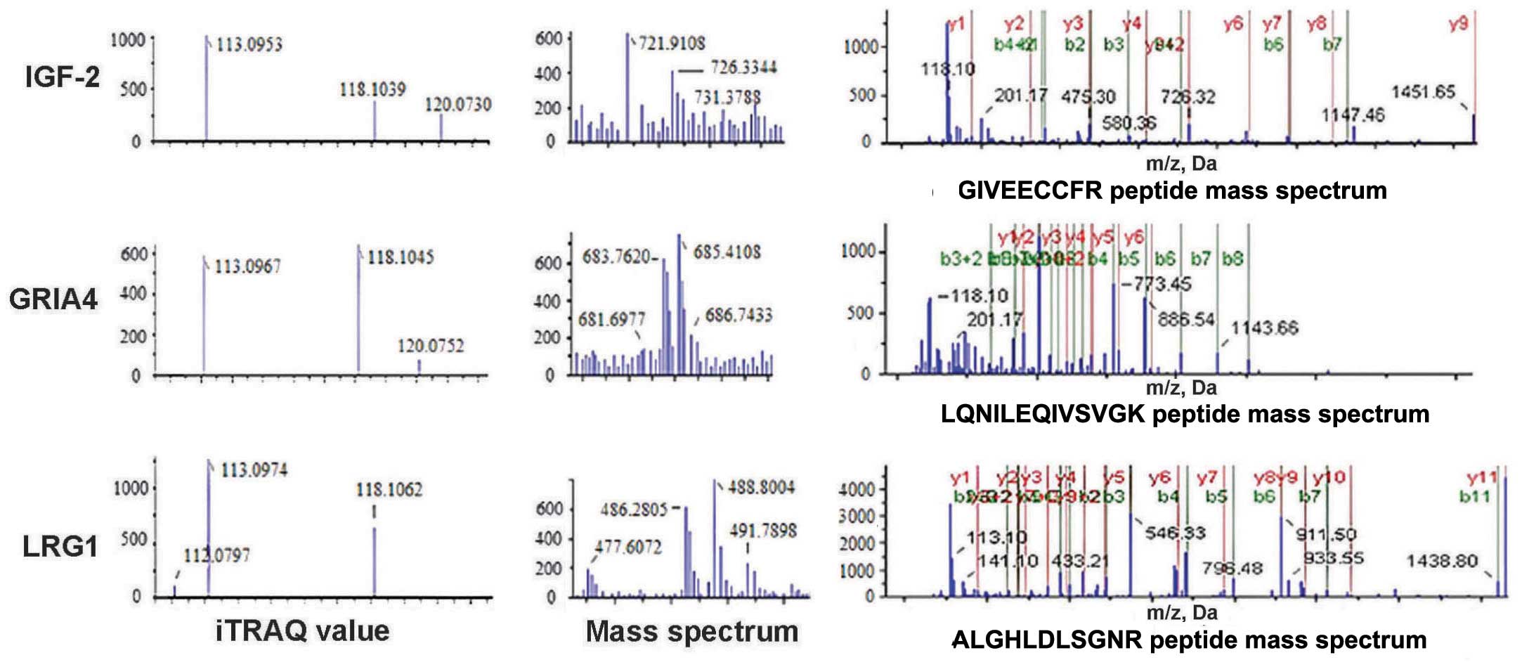
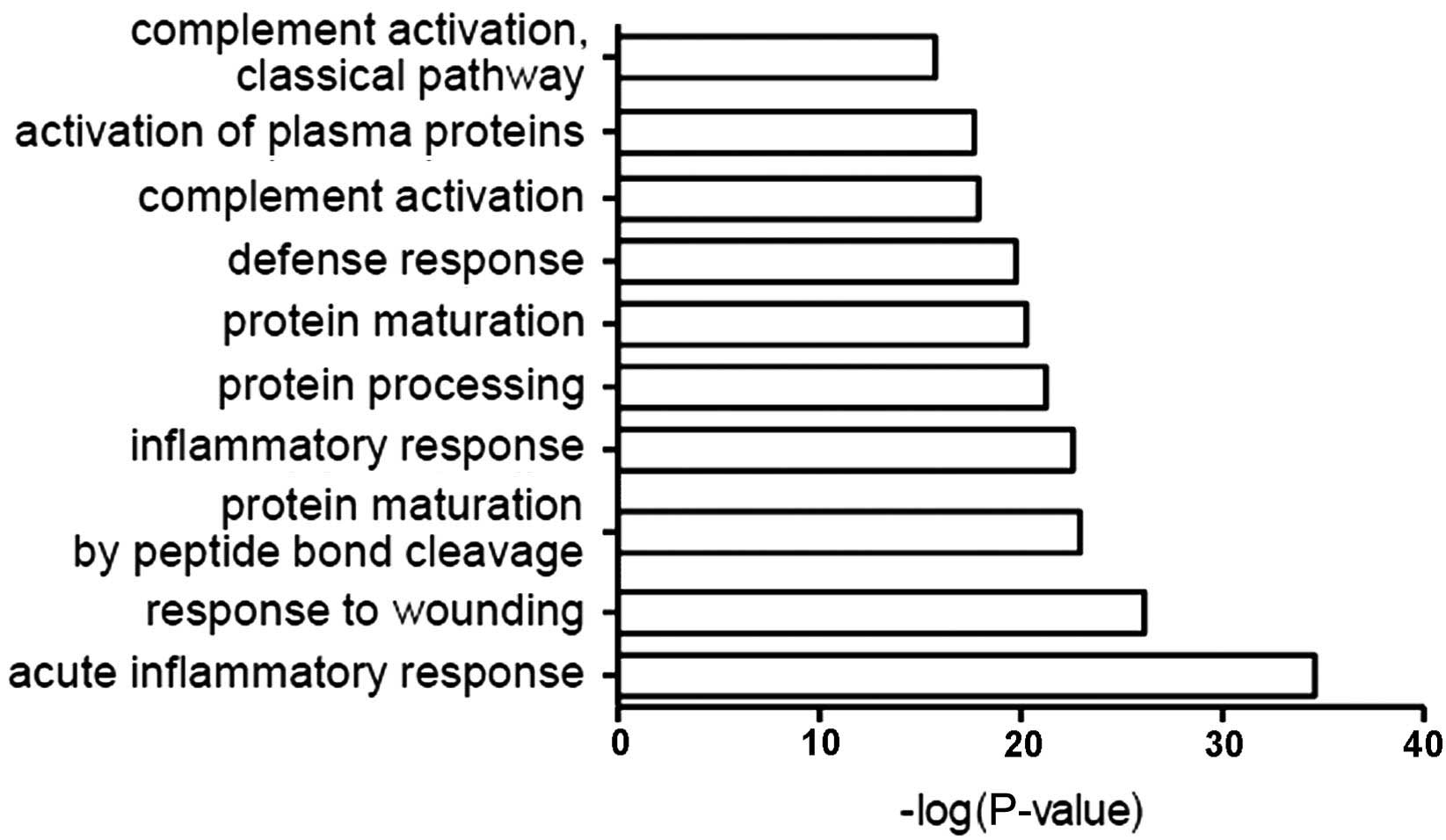
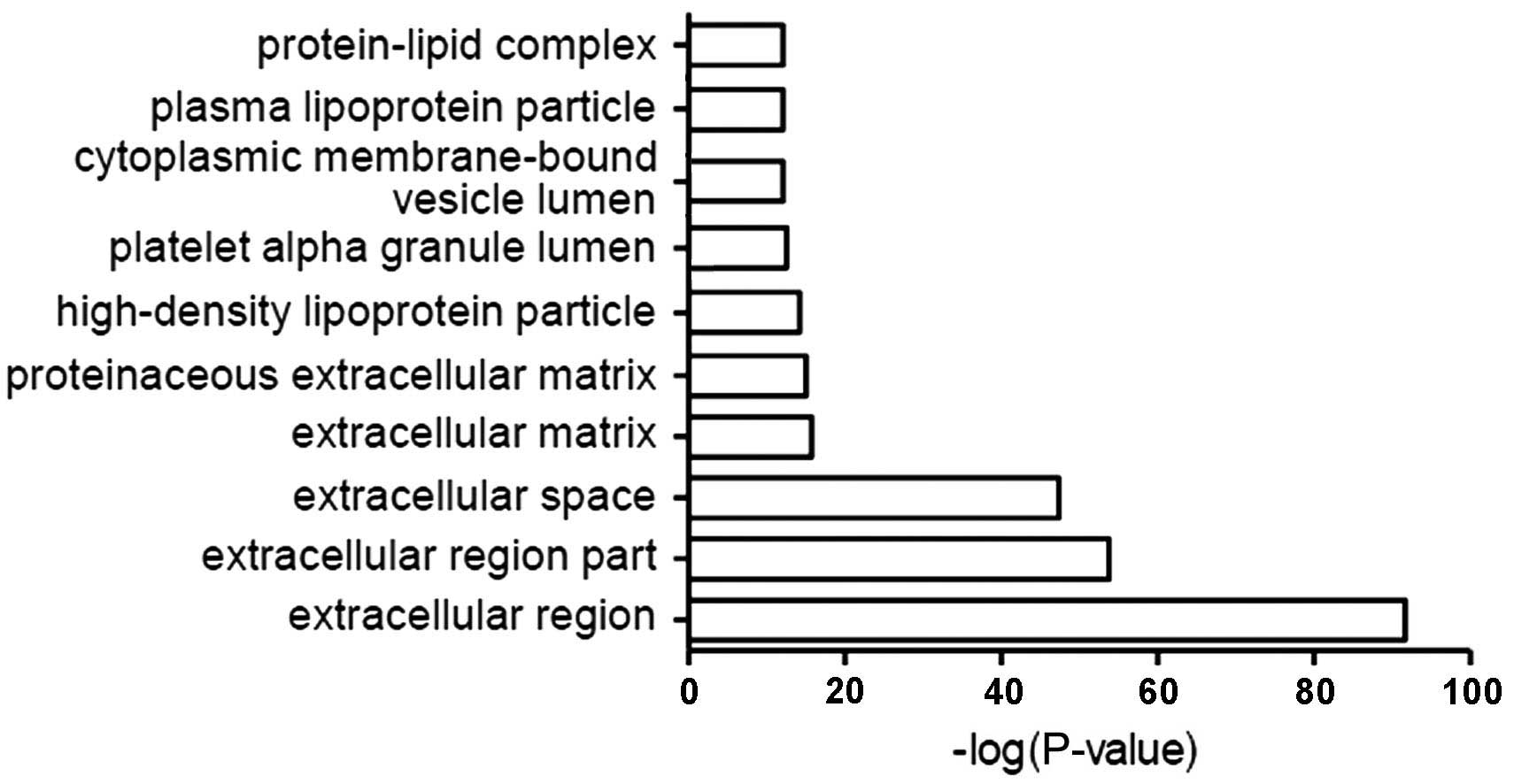
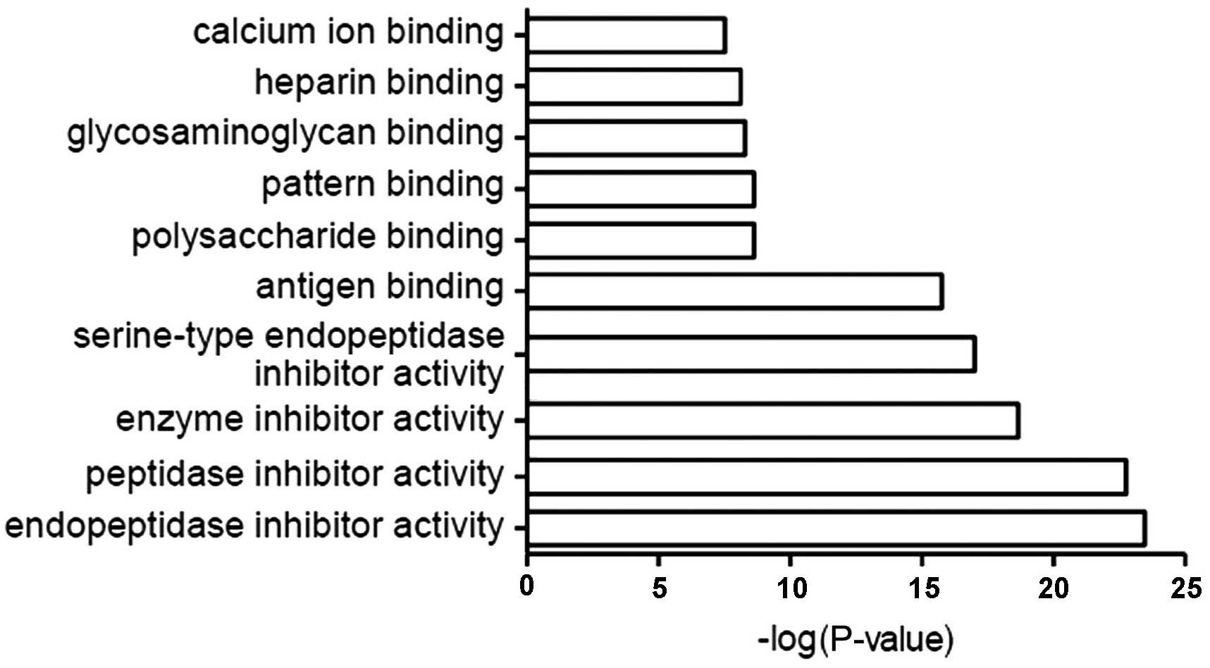
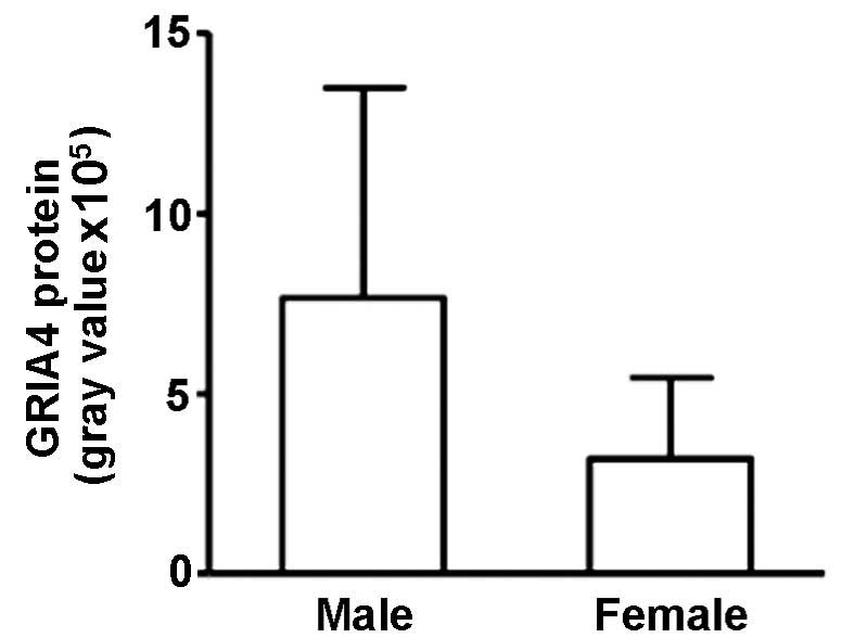
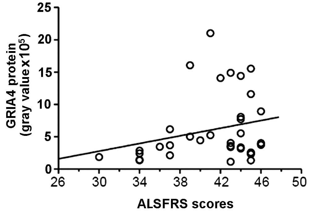
The present study used comparative proteomic analysis of cerebrospinal fluid (CSF) in amyotrophic lateral sclerosis (ALS) patients in order to identify proteins that may act as diagnostic biomarkers and indicators of the pathogenesis of ALS. This analysis was performed using isobaric tags for relative and absolute quantitation (iTRAQ) technology, coupled with 2‑dimensional liquid chromatography/mass spectrometry. Database for Annotation, Visualization and Integrated Discovery software was utilized for bioinformatic analysis of the data. Following this, western blotting was performed in order to examine the expression of 3 candidate proteins in ALS patients compared with healthy individuals [as a normal control (NC) group] or patients with other neurological disease (OND); these proteins were insulin‑like growth factor II (IGF‑2), glutamate receptor 4 (GRIA4) and leucine‑rich α‑2‑glycoprotein 1 (LRG1). Clinical data, including gender, age, disease duration and ALS functional rating scale (ALSFRS‑R) score, were also collected in the ALS patients. Multiple linear regression analysis was performed between the clinical data and the results of western blot analysis. A total of 248 distinct proteins were identified in the ALS and NC groups, amongst which a significant difference could be identified in 35 proteins; of these, 21 proteins were downregulated and 14 were upregulated. These differentially‑expressed proteins were thus revealed to be associated with ALS. The western blot analysis confirmed a proportion of the data attained in the iTRAQ analysis, revealing the differential protein expression of IGF‑2 and GRIA4 between the ALS and NC groups. IGF‑2 was significantly downregulated in ALS patients (P=0.017) and GRIA4 was significantly upregulated (P=0.016). These results were subsequently validated in the 35‑patient ALS and OND groups (P=0.002), but no significant difference was identified in LRG1 expression between these groups. GRIA4 protein expression was higher in male than female patients and was positively correlated with the ALSFRS‑R score, meaning that GRIA4 expression was negatively correlated with the severity of ALS, while IGF‑2 and LRG1 expression did not correlate with any clinical data. The present study thus demonstrated that GRIA4 expression levels, as a marker of severity, may be used as a reference for the timing of treatment, and that IGF‑2 may serve as an effective biomarker of ALS progression.