Open Access

Diagnosis and stage of Graves' ophthalmopathy: Efficacy of quantitative measurements of the lacrimal gland based on 3-T magnetic resonance imaging

  • Authors:
    • Hao Hu
    • Xiao‑Quan Xu
    • Fei‑Yun Wu
    • Huan‑Huan Chen
    • Guo‑Yi Su
    • Jie Shen
    • Xun‑Ning Hong
    • Hai‑Bin Shi
  • View Affiliations

  • Published online on: May 25, 2016     https://doi.org/10.3892/etm.2016.3389
  • Pages: 725-729
  • Copyright: © Hu et al. This is an open access article distributed under the terms of Creative Commons Attribution License.

Metrics: Total Views: 0 (Spandidos Publications: | PMC Statistics: )
Total PDF Downloads: 0 (Spandidos Publications: | PMC Statistics: )


Abstract

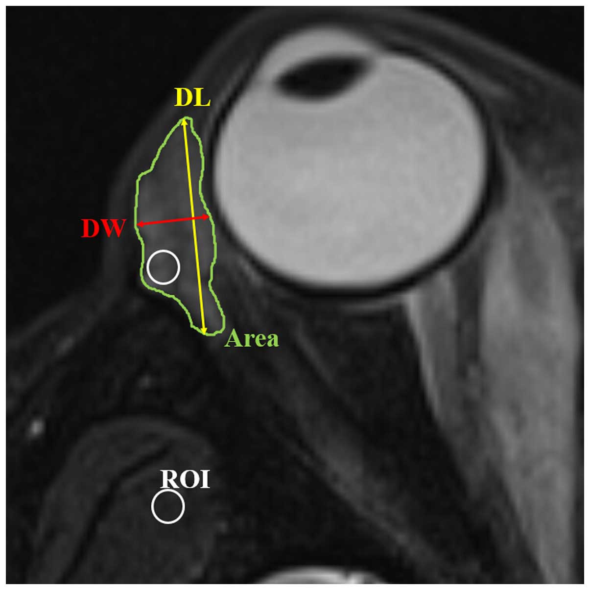
The aim of the present study was to determine the efficacy of quantitative measurements of the lacrimal gland based on 3-T magnetic resonance (MR) imaging in the diagnosis and staging of Graves' ophthalmopathy (GO). The study retrospectively enrolled 33 patients with GO (the GO group) and 24 healthy volunteers [the healthy control (HC) group] with orbit MR imaging performed using a 3‑T MR scanner. Quantitative parameters of the lacrimal gland, including axial length, axial width, axial area, coronal length, coronal width, coronal area, volume and signal intensity ratio (SIR) of the lacrimal gland to the ipsilateral temporal muscle were measured. The difference of quantitative parameters between the GO and HC groups, or between active and inactive GO groups were evaluated using a Student's t-test. Receiver operating characteristic analyses were used to evaluate the diagnostic value of the significant parameters in discriminating patients with GO from healthy controls, or discriminating active from inactive GO. All the quantitative measurements of the GO patients were significantly larger than those of the healthy controls (P<0.05), with the exception of the coronal length (P=0.150). Axial and coronal width had the best efficacy in discriminating patients with GO from healthy controls. Only SIR was found to be different between the active and inactive GO groups (P=0.001). Setting a SIR of 2.57 as the threshold value, the optimal efficacy was achieved (area under the curve, 0.711; sensitivity, 57.7%; specificity, 77.5%) in discriminating between active and inactive GO. Quantitative measurements of the lacrimal gland based on 3‑T MR imaging may assist in the diagnosis and stage of GO.
View Figures
View References

Related Articles

Journal Cover

August-2016
Volume 12 Issue 2

Print ISSN: 1792-0981
Online ISSN:1792-1015

Sign up for eToc alerts

Recommend to Library

Copy and paste a formatted citation
x
Spandidos Publications style
Hu H, Xu XQ, Wu FY, Chen HH, Su GY, Shen J, Hong XN and Shi HB: Diagnosis and stage of Graves' ophthalmopathy: Efficacy of quantitative measurements of the lacrimal gland based on 3-T magnetic resonance imaging. Exp Ther Med 12: 725-729, 2016.
APA
Hu, H., Xu, X., Wu, F., Chen, H., Su, G., Shen, J. ... Shi, H. (2016). Diagnosis and stage of Graves' ophthalmopathy: Efficacy of quantitative measurements of the lacrimal gland based on 3-T magnetic resonance imaging. Experimental and Therapeutic Medicine, 12, 725-729. https://doi.org/10.3892/etm.2016.3389
MLA
Hu, H., Xu, X., Wu, F., Chen, H., Su, G., Shen, J., Hong, X., Shi, H."Diagnosis and stage of Graves' ophthalmopathy: Efficacy of quantitative measurements of the lacrimal gland based on 3-T magnetic resonance imaging". Experimental and Therapeutic Medicine 12.2 (2016): 725-729.
Chicago
Hu, H., Xu, X., Wu, F., Chen, H., Su, G., Shen, J., Hong, X., Shi, H."Diagnosis and stage of Graves' ophthalmopathy: Efficacy of quantitative measurements of the lacrimal gland based on 3-T magnetic resonance imaging". Experimental and Therapeutic Medicine 12, no. 2 (2016): 725-729. https://doi.org/10.3892/etm.2016.3389