Effects of different sutures on fibrosis and wound healing in a rabbit model of corneal wounds
- Authors:
- Ying Li
- Hui J. Chen
- Hua Zhang
- Jian G. Wu
- Yun T. Hu
- Zhi Z. Ma
View Affiliations
Affiliations: Department of Ophthalmology (Eye Center), Peking University Third Hospital, Beijing 100191, P.R. China, Department of Statistics, Peking University Third Hospital, Beijing 100191, P.R. China
- Published online on: September 14, 2016 https://doi.org/10.3892/etm.2016.3703
-
Pages:
2827-2834
-
Copyright: © Li
et al. This is an open access article distributed under the
terms of Creative
Commons Attribution License.
Metrics: Total
Views: 0 (Spandidos Publications: | PMC Statistics: )
Metrics: Total PDF Downloads: 0 (Spandidos Publications: | PMC Statistics: )
This article is mentioned in:
Abstract
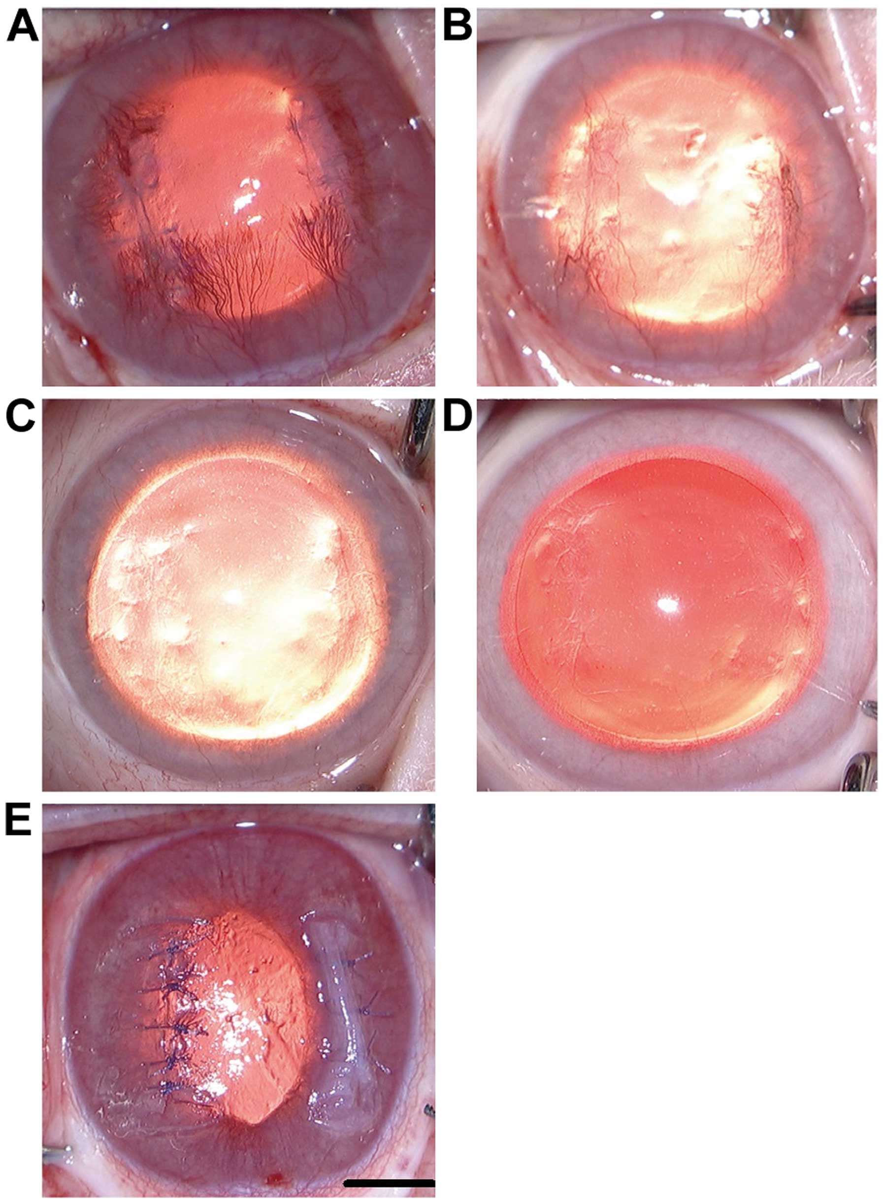
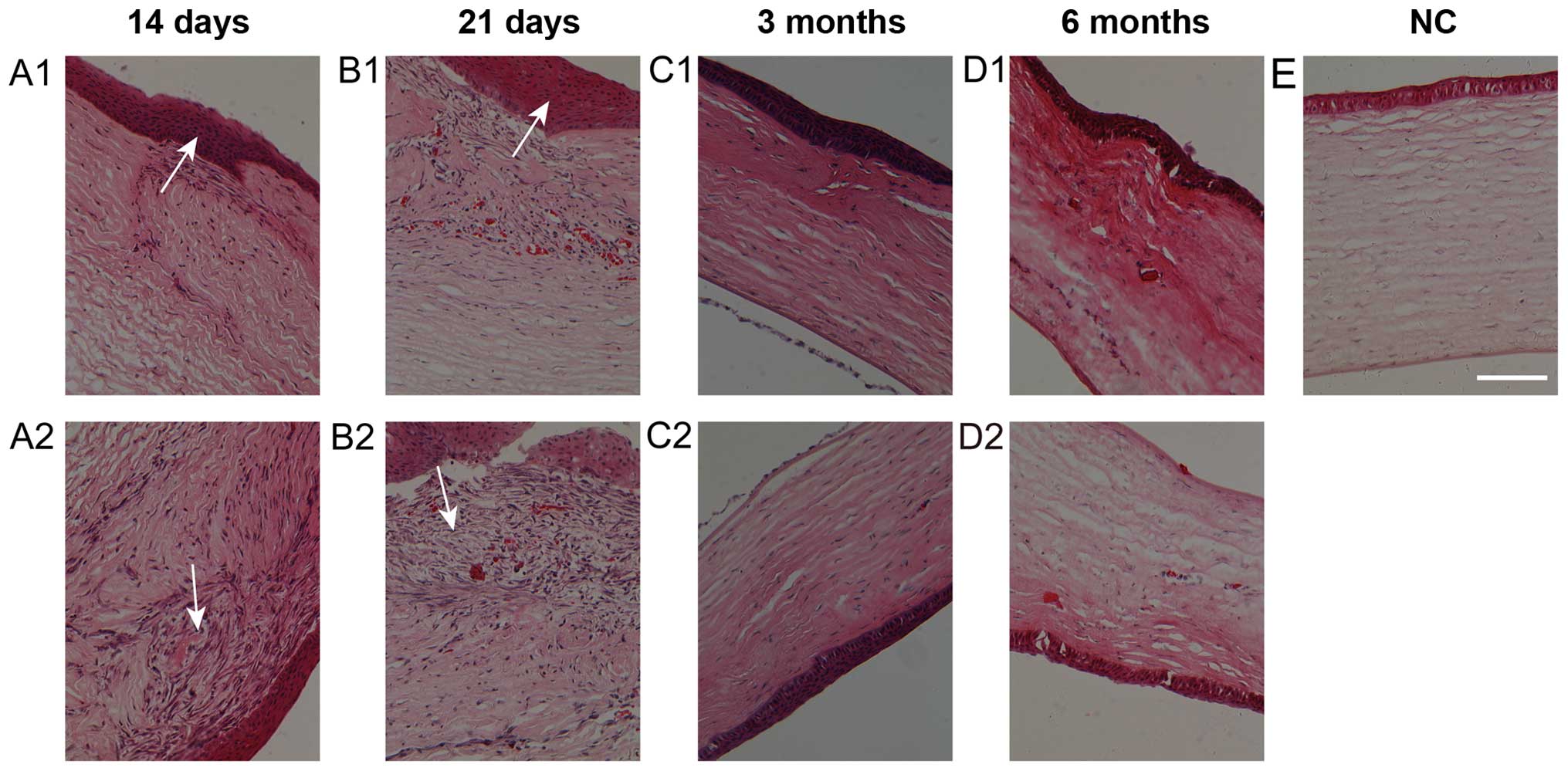
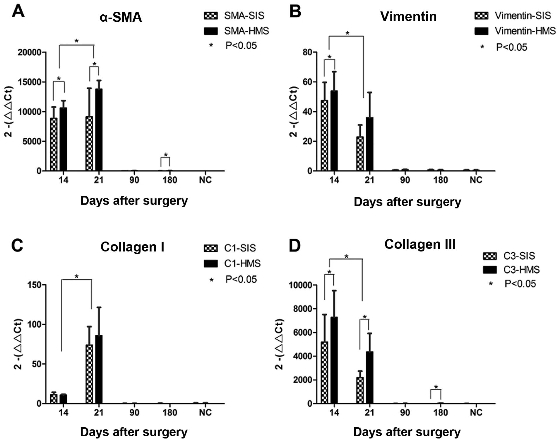
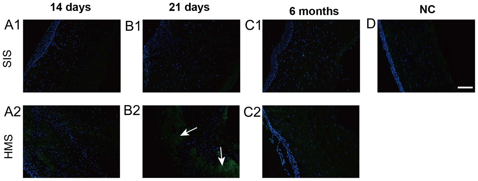
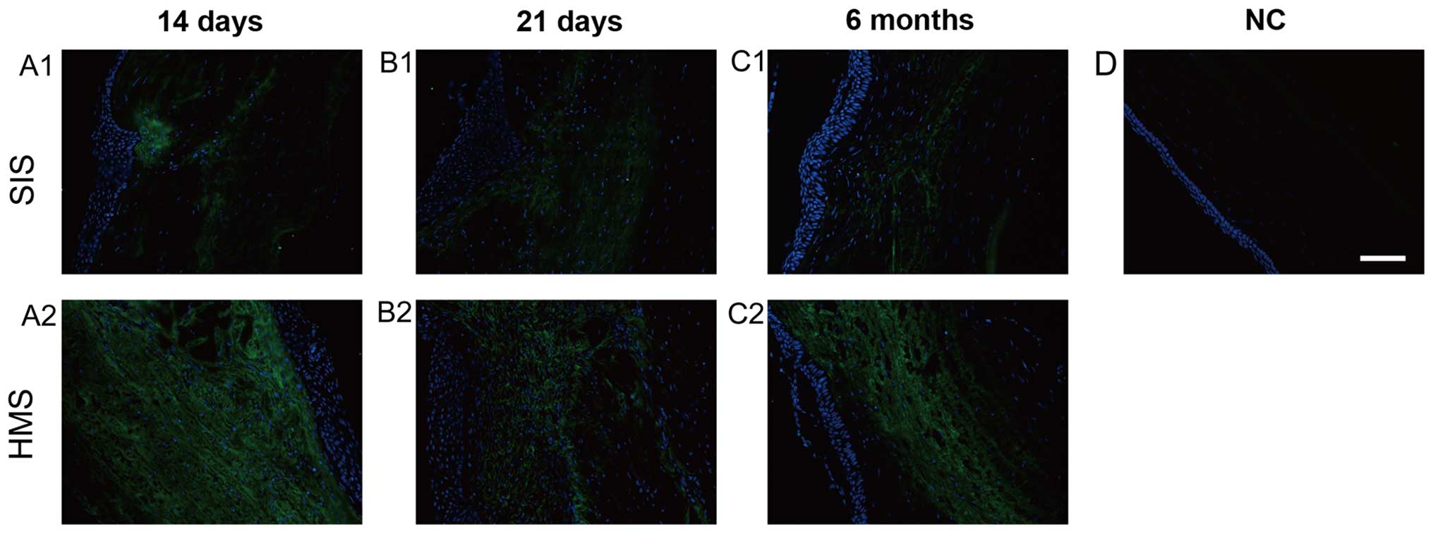
The aim of the study was to investigate wound healing and scar formation in rabbit corneal lamellar wounds repaired with simple interrupted sutures (SIS) or horizontal mattress sutures (HMS). Two parallel ‘I’‑shaped lamellar cornea wounds were created in one eye of 40 white New Zealand rabbits, while 5 uninjured rabbits were sacrificed to serve as normal controls. One side of the wounds, in the test rabbits, was closed with SIS, while the other side was treated with HMS. Ten days later, the stitches were removed under anesthesia. The animals were sacrificed on days 14 and 21, and months 3 and 6 after the suturing surgery, and corneal samples were subjected to histological and immunofluorescent studies: α‑smooth muscle actin (α‑SMA) and vimentin were used to detect myofibroblasts and fibroblasts, respectively, and collagen type I and Ⅲ was used to detect extracellular matrix (ECM) deposition. Relevant mRNA levels were assessed by quantitative polymerase chain reaction (qPCR) to elucidate the differences in wound healing and formation of fibrosis. Macroscopic and hematoxylin and eosin staining observations showed that the two sides of the wounds developed the most prominent fibrotic tissue on day 21. The immunofluorescence and qPCR results showed that HMS wounds produced increased α‑SMA, vimentin and collagen type Ⅲ compared to the SIS wounds on day 14 or 21. The collagen type I expression showed no distinctive difference in SIS and HMS wounds. In conclusion, corneal lamellar wounds treated with SIS developed less fibrotic‑related proteins and related mRNA in the early stages of wound healing than wounds treated with HMS. Although differences were not distinct after 3 months, the results of the present study suggest a benefit in choosing SIS over HMS, as at least the initial fibrotic process seems more benign with SIS. Corneal wounds should be carefully sutured, ensuring the tissue is well aligned.
View Figures |
Figure 1
|
|
Figure 2
|
|
Figure 3
|
|
Figure 4
|
|
Figure 5
|
|
Figure 6
|
|
Figure 7
|
View References
1
|
Thomas JR and Somenek M: Scar revision
review. Arch Facial Plast Surg. 14:162–174. 2012. View Article : Google Scholar : PubMed/NCBI
|
2
|
Henderson J, Sutcliffe M and Gillespie P:
Epitendinous suture techniques in extensor tendon repairs - an
experimental evaluation. J Hand Surg Am. 36:1968–1973. 2011.
View Article : Google Scholar : PubMed/NCBI
|
3
|
Kan CD and Yang YJ: Double telescopic
anastomosis with interrupted suture technique in acute aortic
dissection. Ann Thorac Surg. 91:1630–1631. 2011. View Article : Google Scholar : PubMed/NCBI
|
4
|
Fini ME and Stramer BM: How the cornea
heals: Cornea-specific repair mechanisms affecting surgical
outcomes. Cornea. 24(Suppl 8): S2–S11. 2005. View Article : Google Scholar : PubMed/NCBI
|
5
|
Myrna KE, Pot SA and Murphy CJ: Meet the
corneal myofibroblast: The role of myofibroblast transformation in
corneal wound healing and pathology. Vet Ophthalmol. 12(Suppl 1):
25–27. 2009. View Article : Google Scholar : PubMed/NCBI
|
6
|
Klingberg F, Hinz B and White ES: The
myofibroblast matrix: Implications for tissue repair and fibrosis.
J Pathol. 229:298–309. 2013. View Article : Google Scholar : PubMed/NCBI
|
7
|
Weis AJ, Huxlin KR, Callan CL, DeMagistris
MA and Hindman HB: Keratocyte apoptosis and not myofibroblast
differentiation mark the graft/host interface at early time-points
post-DSAEK in a cat model. PLoS One. 8:e756232013. View Article : Google Scholar : PubMed/NCBI
|
8
|
Chaurasia SS, Kaur H, de Medeiros FW,
Smith SD and Wilson SE: Dynamics of the expression of intermediate
filaments vimentin and desmin during myofibroblast differentiation
after corneal injury. Exp Eye Res. 89:133–139. 2009. View Article : Google Scholar : PubMed/NCBI
|
9
|
Bargagna-Mohan P, Paranthan RR, Hamza A,
Zhan CG, Lee DM, Kim KB, Lau DL, Srinivasan C, Nakayama K, Nakayama
KI, et al: Corneal antifibrotic switch identified in genetic and
pharmacological deficiency of vimentin. J Biol Chem. 287:989–1006.
2012. View Article : Google Scholar : PubMed/NCBI
|
10
|
Karamichos D, Guo XQ, Hutcheon AE and
Zieske JD: Human corneal fibrosis: An in vitro model. Invest
Ophthalmol Vis Sci. 51:1382–1388. 2010. View Article : Google Scholar : PubMed/NCBI
|
11
|
Volk SW, Wang Y, Mauldin EA, Liechty KW
and Adams SL: Diminished type III collagen promotes myofibroblast
differentiation and increases scar deposition in cutaneous wound
healing. Cells Tissues Organs. 194:25–37. 2011. View Article : Google Scholar : PubMed/NCBI
|
12
|
Boote C, Du Y, Morgan S, Harris J,
Kamma-Lorger CS, Hayes S, Lathrop KL, Roh DS, Burrow MK, Hiller J,
et al: Quantitative assessment of ultrastructure and light scatter
in mouse corneal debridement wounds. Invest Ophthalmol Vis Sci.
53:2786–2795. 2012. View Article : Google Scholar : PubMed/NCBI
|
13
|
Guo S and Dipietro LA: Factors affecting
wound healing. J Dent Res. 89:219–229. 2010. View Article : Google Scholar : PubMed/NCBI
|
14
|
Zuber TJ: The mattress sutures: Vertical,
horizontal, and corner stitch. Am Fam Physician. 66:2231–2236.
2002.PubMed/NCBI
|