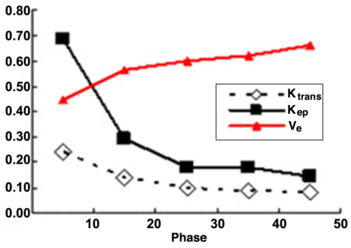
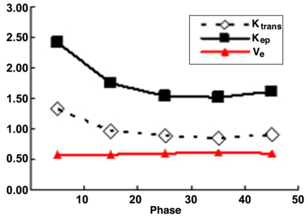
|
1
|
Aberle DR, Chiles C, Gatsonis C, Hillman
BJ, Johnson CD, McClennan BL, Mitchell DG, Pisano ED, Schnall MD
and Sorensen AG: American College of Radiology Imaging Network:
Imaging and cancer: Research strategy of the American College of
Radiology Imaging Network. Radiology. 235:741–751. 2005. View Article : Google Scholar : PubMed/NCBI
|
|
2
|
Leung JW: Screening mammography reduces
morbidity of breast cancer treatment. AJR Am J Roentgenol.
184:1508–1509. 2005. View Article : Google Scholar : PubMed/NCBI
|
|
3
|
Peters NH, Rinkes IH Borel, Zuithoff NP,
Mali WP, Moons KG and Peeters PH: Meta-analysis of MR imaging in
the diagnosis of breast lesions. Radiology. 246:116–124. 2008.
View Article : Google Scholar : PubMed/NCBI
|
|
4
|
Jones EF, Sinha SP, Newitt DC, Klifa C,
Kornak J, Park CC and Hylton NM: MRI enhancement in stromal tissue
surrounding breast tumors: Association with recurrence free
survival following neoadjuvant chemotherapy. PloS One.
8:e619692013. View Article : Google Scholar : PubMed/NCBI
|
|
5
|
Li X, Arlinghaus LR, Ayers GD,
Chakravarthy AB, Abramson RG, Abramson VG, Atuegwu N, Farley J,
Mayer IA, Kelley MC, et al: DCE-MRI analysis methods for predicting
the response of breast cancer to neoadjuvant chemotherapy: Pilot
study findings. Magn Reson Med. 71:1592–1602. 2104. View Article : Google Scholar
|
|
6
|
Baek HM, Chen JH, Nie K, Yu HJ, Bahri S,
Mehta RS, Nalcioglu O and Su MY: Predicting pathologic response to
neoadjuvant chemotherapy in breast cancer by using MR imaging and
quantitative 1H MR spectroscopy. Radiology. 251:653–662.
2009. View Article : Google Scholar : PubMed/NCBI
|
|
7
|
Magometschnigg HF, Helbich T, Brader P,
Abeyakoon O, Baltzer P, Füger B, Wengert G, Polanec S, Bickel H and
Pinker K: Molecular imaging for the characterization of breast
tumors. Expert Rev Anticancer Ther. 14:711–722. 2014. View Article : Google Scholar : PubMed/NCBI
|
|
8
|
Rahbar H and Partridge SC: Multiparametric
MR Imaging of Breast Cancer. Magn Reson Imaging Clin N Am.
24:223–238. 2016. View Article : Google Scholar : PubMed/NCBI
|
|
9
|
Petralia G, Bonello L, Priolo F, Summers P
and Bellomi M: Breast MR with special focus on DW-MRI and DCE-MRI.
Cancer Imaging. 11:76–90. 2011. View Article : Google Scholar : PubMed/NCBI
|
|
10
|
Koo HR, Cho N, Song IC, Kim H, Chang JM,
Yi A, Yun BL and Moon WK: Correlation of perfusion parameters on
dynamic contrast-enhanced MRI with prognostic factors and subtypes
of breast cancers. J Magn Reson Imaging. 36:145–151. 2012.
View Article : Google Scholar : PubMed/NCBI
|
|
11
|
El Khouli RH, Macura KJ, Kamel IR, Jacobs
MA and Bluemke DA: 3-T dynamic contrast-enhanced MRI of the breast:
Pharmacokinetic parameters versus conventional kinetic curve
analysis. AJR Am J Roentgenol. 197:1498–1505. 2011. View Article : Google Scholar : PubMed/NCBI
|
|
12
|
Ma ZS, Wang DW, Sun XB, Shi H, Pang T,
Dong GQ and Zhang CQ: Quantitative analysis of 3-Tesla magnetic
resonance imaging in the differential diagnosis of breast lesions.
Exp Ther Med. 9:913–918. 2015.PubMed/NCBI
|
|
13
|
Yim H, Kang DK, Jung YS, Jeon GS and Kim
TH: Analysis of kinetic curve and model-based perfusion parameters
on dynamic contrast enhanced MRI in breast cancer patients:
Correlations with dominant stroma type. Magn Reson Imaging.
34:60–65. 2016. View Article : Google Scholar : PubMed/NCBI
|
|
14
|
Li J, Yu Y, Zhang Y, Bao S, Wu C, Wang X,
Li J, Zhang X and Hu J: A clinically feasible method to estimate
pharmacokinetic parameters in breast cancer. Med Phys.
36:3786–3794. 2009. View Article : Google Scholar : PubMed/NCBI
|
|
15
|
Amarnath J, Sangeeta T and Mehta SB: Role
of quantitative pharmacokinetic parameter (transfer constant:
K(trans)) in the characterization of breast lesions on MRI. Indian
J Radiol Imaging. 23:19–25. 2013. View Article : Google Scholar : PubMed/NCBI
|
|
16
|
Li L, Wang K, Sun X, Wang K, Sun Y, Zhang
G and Shen B: Parameters of dynamic contrast-enhanced MRI as
imaging markers for angiogenesis and proliferation in human breast
cancer. Med Sci Monit. 21:376–382. 2015. View Article : Google Scholar : PubMed/NCBI
|
|
17
|
Ocak I, Bernardo M, Metzger G, Barrett T,
Pinto P, Albert PS and Choyke PL: Dynamic contrast-enhanced MRI of
prostate cancer at 3 T: A study of pharmacokinetic parameters. AJR
Am J Roentgenol. 189:8492007. View Article : Google Scholar : PubMed/NCBI
|