First branchial cleft anomalies in children: Experience with 30 cases
- Authors:
- Wanpeng Li
- Liming Zhao
- Hongming Xu
- Xiaoyan Li
View Affiliations
Affiliations: Department of Otolaryngology‑Head and Neck Surgery, Shanghai Children's Hospital, Shanghai Jiao Tong University, Shanghai 200062, P.R. China
- Published online on: May 24, 2017 https://doi.org/10.3892/etm.2017.4511
-
Pages:
333-337
-
Copyright: © Li
et al. This is an open access article distributed under the
terms of Creative
Commons Attribution License.
Metrics: Total
Views: 0 (Spandidos Publications: | PMC Statistics: )
Metrics: Total PDF Downloads: 0 (Spandidos Publications: | PMC Statistics: )
This article is mentioned in:
Abstract
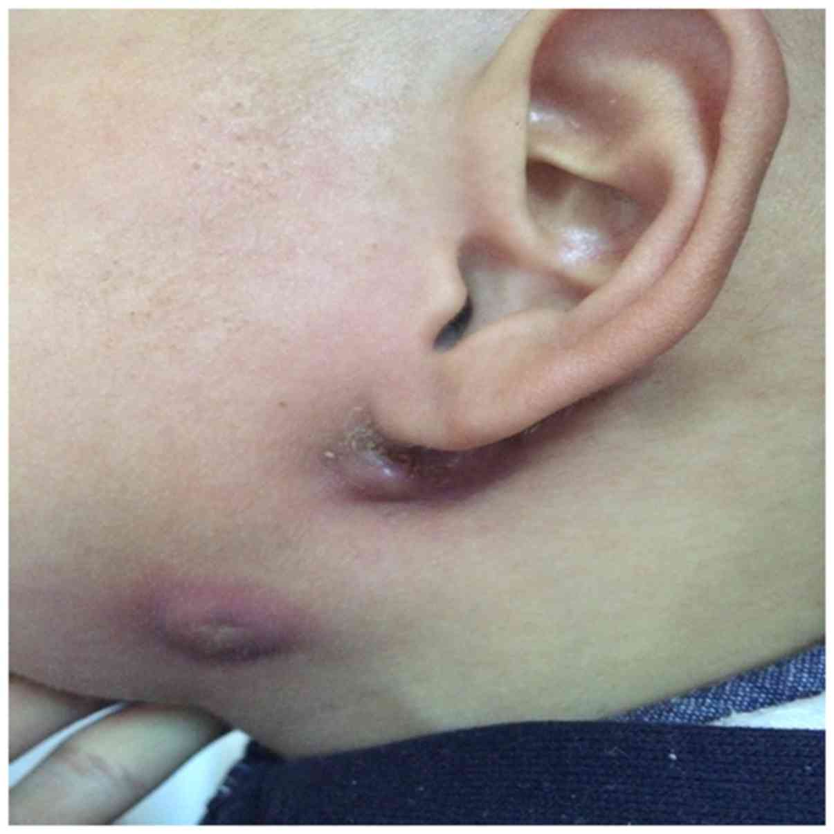
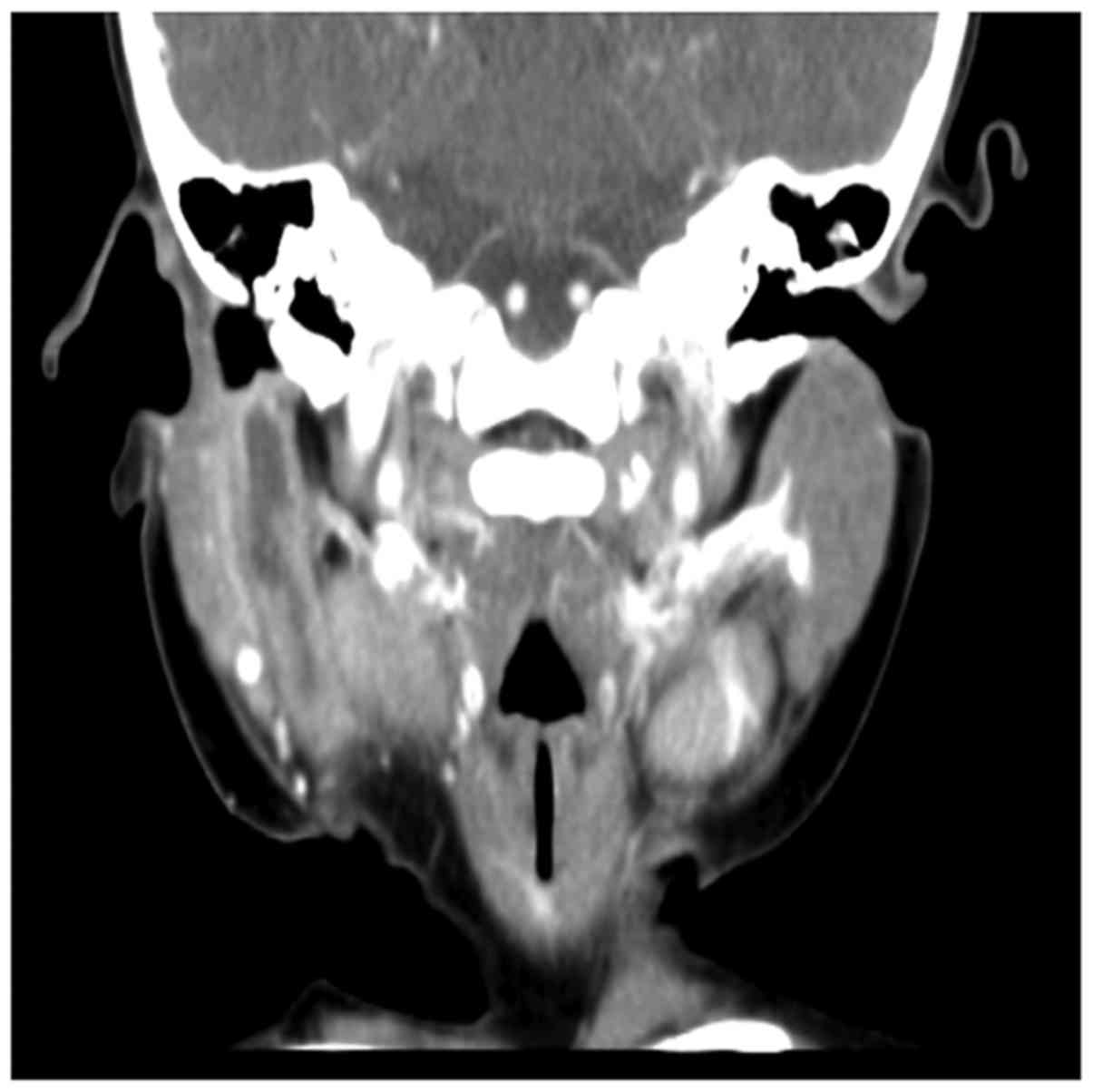
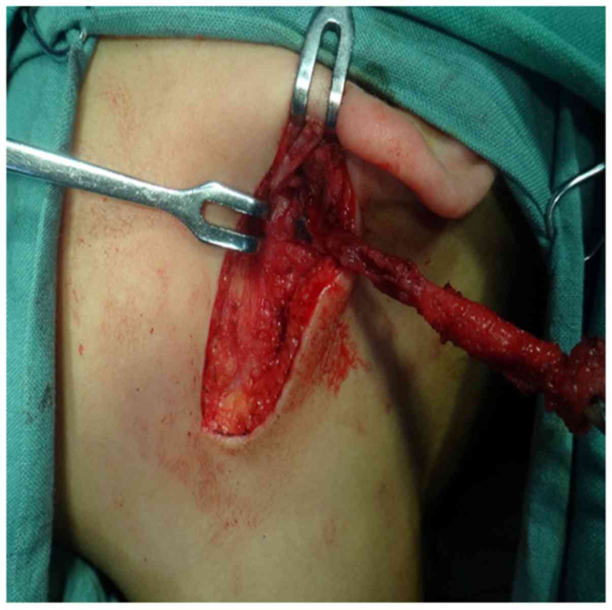
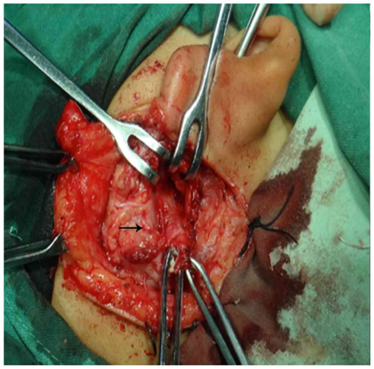
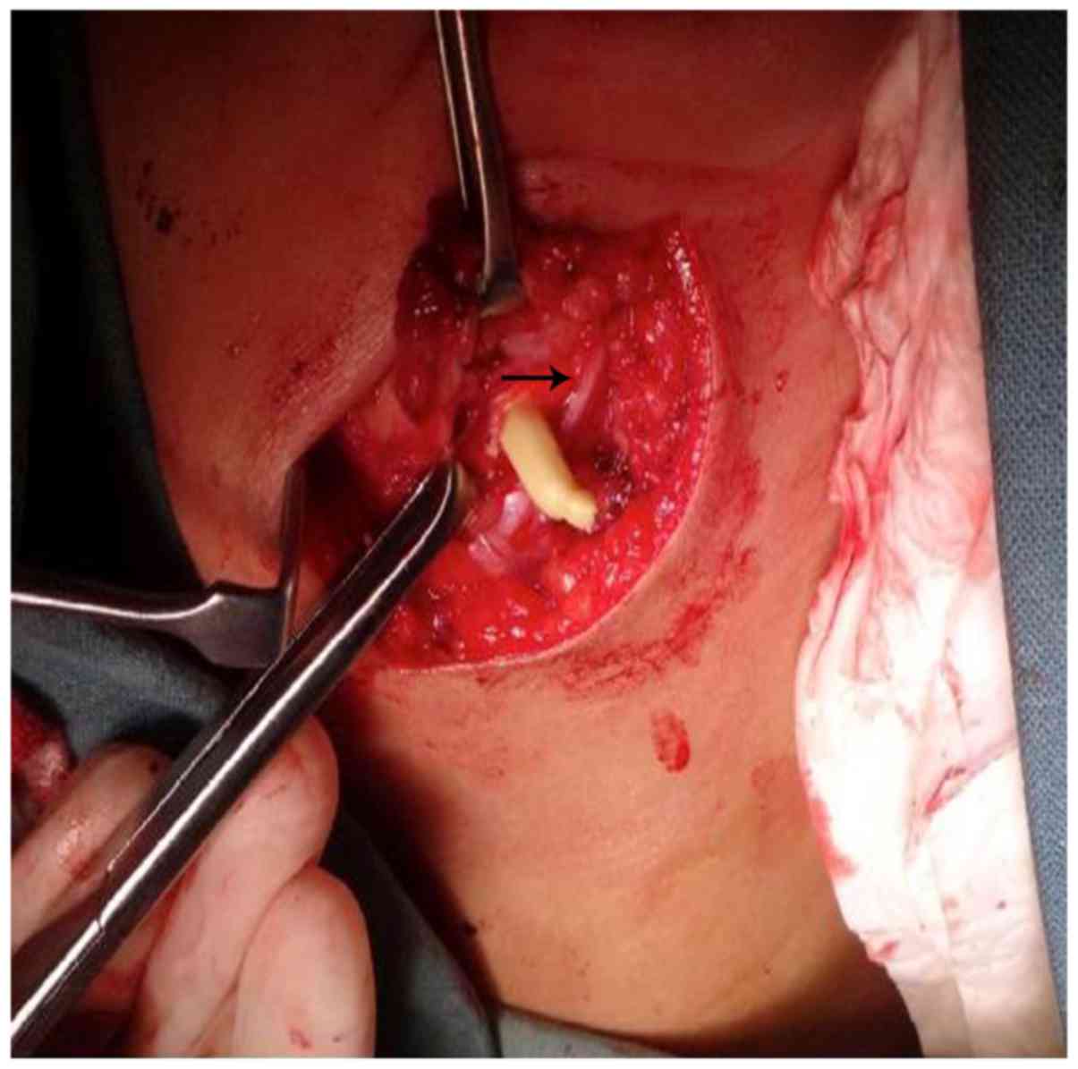
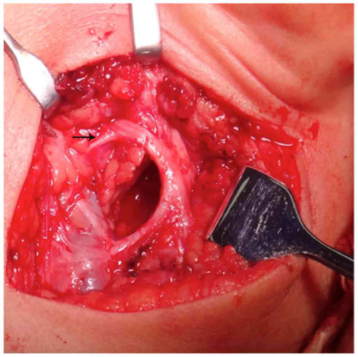
First branchial cleft anomalies (FBCA) are rare in the clinical setting, as they account for 1 to 8% of all branchial abnormalities. The purpose of this study is to explore the relationship between the fistula tract and facial nerve and the surgical method of FBCA. This retrospective study included 30 cases of FBCA in children managed from 2009 to 2016. All patients underwent surgery to remove the tract of the FBCA. We reviewed the clinical data of the patients to obtain their demographics and management. Thirty patients (11 male and 19 female) with anomalies of FBCA were diagnosed. The ages ranged from 1 to 13 years (median, 3 years). Twenty cases had a close relationship with the parotid gland. The facial nerve was identified in 20 of the 30 patients. The tract ran deep to the facial nerve in 3 cases, superficial to it in 21 cases, and passed between the branches of the nerve in 6 cases. The facial nerve was not identified in ten patients, as the tract was superficial to it. There were 2 cases of postoperative temporary facial paralysis (2/30, 6.7%). The symptoms gradually improved after one month, 1 case had permanent facial paralysis (1/30, 3.3%), and 1 case had postoperative recurrence. Complete excision of the tract is the only way to manage FBCA, and the course of the tracts vary and have different relationships with the facial nerve. There are 3 types: Superficial, deep to the facial nerve, and between the branches of the nerve. Therefore, surgical approaches differ among the various types, and careful preoperative planning and protecting the facial nerve during resection of the tract are essential.
View Figures |
Figure 1
|
|
Figure 2
|
|
Figure 3
|
|
Figure 4
|
|
Figure 5
|
|
Figure 6
|
View References
1
|
Bajaj Y, Ifeacho S, Tweedie D, Jephson CG,
Albert DM, Cochrane LA, Wyatt ME, Jonas N and Hartley BE: Branchial
anomalies in children. Int J Pediatr Otorhinolaryngol.
75:1020–1023. 2011. View Article : Google Scholar : PubMed/NCBI
|
2
|
Mitroi M, Dumitrescu D, Simionescu C,
Popescu C, Mogoantă C, Cioroianu L, Surlin C, Căpitănescu A and
Georgescu M: Management of second branchial cleft anomalies. Rom J
Morphol Embryol. 49:69–74. 2008.PubMed/NCBI
|
3
|
Chandler RJ and Mitchell B: Branchial
cleft cysts, sinuses, and fistulas. Otolaryngol Clin North Am.
14:175–186. 1981.PubMed/NCBI
|
4
|
Benson MT, Dalen K, Mancuso AA, Kerr HH,
Cacciarelli AA and Mafee MF: Congenital anomalies of the branchial
apparatus: Embryology and pathologic anatomy. Radiographics.
12:943–960. 1992. View Article : Google Scholar : PubMed/NCBI
|
5
|
D'Souza AR, Uppal HS, De R and Zeitoun H:
Updating concepts of first branchial cleft defects: A literature
review. Int J Pediatr Otorhinolaryngol. 62:103–109. 2002.
View Article : Google Scholar : PubMed/NCBI
|
6
|
Rattan KN, Rattan S, Parihar D, Gulia JS
and Yadav SP: Second branchial cleft fistula: Is fistulogram
necessary for complete excision. Int J Pediatr Otorhinolaryngol.
70:1027–1030. 2006. View Article : Google Scholar : PubMed/NCBI
|
7
|
Quintanilla-Dieck L, Virgin F, Wootten C,
Goudy S and Penn E Jr: Surgical approaches to first branchial cleft
anomaly excision: A case series. Case Rep Otolaryngol.
2016:39029742016.PubMed/NCBI
|
8
|
Olsen KD, Maragos NE and Weiland LH: First
branchial cleft anomalies. Laryngoscope. 90:423–436. 1980.
View Article : Google Scholar : PubMed/NCBI
|
9
|
Tham YS and Low WK: First branchial cleft
anomalies have relevance in otology and more. Ann Acad Med
Singapore. 34:335–338. 2005.PubMed/NCBI
|
10
|
Ford GR, Balakrishnan A, Evans JN and
Bailey CM: Branchial cleft and pouch anomalies. J Laryngol Otol.
106:137–143. 1992. View Article : Google Scholar : PubMed/NCBI
|
11
|
Arnot RS: Defects of the first branchial
cleft. S Afr J Surg. 9:93–98. 1971.PubMed/NCBI
|
12
|
Work WP: Newer concepts of first branchial
cleft defects. Laryngoscope. 82:1581–1593. 1972. View Article : Google Scholar : PubMed/NCBI
|
13
|
Triglia JM, Nicollas R, Ducroz V, Koltai
PJ and Garabedian EN: First branchial cleft anomalies: A study of
39 cases and a review of the literature. Arch Otolaryngol Head Neck
Surg. 124:291–295. 1998. View Article : Google Scholar : PubMed/NCBI
|
14
|
Shinn JR, Purcell PL, Horn DL, Sie KC and
Manning SC: First branchial cleft anomalies: Otologic
manifestations and treatment outcomes. Otolaryngol Head Neck Surg.
152:506–512. 2015. View Article : Google Scholar : PubMed/NCBI
|
15
|
Guo YX and Guo CB: Relation between a
first branchial cleft anomaly and the facial nerve. Br J Oral
Maxillofac Surg. 50:259–263. 2012. View Article : Google Scholar : PubMed/NCBI
|
16
|
Piccioni M, Bottazzoli M, Nassif N,
Stefini S and Nicolai P: Intraoperative use of fibrin glue dyed
with methylene blue in surgery for branchial cleft anomalies.
Laryngoscope. 126:2147–2150. 2016. View Article : Google Scholar : PubMed/NCBI
|