Introduction
Autoimmune thyroiditis (AIT) is also known as
Hashimoto's thyroiditis or chronic lymphocytic thyroiditis, and is
a relatively common specific immunity disease of the organs
(1). AIT is the second most common
thyroid disorder and accounts for 21% of all thyroid disorders
(2). Recently, due to environmental
and multiple other factors, the morbidity of the disease has been
increasing year by year (3).
Traditional Chinese medicine (TCM) considers that the etiology of
the disease is closely associated with internal injury and eating
disorders (4). Due to the disease
not having clear clinical symptoms at an early stage, the majority
of patients only present increase of thyroid autoantibodies
(4). AIT is often diagnosed as a
subclinical stage of chronic lymphocytic thyroiditis. Currently,
there are no specific conventional medicine strategies for
effective treatment of AIT. TCM applies the therapeutic principle
of treatment based on syndrome differentiation and specimen
consideration, so as to improve immunocompetence and develop an
effective therapy (5). However, no
drugs are currently available in clinical practice for the
effective treatment of the disease (5). At present, medical treatment for AIT
mainly adopts the replacement of thyroid hormones, immunization
therapy and surgical treatment; however, toxicity, side effects and
recurrence restrain the development of an effective strategy
(6). Therefore, alternative
therapies have certain superiority with regards to safety and
curative stability in the treatment of AIT (7).
Apoptosis is a spontaneous and programmed cell death
process that occurs under the induction of specific physiological
or pathological factors in cells (8). Morphological characteristics of
apoptosis are mainly reflected in the following five aspects:
Vesicles on cytomembrane, concentration of chromatin, release of
cytochrome c, potential reduction of mitochondrial membrane,
and DNA bond fission and loss of contact with neighboring cells
(8). The role of apoptosis is mainly
reflected in the normal differentiation of cell (9). In addition, the immune response of
autoimmune T and B lymphocytes also depends on apoptosis (10).
Polyphenols are components of green tea, accounting
for 22–30% of the tea dry weight, and are mainly composed of
catechins, flavonoid, phenolic acid and anthocyanidins (11). As the key components of green tea,
catechins account for 60–80% of the total tea polyphenol content.
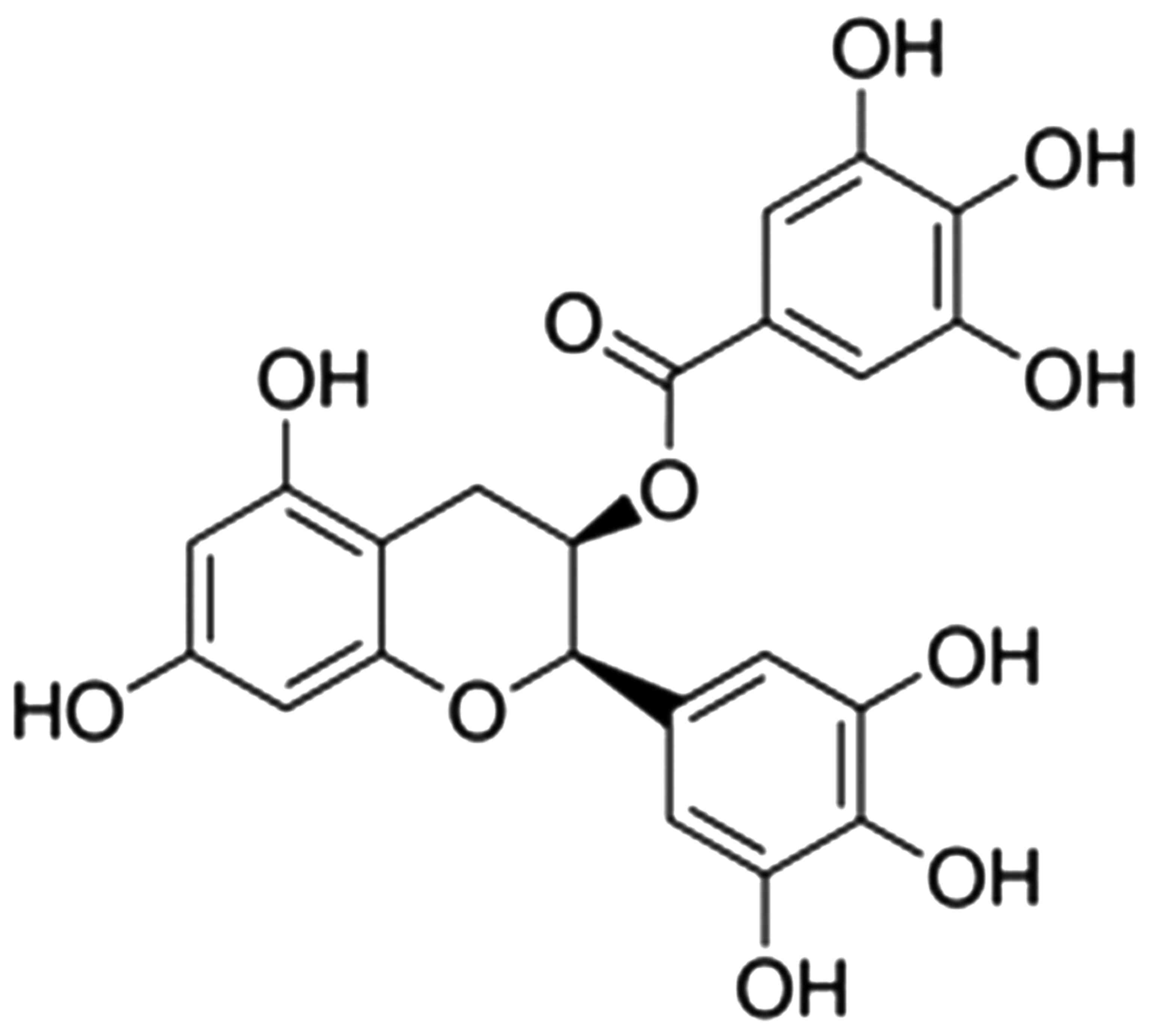
(−)-Epigallocatechin-3-gallate (EGCG) is a catechin present in
green tea that can relieve the inflammatory process in skin
diseases associated with blood vessels, and its structural formula
is shown in Fig. 1. In dermatitis,
vascular endothelial growth factor (VEGF) and interleukin-8 (IL-8)
serve an important role (12), EGCG
and soy isoflavone inhibit NHK proliferation, but do not induce
apoptosis. EGCG can also inhibit the IL-8 release in NHK that is
induced by preventing TNF-α expression, while isoflavone only has a
weak effect (11). Green tea
contains various natural active materials, and as the catechin
compound with the highest content in green tea, EGCG has multiple
important physiological and pharmacologic activities (13). In the current study, it was
investigated whether EGCG exerts a neuroprotective effect on AIT
and examined the possible underlying mechanism.
Materials and methods
Animals and chemicals
A total of 30 female Lewis rats (4-weeks-old;
weight, 80±2 g) were purchased from Beijing Vital River Laboratory
Animal Technology Co., Ltd. (Beijing, China). Animals were kept
under a temperature of 24±2°C and humidity of 55±5% with a 12 h
light/dark cycle, and were allowed free access to food and water.
The rats were randomly divided into three groups (n=10 rats per
group): Sham control, AIT model and EGCG groups.
Next, rats in the AIT model group and EGCG group
were immunized by subcutaneous injection of 0.1 ml pig
thyroglobulin (8 mg/ml) in incomplete Freund's adjuvant (both from
Sigma-Aldrich; Merck, Darmstadt, Germany) once every 2 weeks for 6
weeks. Rats in the EGCG group were injected intraperitoneally with
0.5 mg/kg EGCG three times with a 1-h interval at 3 h. Rats in the
Sham control or AIT group were injected intraperitoneally with 100
µl of PBS. The animal experiments were conducted according to the
ethical guidelines approved by the Medical Ethics Review Committee
of the First Center Hospital of Tianjin (Tianjin, China).
Iodine intake level
Prior to euthanasia, urine was collected from the
rats. Urine iodine levels were determined by arsenic cerium
catalytic spectrophotometry method (Victor 3 Multilabel Counter
system; PerkinElmer, Inc., Waltham, MA, USA) as mentioned
previously (14).
Body and thyroid weight
measurement
The weights of the body and thyroid of the rats were
measured following euthanasia. Next, the relative thyroid weight
was calculated as the thyroid wet weight (mg)/rat body weight
(g).
Thyroid pathological features
measurement
The thyroid was fixed using 4% paraformaldehyde for
24 h at room temperature, embedded into paraffin, and sectioned
into 0.5-µm thick sections. The thyroid pathological features were
classified into four grades, as follows: 0, indicating normal
physiology; 1, slight disruption of thyroid follicles (two or more
thyroid follicles); 2, moderate destruction of thyroid follicles
(10–40% of thyroid sections); 3, severe disruption of thyroid
physiology (>40% of thyroid sections) (14).
Antibody-capture ELISA for detection
of IL-1β, interferon-γ (IFN-γ) and TNF-α levels
Thyroid tissue samples (20 mg) were collected from
rats after euthanasia and homogenized in radioimmunoprecipitation
assay (RIPA) buffer (Sigma-Aldrich; Merck). IL-1β (EK-R30172),
IFN-γ (EK-R30149) and TNF-α (EK-R31092) levels were determined with
sandwich ELISA kits (Shanghai BlueGene Biotech Co., Ltd., Shanghai,
China). The absorbance of samples was determined using an ELISA
reader (Model ELX800, BioTek Instruments, Inc., Winooski, VT, USA)
at 450 nM.
Western blot analysis
Thyroid tissue samples were collected from rats
following euthanasia and homogenized in RIPA buffer
(Sigma-Aldrich). Total protein was acquired following
centrifugation at 12,000 × g for 10 min at 4°C, and protein
concentration was measured using a bicinchoninic acid assay method
(Beyotime Institute of Biotechnology, Haimen, China). Equal amounts
of protein (50 µg) were subjected to 10–15% SDS-polyacrylamide gel
electrophoresis and then transferred to a nitrocellulose membrane
(EMD Millipore, Bedford, MA, USA). The membranes were blocked with
5% skimmed milk powder in Tris-buffered saline with Tween-20 for 1
h at 37°C and incubated with primary antibodies against nuclear
factor-κB (NF-κB; cat. no. sc-71675; dilution, 1:500; Santa Cruz
Biotechnology, Inc., Dallas, TX, USA), B-cell lymphoma-2 (Bcl-2;
cat. no. sc-7382; dilution, 1:500; Santa Cruz Biotechnology, Inc.),
TNF-α-related apoptosis-inducing ligand (TRAIL; cat. no. sc-8440;
dilution, 1:500, Santa Cruz Biotechnology, Inc.) and GAPDH (cat.
no. B661204; dilution, 1:500; Sangon Biotech Co., Ltd., Shanghai,
China) at 4°C overnight. The blots were then washed with
Tris-buffered saline/Tween-20 and incubated with a horseradish
peroxidase-conjugated secondary antibody (cat. no. sc-2314;
dilution, 1:2,000; Sangon Biotech Co., Ltd.). Protein was detected
with BeyoECL Star Plus reagent (Beyotime Institute of
Biotechnology) and analyzed using Bio-Rad Laboratories Q 3.0
software (Bio-Rad Laboratories, Inc., Hercules, CA, USA).
Statistical analysis
Results are expressed as the mean ± standard error
of the mean using SPSS 17.0 (SPSS, Inc., Chicago, IL, USA).
Statistical analyses were performed using one-way analysis of
variance followed by a Student-Newman-Keuls test for multiple
comparisons. Differences with P<0.05 were considered as
statistically significant.
Results
Effect of EGCG treatment on urinary
iodine values in AIT rats
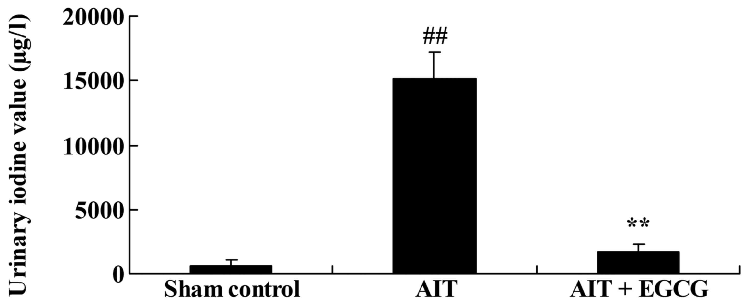
The neuroprotective effect of EGCG in AIT rats was
initially evaluated by investigating the urinary iodine values.
Compared with the sham control group, urinary iodine values of the
AIT rat model were significantly increased (Fig. 2). However, pretreatment with EGCG
significantly inhibited the AIT-induced urinary iodine values,
compared with those in the untreated AIT model group (Fig. 2).
Body and thyroid weights in AIT
rats
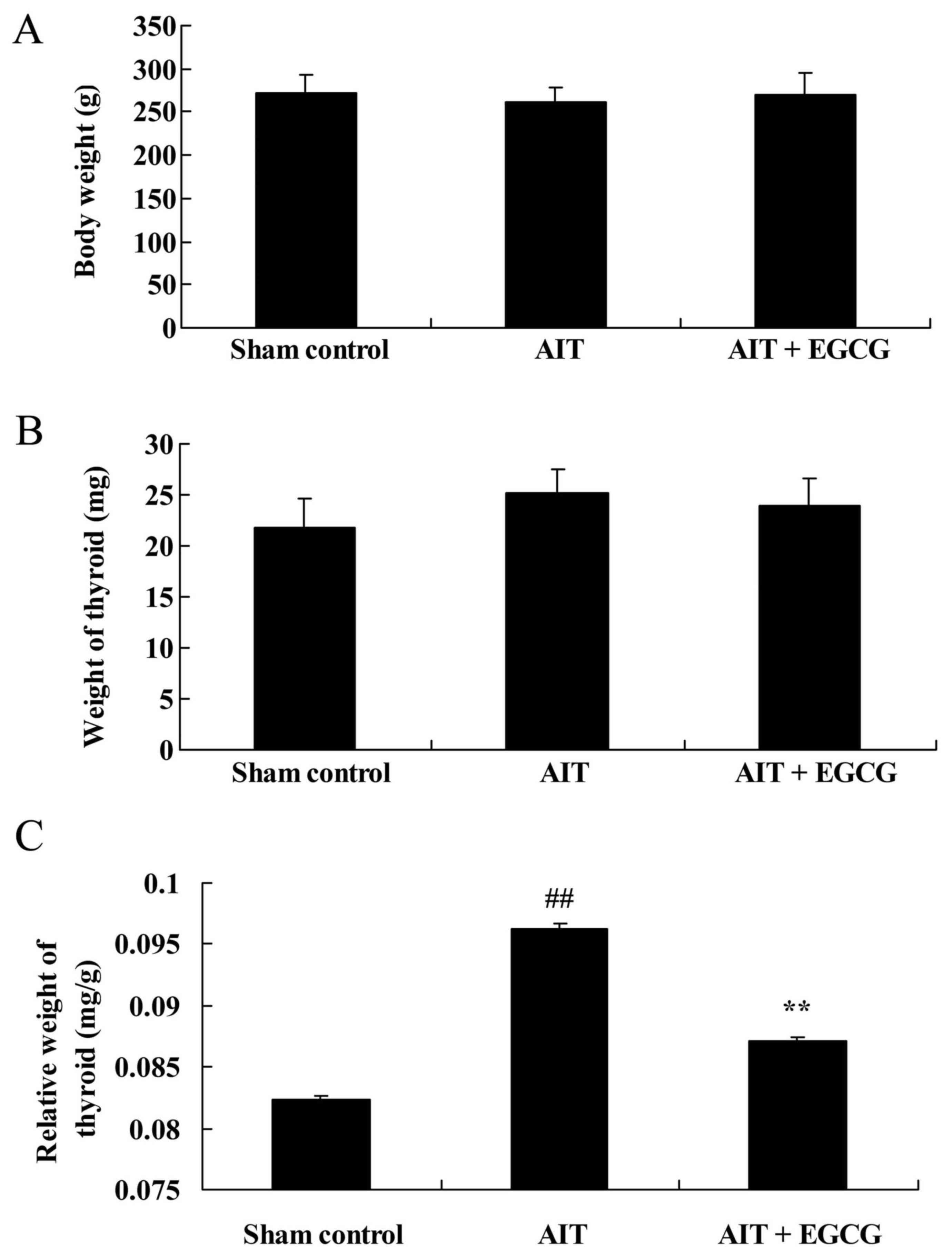
The study also examined the effect of EGCG on the
body and thyroid weights in AIT rats. There was no significant
difference in the body and thyroid weights among the three
experimental groups (Fig. 3A and B).
However, AIT significantly increased the relative thyroid weight in
model rats as compared with the sham control group. Pretreatment
with EGCG markedly reduced the relative thyroid weight of AIT rats,
as compared with the untreated AIT model group (Fig. 3C).
Thyroid pathological features in AIT
rats
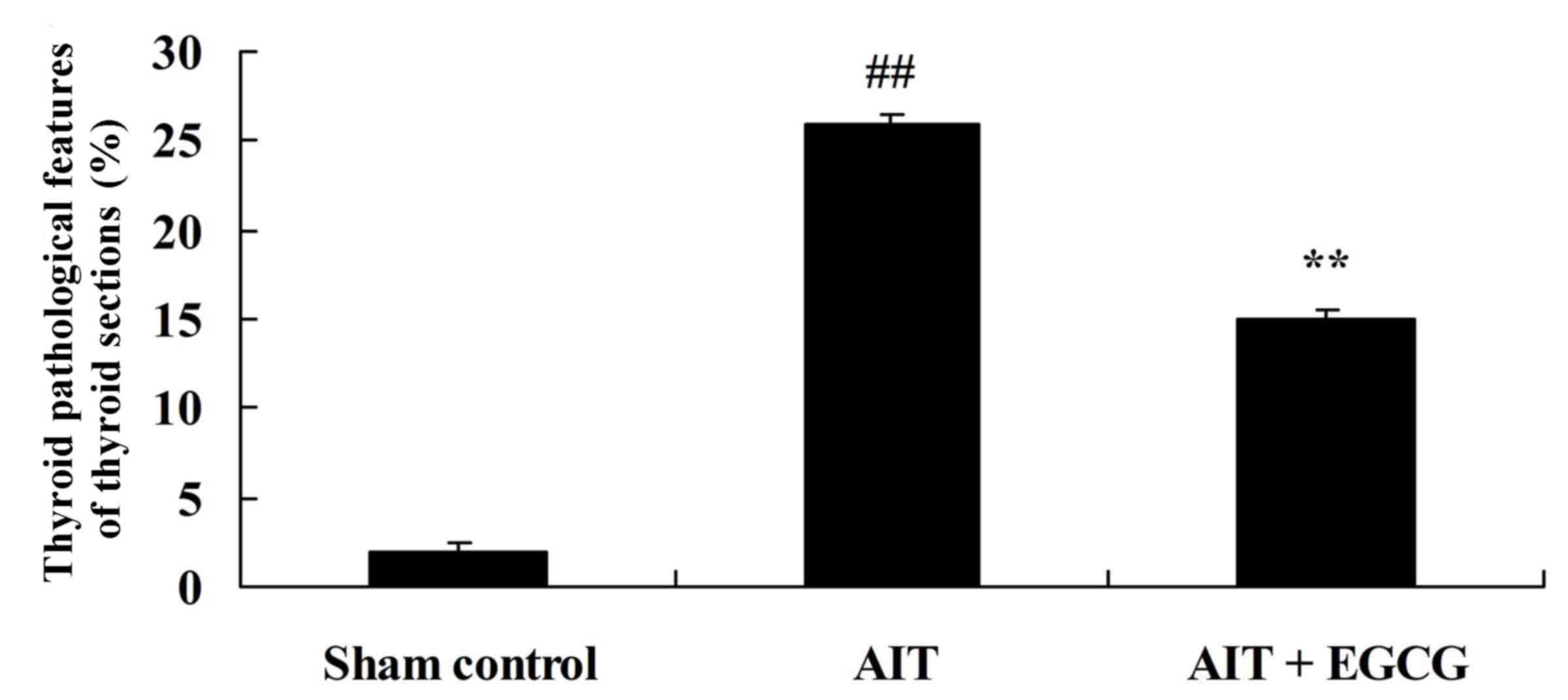
The present study further examined whether EGCG
treatment affected the thyroid pathological features in AIT rats.
As shown in Fig. 4, thyroid
pathological features of AIT model rat were significantly more
severe in comparison with those of the control sham group. However,
the AIT-induced thyroid pathological features were significantly
suppressed by treatment with EGCG, as compared with the AIT model
group (Fig. 4).
IL-1β, IFN-γ and TNF-α protein levels
in AIT rats
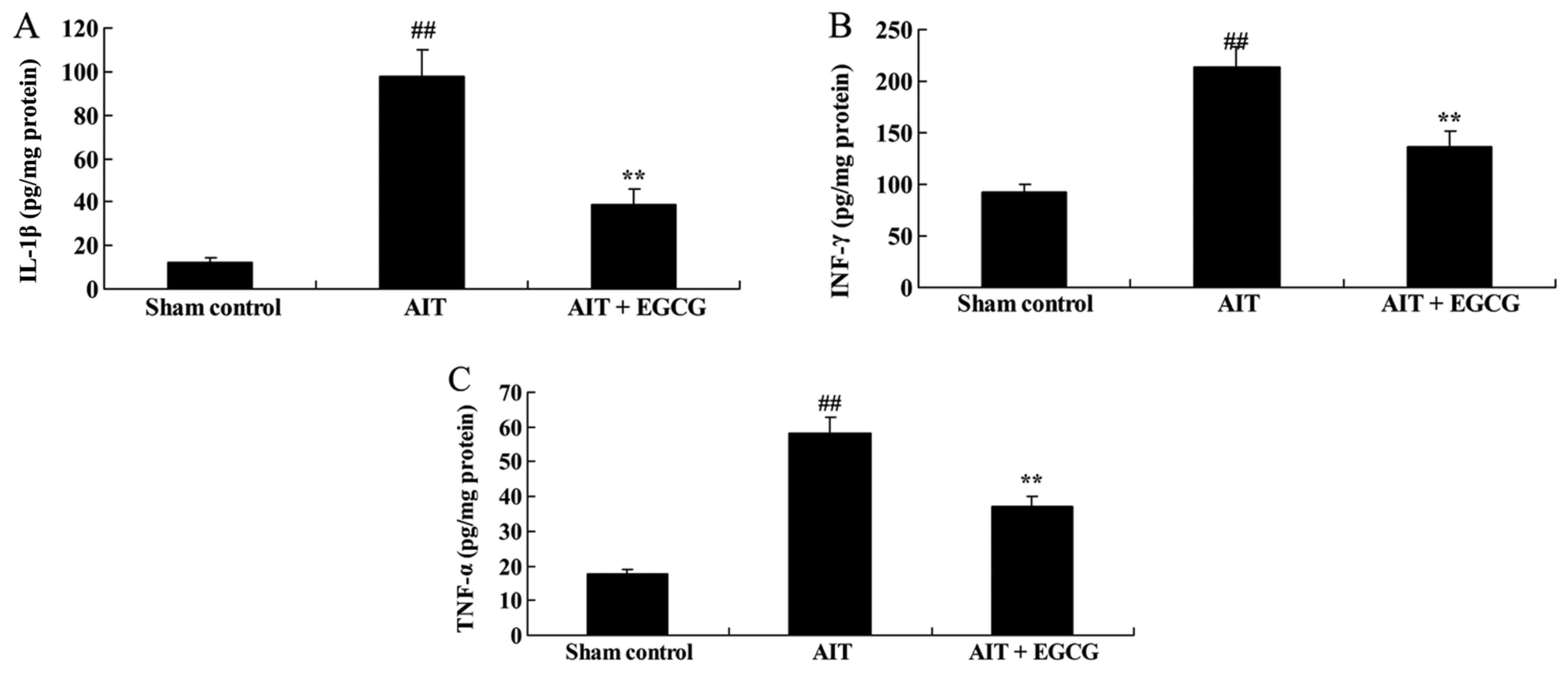
The thyroid tissue protein levels of IL-1β, IFN-γ
and TNF-α were measured using ELISA kits. The results indicated
that IL-1β (Fig. 5A), IFN-γ
(Fig. 5B) and TNF-α (Fig. 5C) levels in the thyroid tissue of AIT
rats were significantly enhanced, compared with the sham control
group. However, treatment with EGCG significantly inhibited the
AIT-induced IL-1β, IFN-γ and TNF-α levels in AIT rats, as compared
with the untreated AIT model group (Fig.
5).
NF-κB protein expression in AIT
rats
The mechanism underlying the anti-inflammatory
effect of EGCG in AIT rats, NF-κB protein expression in thyroid
tissue was measured using western blot analysis. Compared with the
sham control group, NF-κB/p65 protein expression of AIT rats was
significantly promoted, whereas pretreatment with EGCG
significantly suppressed the AIT-induced NF-κB/p65 protein
expression in rats (Fig. 6).
Bcl-2 protein expression in AIT
rats
The role of Bcl-2 in the neuroprotective effect of
EGCG on AIT rats was examined using western blot analysis. As
illustrated in Fig. 7, Bcl-2 protein
expression of AIT rats was significantly decreased, compared with
the sham control group. However, the AIT-induced Bcl-2 protein
expression was significantly increased by treatment with EGCG, as
compared with untreated AIT model group (Fig. 7).
Caspase-3 activity in AIT rats
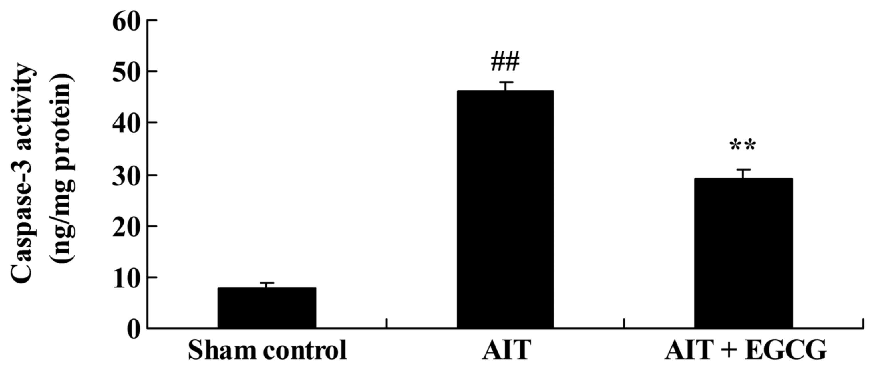
To investigate the effect of EGCG on apoptosis in
AIT rats, caspase-3 activity was measured using ELISA. As expected,
the caspase-3 activity of the AIT rat model group was evidently
higher in comparison to that of the sham control group (Fig. 8). However, treatment with EGCG
significantly reduced the AIT-induced caspase-3 activity in the
rats (Fig. 8).
TRAIL protein expression in AIT
rats
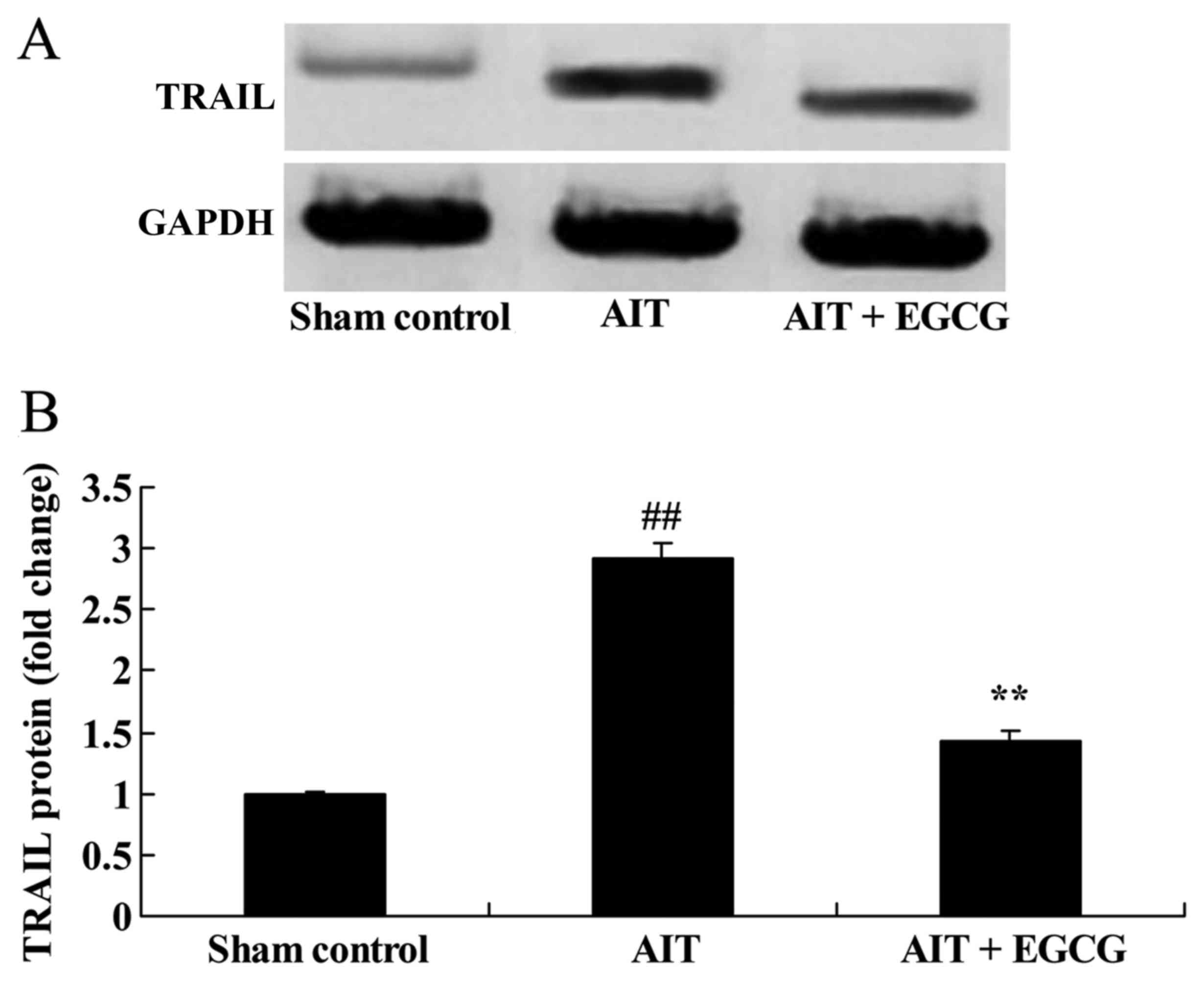
To further investigate the mechanism underlying the
neuroprotective effect of EGCG in AIT rats, TRAIL expression was
detected by western blot analysis. As demonstrated in Fig. 9, AIT significantly induced the TRAIL
protein expression in AIT rats, as compared with the sham control
group. By contrast, treatment with EGCG significantly suppressed
the AIT-induced TRAIL protein expression in AIT rats, as compared
with the AIT model group (Fig.
9).
Discussion
NF-κB is a nucleoprotein factor with pleiotropy
transcription regulators, which participates in the expression and
regulation of multiple genes, particularly genes associated with
the immune and inflammatory responses, while it also serves an
important role on the body inflammatory reaction process (15). In the present study, treatment with
EGCG significantly inhibited the AIT-induced urinary iodine values,
the relative thyroid weight and the thyroid pathological features
in the AIT rats, while it suppressed IL-1β, IFN-γ and TNF-α levels,
as well as NF-κB protein expression, which showed that EGCG had a
neuroprotective effect against AIT (16). These results are in accordance with
the findings of Liu et al, which suggested that EGCG
prevents PCB-126-induced endothelial cell inflammation through the
inhibition of NF-κB in human endothelial cells (12).
The Bcl-2 gene family is a group of genes that have
a close association with apoptosis, and induce or inhibit the role
of apoptosis by coding proteins (17). Bcl-2 and Bcl-2-associated X protein
(Bax) are genes regulating apoptosis, positively or negatively. Bax
gene serves a role in promoting apoptosis, whereas the Bcl-2 gene
inhibits apoptosis (18). Therefore,
the ratio of relative Bcl-2 and Bax levels (namely Bax/Bcl-2) is an
important indicator of the apoptosis degree. When the Bax/Bcl-2
ratio value is higher (indicating increased Bax expression), then
apoptosis is promoted (18). By
contrast, a lower Bax/Bcl-2 ratio indicates reduced apoptosis
(19). In the present study, EGCG
treatment significantly increased Bcl-2 protein expression in AIT
rats, and may reduce the AIT-induced apoptosis. Similarly,
Adikesavan et al reported that EGCG stabilizes the
mitochondrial enzymes and inhibits the apoptosis through
upregulation of Bcl-2 in cigarette smoke-induced myocardial
dysfunction (20).
The caspase family serves a main executer role in
the process of apoptosis, with the main function of inactivating
inhibitors of apoptosis (such as apoptin and Bcl-2), ultimately
resulting in degradation of cell components and formation of
apoptotic bodies by hydrolyzing the cell protein structure
(21). Caspase-3 is an important
effector molecule in apoptosis, which can activate DNA enzyme,
degrade chromosome and ultimately cause apoptosis (22). Therefore, the present study
investigated caspase-3 activity, and the results demonstrated that
EGCG significantly reduced the AIT-induced caspase-3 activity in
rats.
TRAIL selectively induces the apoptosis of various
tumor cells; however, it does not have an impact on cell growth,
development and differentiation under normal conditions (23). The TRAIL gene group mRNA is widely
detected in various tissues, while regions of its cytomembrane
present a homologous consistency with members of the TNF family
(24). However, TRAIL differs from
other members of the TNF family in that it is much shorter and only
has 14 amino acid residues as the support (24). In addition, the broad role of TRAIL
in inducing apoptosis can only be compared with Fas-L (25).
Apoptosis is caused by TRAIL by Fas and TNF receptor
(TNFR) in AIT (26). At present,
TRAIL is considered to result in apoptosis through activation of
immune cells, leading to generation of TNF-α and IL-1 (24). TNF-α and IL-1 have a synergistic
effect that improves thyroid cell sensitivity of TRAIL, resulting
in the death of thyroid cells (27).
TRAIL expression activates immune cells through thyroid autoantigen
generation, in order to generate IFN-γ and TNF-α. The synergic
induction of IFN-γ and TNF-α results in the expression of TRAIL by
thyroid cells, increasing the sensitivity of TRAIL on immune cells,
and ultimately resulting in apoptosis (27). Furthermore, TNF-α and IL-1 may result
in the destruction of thyroid cells. IFN-γ, TNF-α and IL-1 inhibit
TRAIL expression and thus induce the death of thyroid cells
(28). The results of the present
study indicated that EGCG treatment significantly suppressed the
AIT-induced TRAIL protein expression in AIT rats. These results
showed that EGCG reduced TRAIL expression, and may therefore reduce
apoptosis in AIT. Similarly, Aktas et al (29) reported that EGCG mediates T cellular
NF-κB inhibition and suppression of TRAIL in autoimmune
encephalomyelitis.
In conclusion, the results of the present study
indicated that EGCG exerted a potent neuroprotective effect in an
AIT rat model, since it inhibited the AIT-induced urinary iodine
values, the relative thyroid weight and the thyroid pathological
features of the rats. Furthermore, it was demonstrated the EGCG
suppressed IL-1β, IFN-γ, TNF-α and NF-κB protein expression levels,
while it upregulated Bcl-2 expression, and reduced caspase-3
activity and TRAIL protein expression in AIT rats. Therefore, EGCG
may be a potential novel drug for neuroinflammatory diseases.
Acknowledgements
The present study was supported by Scientific
Research Task of Chinese Medicine and Chinese and Western Medicine
Combined (grant no. 2015048).
References
|
1
|
Nordio M and Basciani S: Efficacy of a
food supplement in patients with hashimoto thyroiditis. J Biol
Regul Homeost Agents. 29:93–102. 2015.PubMed/NCBI
|
|
2
|
Krysiak R and Okopien B: The effect of
levothyroxine and selenomethionine on lymphocyte and monocyte
cytokine release in women with Hashimoto's thyroiditis. J Clin
Endocrinol Metab. 96:2206–2215. 2011. View Article : Google Scholar : PubMed/NCBI
|
|
3
|
Hofling DB, Chavantes MC, Juliano AG,
Cerri GG, Romão R, Yoshimura EM and Chammas MC: Low-level laser
therapy in chronic autoimmune thyroiditis: A pilot study. Lasers
Surg Med. 42:589–596. 2010. View Article : Google Scholar : PubMed/NCBI
|
|
4
|
Sugiyama A, Zhu BM, Takahara A, Satoh Y
and Hashimoto K: Cardiac effects of salvia miltiorrhiza/dalbergia
odorifera mixture, an intravenously applicable Chinese medicine
widely used for patients with ischemic heart disease in China. Circ
J. 66:182–184. 2002. View Article : Google Scholar : PubMed/NCBI
|
|
5
|
Iwasaki K, Kosaka K, Mori H, Okitsu R,
Furukawa K, Manabe Y, Yoshita M, Kanamori A, Ito N, Wada K, et al:
Open label trial to evaluate the efficacy and safety of Yokukansan,
a traditional Asian medicine, in dementia with Lewy bodies. J Am
Geriatr Soc. 59:936–938. 2011. View Article : Google Scholar : PubMed/NCBI
|
|
6
|
Kristensen B, Hegedüs L, Madsen HO, Smith
TJ and Nielsen CH: Altered balance between self-reactive T helper
(Th)17 cells and Th10 cells and between full-length forkhead box
protein 3 (FoxP3) and FoxP3 splice variants in Hashimoto's
thyroiditis. Clin Exp Immunol. 180:58–69. 2015. View Article : Google Scholar : PubMed/NCBI
|
|
7
|
Scarpa V, Kousta E, Tertipi A, Vakaki M,
Fotinou A, Petrou V, Hadjiathanasiou C and Papathanasiou A:
Treatment with thyroxine reduces thyroid volume in euthyroid
children and adolescents with chronic autoimmune thyroiditis. Horm
Res Paediatr. 73:61–67. 2010. View Article : Google Scholar : PubMed/NCBI
|
|
8
|
Vecchiatti SM, Lin CJ, Capelozzi VL,
Longatto-Filho A and Bisi H: Prevalence of thyroiditis and
immunohistochemistry study searching for a morphologic consensus in
morphology of autoimmune thyroiditis in a 4613 autopsies series.
Appl Immunohistochem Mol Morphol. 23:402–408. 2015. View Article : Google Scholar : PubMed/NCBI
|
|
9
|
Wang SH, Cao Z, Wolf JM, Van Antwerp M and
Baker JR Jr: Death ligand tumor necrosis factor-related
apoptosis-inducing ligand inhibits experimental autoimmune
thyroiditis. Endocrinology. 146:4721–4726. 2005. View Article : Google Scholar : PubMed/NCBI
|
|
10
|
Liu H, Zheng T, Mao Y, Xu C, Wu F, Bu L,
Mou X, Zhou Y, Yuan G, Wang S, et al: γδ T cells enhance B cells
for antibody production in Hashimoto's thyroiditis, and retinoic
acid induces apoptosis of the γδ T cell. Endocrine. 51:113–122.
2016. View Article : Google Scholar : PubMed/NCBI
|
|
11
|
Lee JY, Paik JS, Yun M, Lee SB and Yang
SW: The effect of (−)-epigallocatechin-3-gallate on IL-1β induced
IL-8 expression in orbital fibroblast from patients with
thyroid-associated ophthalmopathy. PLoS One. 11:e01486452016.
View Article : Google Scholar : PubMed/NCBI
|
|
12
|
Liu D, Perkins JT and Hennig B: EGCG
prevents PCB-126- induced endothelial cell inflammation via
epigenetic modifications of NF-κB target genes in human endothelial
cells. J Nutr Biochem. 28:164–170. 2016. View Article : Google Scholar : PubMed/NCBI
|
|
13
|
Liu Q, Qian Y, Chen F, Chen X, Chen Z and
Zheng M: EGCG attenuates pro-inflammatory cytokines and chemokines
production in LPS-stimulated L02 hepatocyte. Acta Biochim Biophys
Sin (Shanghai). 46:31–39. 2014. View Article : Google Scholar : PubMed/NCBI
|
|
14
|
Cui SL, Yu J and Shoujun L: Iodine intake
increases IP-10 expression in the serum and thyroids of rats with
experimental autoimmune thyroiditis. Int J Endocrinol.
2014:5810692014. View Article : Google Scholar : PubMed/NCBI
|
|
15
|
Eguchi K: Apoptosis in autoimmune
diseases. Intern Med. 40:275–284. 2001. View Article : Google Scholar : PubMed/NCBI
|
|
16
|
Arslan S, Korkmaz Ö, Özbilüm N and Berkan
Ö: Association between NF-κBI and NF-κBIA polymorphisms and
coronary artery disease. Biomed Rep. 3:736–740. 2015. View Article : Google Scholar : PubMed/NCBI
|
|
17
|
Kaczmarek E, Lacka K,
Jarmolowska-Jurczyszyn D, Sidor A and Majewski P: Changes of B and
T lymphocytes and selected apopotosis markers in Hashimoto's
thyroiditis. J Clin Pathol. 64:626–630. 2011. View Article : Google Scholar : PubMed/NCBI
|
|
18
|
Myśliwiec J, Okota M, Nikołajuk A and
Górska M: Soluble Fas, Fas ligand and Bcl-2 in autoimmune thyroid
diseases: Relation to humoral immune response markers. Adv Med Sci.
51:119–122. 2006.
|
|
19
|
Mysliwiec J, Okłota M, Nikołajuk A and
Górska M: Age related changes of soluble Fas, Fas ligand and Bcl-2
in autoimmune thyroid diseases. Endokrynol Pol. 58:492–495.
2007.(In Polish). PubMed/NCBI
|
|
20
|
Adikesavan G, Vinayagam MM, Abdulrahman LA
and Chinnasamy T: (−)-Epigallocatechin-gallate (EGCG) stabilize the
mitochondrial enzymes and inhibits the apoptosis in cigarette
smoke-induced myocardial dysfunction in rats. Mol Biol Rep.
40:6533–6545. 2013. View Article : Google Scholar : PubMed/NCBI
|
|
21
|
Bossowski A, Czarnocka B, Bardadin K,
Stasiak-Barmuta A, Urban M, Dadan J, Ratomski K and Bossowska A:
Identification of apoptotic proteins in thyroid gland from patients
with Graves' disease and Hashimoto's thyroiditis. Autoimmunity.
41:163–173. 2008. View Article : Google Scholar : PubMed/NCBI
|
|
22
|
Minatoguchi S, Kariya T, Uno Y, Arai M,
Nishida Y, Hashimoto K, Wang N, Aoyama T, Takemura G, Fujiwara T
and Fujiwara H: Caspase-dependent and serine protease-dependent DNA
fragmentation of myocytes in the ischemia-reperfused rabbit heart:
These inhibitors do not reduce infarct size. Jpn Circ J.
65:907–911. 2001. View Article : Google Scholar : PubMed/NCBI
|
|
23
|
Zhitao J, Long L, Jia L, Yunchao B and
Anhua W: Temozolomide sensitizes stem-like cells of glioma spheres
to TRAIL-induced apoptosis via upregulation of casitas B-lineage
lymphoma (c-Cbl) protein. Tumour Biol. 36:9621–9630. 2015.
View Article : Google Scholar : PubMed/NCBI
|
|
24
|
Zauli G, Tisato V, Melloni E, Volpato S,
Cervellati C, Bonaccorsi G, Radillo O, Marci R and Secchiero P:
Inverse correlation between circulating levels of TNF-related
apoptosis-inducing ligand and 17β-estradiol. J Clin Endocrinol
Metab. 99:E659–E664. 2014. View Article : Google Scholar : PubMed/NCBI
|
|
25
|
Ge Y, Yan D, Deng H, Chen W and An G:
Novel molecular regulators of tumor necrosis factor-related
apoptosis-inducing ligand (TRAIL)-induced apoptosis in NSCLC cells.
Clin Lab. 61:1855–1863. 2015. View Article : Google Scholar : PubMed/NCBI
|
|
26
|
El-Karaksy SM, Kholoussi NM, Shahin RM,
El-Ghar MM and Gheith Rel-S: TRAIL mRNA expression in peripheral
blood mononuclear cells of Egyptian SLE patients. Gene.
527:211–214. 2013. View Article : Google Scholar : PubMed/NCBI
|
|
27
|
Zhang N, Wang X, Huo Q, Li X, Wang H,
Schneider P, Hu G and Yang Q: The oncogene metadherin modulates the
apoptotic pathway based on the tumor necrosis factor superfamily
member TRAIL (tumor necrosis factor-related apoptosis-inducing
ligand) in breast cancer. J Biol Chem. 288:9396–9407. 2013.
View Article : Google Scholar : PubMed/NCBI
|
|
28
|
Ramamurthy V, Yamniuk AP, Lawrence EJ,
Yong W, Schneeweis LA, Cheng L, Murdock M, Corbett MJ, Doyle ML and
Sheriff S: The structure of the death receptor 4-TNF-related
apoptosis-inducing ligand (DR4-TRAIL) complex. Acta Crystallogr F
Struct Biol Commun. 71:1273–1281. 2015. View Article : Google Scholar : PubMed/NCBI
|
|
29
|
Aktas O, Prozorovski T, Smorodchenko A,
Savaskan NE, Lauster R, Kloetzel PM, Infante-Duarte C, Brocke S and
Zipp F: Green tea epigallocatechin-3-gallate mediates T cellular
NF-kappa B inhibition and exerts neuroprotection in autoimmune
encephalomyelitis. J Immunol. 173:5794–5800. 2004. View Article : Google Scholar : PubMed/NCBI
|


