Introduction
Hypertension is a common chronic cardiovascular
disease clinically characterized by an elevated blood pressure and
is an independent risk factor for cardiac and cerebral vascular
diseases (1–3). The incidence of hypertension increases
each year, particularly among young people (4); however, its molecular mechanism of
action remains unclear. As an important component of the blood
circulation and immune system, vascular endothelial cells are
widely involved in biological processes, including blood pressure
regulation, blood vessel formation, anticoagulation, fibrinolysis
and inflammation (5–7). Previous studies have demonstrated that
vascular endothelial cell dysfunction serves an important role in
the onset and development of hypertension (8,9).
Endothelial cells not only act as a physical barrier but also
secrete a variety of substances that affect the function of
relaxation and contraction of blood vessels. During sustained
hypertension, endothelial cells are damaged and the expression of
inflammatory factors is increased (10,11).
Therefore, investigating the molecular mechanism of vascular
endothelial cells in the onset and development of hypertension may
help to improve the diagnosis and treatment of hypertension.
MicroRNA (miRNA) are a class of non-coding small RNA
molecules consisting of 18–23 nucleotides. miRNA regulates the
expression of target mRNA at the post-transcriptional level by
binding to the 3′-untranslated region (UTR) of target gene mRNA
(12,13). miRNA participates in a wide variety
of biological processes, including cell proliferation,
differentiation, apoptosis, energy metabolism, hormone secretion
and development (14,15). miRNA serves an important biological
role in cardiovascular diseases and affects the progression of
hypertension by regulating a variety of tissues and organs,
including myocardial tissue, vascular wall tissue, fundus tissue
and kidney (16). Furthermore, miRNA
serves important regulatory roles in cardiac hypertrophy and heart
failure (17,18) and its expression is closely
associated with arrhythmia and atherosclerotic plaque formation. As
miRNA expression is stable in peripheral blood, it may act as a
potential diagnostic marker for various diseases, including
myocardial infarction and cancer (19). The upregulation of miR-1 levels in
the peripheral blood is closely associated with myocardial injury
(20). However, the mechanism by
which miRNA induces endothelial injury in hypertension remains
unclear.
It has been demonstrated that the recently
identified miRNA molecule miR-506 is closely associated with
proliferation, drug resistance, invasion and metastasis in tumors.
The upregulation of miR-506 increases drug resistance in colon
cancer cells (21) and in
neuroglioma, miR-506 inhibits the proliferation and metastasis of
tumor cells by targeting signal transducer and activator of
transcription 3 (22). In addition,
miR-506 participates in angiogenesis, suggesting that it may serve
a role in cardiovascular disease (23). Beclin1 (BECN1) is a key gene in the
regulation of autophagy activity and participates in the onset and
development of different tumors by directly regulating autophagy
activity (24). In most tumors,
BECN1 expression is downregulated and BECN1 overexpression inhibits
tumor growth (25,26). It remains unclear whether miR-506-3p
is associated with BECN1 expression. Therefore, the aim of the
present study was to investigate the effect of miR-506-3p in the
onset and development of hypertension at the clinical and cellular
levels.
Materials and methods
Patients
A total of 61 patients with primary hypertension
admitted to Zaozhuang Municipal Hospital (Zaozhuang, China) between
December 2014 and January 2016 were included in the present study.
Peripheral blood was collected from all patients, as well as 31
healthy subjects who were included in a control group. The 61
patients included 34 males and 27 females, with a mean age of 62.5
years (median, 61 years; range, 46–81 years). Diagnosis of
hypertension was determined when systolic blood pressure ≥160 mmHg
and/or diastolic blood pressure ≥100 mmHg without the use of
antihypertensive drugs, in accordance with the 2010 Chinese
Guidelines for the Management of Hypertension (27). The 61 patients were divided into
grades I, II and III according to the severity of hypertension
(27). Patients with grade I had
mild hypertension (systolic blood pressure of 140–159 mmHg and/or
diastolic blood pressure of 90–99 mmHg), patients with grade II had
moderate hypertension (systolic blood pressure of 160–179 mmHg
and/or diastolic blood pressure of 100–109 mmHg) and patients with
grade III had severe hypertension (systolic blood pressure of ≥180
mmHg and/or diastolic blood pressure of ≥110 mmHg). There were 23,
27 and 11 patients with grades I, II and III, respectively. The
duration of hypertension for each patient was >5 years. All
procedures were approved by the Ethics Committee of Zaozhuang
Municipal Hospital (Zaozhuang, China) and written informed consent
was obtained from all patients or their families.
Cells
Human umbilical vein endothelial cells (HUVECs; Cell
Bank; Chinese Academy of Sciences, Shanghai, China) and HEK293T
cells (Cell Bank; Chinese Academy of Sciences) were cultured in
Dulbecco's Modified Eagle's medium (DMEM) supplemented with 10%
fetal bovine serum (Thermo Fisher Scientific, Inc., Waltham, MA,
USA) at 37°C and 5% CO2. Upon reaching 70–90%
confluence, cells were seeded into 24-well plates at
1×10⁵ cells/well. HUVECs were divided into three groups:
A negative control (NC) group, a miR-506-3p mimics group
(transfection with miR-506-3p mimics) and a miR-506-3p inhibitor
group (co-transfected with miR-506-3p mimics and inhibitor).
Transfection of cells
Prior to transfection, HUVECs were cultured in
serum-free DMEM until they reached 90% confluence. In the first
vial, 2.5 µl miR-506-3p mimics (25 pmol/µl; miR-506-3p mimics
group; HanBio, Shanghai, China; sequences not supplied), or 2.5 µl
miR-506-3p mimics (25 pmol/µl; HanBio; sequences not supplied) and
2.5 µl miR-506-3p inhibitor (25 pmol/µl; miR-506-3p inhibitor
group; HanBio; sequences not supplied) were mixed with 50 µl Opti
Mem I medium (Thermo Fisher Scientific, Inc.). In the second vial,
1 µl Lipofectamine 2000 (Thermo Fisher Scientific, Inc.) was mixed
with 50 µl Opti Mem I medium. Vials were left to stand for 5 min,
the two vials were then combined and left to stand at room
temperature for a further 20 min. Subsequently, mixtures were added
to cells in each respective group. After 6 h, the medium was
replaced with fresh DMEM containing 10% fetal bovine serum.
Following cultivation for 48 h, cells were collected for further
assays.
Reverse transcription-quantitative
polymerase chain reaction (RT-qPCR)
Cells (1×10⁵) were trypsinized and mixed
with 0.5 ml TRIzol (Thermo Fisher Scientific, Inc.) for lysis.
Total RNA was then extracted using the phenol chloroform method
(28). The purity of RNA was
determined by A260/A280 using ultraviolet spectrophotometry
(Nanodrop ND2000, Thermo Fisher Scientific, Inc.). Subsequently,
cDNA was obtained by reverse transcription using the Reverse
Transcription System (Takara Biotechnology, Co., Ltd., Dalian,
China) using 0.5 µg RNA and stored at −20°C. The expression of
miR-506-3p was determined using a SYBR PrimeScript miRNA RT-PCR kit
(Takara Biotechnology, Co., Ltd.) and U6 (forward,
5′-CTCGCTTCGGCAGCACA-3′; reverse, 5′-AACGCTTCACGAATTTGCGT-3′) was
used as an internal reference. The reaction system (20 µl)
contained 10 µl qRT-PCR-Mix, 0.5 µl upstream primer
(5′-GGCACCCTTCTGAGTAGA−3′), 0.5 µl downstream universal primer
(provided by the kit), 2 µl cDNA and 7 µl ddH2O. The
reaction protocol was: Initial denaturation at 95°C for 10 min,
followed by 40 cycles of denaturation 95°C for 1 min and annealing
at 60°C for 30 sec. The reaction was performed on ABI StepOne Plus
Real-Time PCR system (Thermo Fisher Scientific, Inc.). The
2−ΔΔCq method (29) was
used to calculate the relative expression of miR-506-3p against U6
and each sample was measured in triplicate.
Cell counting kit-8 (CCK-8) assay
At 24 h after transfection, HUVECs were trypsinized
and seeded into 96-well plates at a density of
1×10³/well. At 24, 48 and 72 h, the medium was discarded
and the cells were washed with sterile phosphate-buffered saline
twice. Subsequently, DMEM and 10% CCK-8 reaction reagent (Beyotime
Institute of Biotechnology, Haimen, China) were added. Following
incubation at 37°C for 1 h, the absorbance of each well was
measured at 490 nm and cell proliferation curves were plotted.
Flow cytometry
At 48 h after transfection, HUVECs were trypsinized,
collected and washed twice with precooled phosphate-buffered
saline. Cellular apoptosis was measured by flow cytometry using a
FITC Annexin V apoptosis detection kit I (BD Biosciences, Franklin
Lakes, NJ, USA), following the manufacturer's instructions. Early
apoptotic cells were identified by single-positive staining with
Annexin V, necrotic cells were identified by single-positive
staining with propidium iodide and late apoptotic cells were
identified by double-positive staining with propidium iodide and
Annexin V. To determine the cell cycle, a cell cycle assay kit (BD
Biosciences) was employed. Cells were incubated with 200 µl liquid
A at room temperature for 10 min, followed by 150 µl liquid B at
room temperature for 10 min and finally 120 µl liquid C at room
temperature in the dark for 10 min. A flow cytometer was used for
detection and the results were analyzed using ModFit v3.2 software
(http://www.vsh.com).
Transwell assay
Transwell chambers (8 µm; 24-wells; Corning Inc.,
Corning, NY, USA) were used to evaluate the migration ability of
HUVECs. Cells were collected by trypsin digestion and resuspended
to a density of 5×10⁵ cells/ml using DMEM containing
0.1% bovine serum albumin (Beyotime Institute of Biotechnology).
The cell suspension (200 µl) was added to the upper chamber. In the
lower chamber, 500 µl DMEM supplemented with 10% fetal bovine serum
was added. Following 24 h cultivation, cells in the upper chamber
were wiped using a cotton swab. Cells that moved to the other side
of the chamber were fixed with 4% formaldehyde at room temperature
for 10 min. Following staining with Giemsa at room temperature for
1 min, the number of cells in five randomly selected in fields were
counted under a light microscope (magnification, ×200) and
averaged.
Western blot analysis
HUVECs were trypsinized and collected prior to
mixing with 100 µl precooled radioimmunoprecipitation assay lysis
buffer (Beyotime Institute of Biotechnology) containing 1%
phenylmethylsulfonyl fluoride for 15 min lysis at 4°C.
Subsequently, cells were centrifuged at 12,000 × g and 4°C for 10
min. The supernatant was used to determine protein concentration
using a bicinchoninic acid protein concentration determination kit
[RTP7102, Real-Times (Beijing) Biotechnology Co., Ltd., Beijing,
China]. Subsequently, samples were mixed with 5X loading buffer
prior to denaturation in a boiling water bath for 10 min. Protein
samples (20 µg) were subjected to 10% sodium dodecyl
sulfate-polyacrylamide gel electrophoresis at 100 V. The resolved
proteins were transferred to polyvinylidene difluoride membranes on
ice (300 mA, 1 h) and blocked with 5% skimmed milk at room
temperature for 1 h. Then, membranes were incubated with rabbit
anti-human polyclonal primary antibodies for BECN1 (1:1,000; cat.
no. ab62557) and GAPDH (1:1,000; cat. no. ab8245; both Abcam,
Cambridge, UK) at 4°C overnight. Following six washes with
phosphate-buffered saline and Tween-20 (5 min each), membranes were
incubated with polyclonal goat anti-rabbit horseradish
peroxidase-conjugated secondary antibody (1:4,000; cat. no. ab6721;
Abcam) for 1 h at room temperature prior to six washes with
phosphate-buffered saline and Tween-20 (5 min each). Subsequently,
membranes were developed using an enhanced chemiluminescence
detection kit (Sigma-Aldrich; Merck KGaA, Darmstadt, Germany).
Image lab v3.0 software (Bio-Rad Laboratories, Inc., Hercules, CA,
USA) was used to acquire and analyze imaging signals. Relative
protein content was expressed against GAPDH.
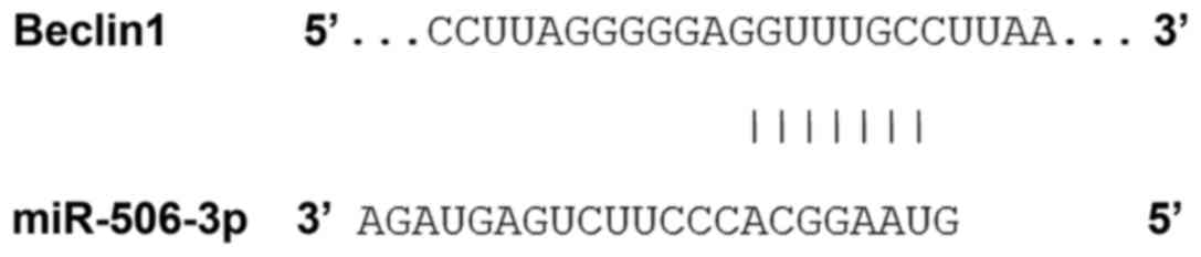
Bioinformatics prediction
Bioinformatics prediction is a powerful tool for the
study of the functions of miRNA. To determine the regulatory
mechanism of BECN1, miRanda (http://www.microrna.org/microrna/home.do), TargetScan
(http://www.targetscan.org), PiTa
(http://genie.weizmann.ac.il/pubs/mir07/mir07_data.html),
RNAhybrid (http://bibiserv.techfak.uni-bielefeld.de/rnahybrid/)
and PICTA (http://pictar.mdc-berlin.de/) were used to predict the
miRNA molecules that may regulate BECN1. The results indicated that
miR-506-3p may potentially regulate BECN1 (Fig. 1).
Dual-luciferase reporter assay
Using the results from bioinformatics, wild-type
(WT) and mutant seed regions of miR-506-3p in the 3′-UTR of BECN1
gene were chemically synthesized in vitro. SpeI and
HindIII restriction sites were added and subsequently cloned
into pMIR-REPORT luciferase reporter plasmids (Thermo Fisher
Scientific, Inc.). Plasmids (1 µg) with WT or mutant 3′-UTR DNA
sequences were co-transfected with miR-506-3p mimics (100 nM;
Sangon Biotech, Shanghai, China) into HEK293T cells. Following
cultivation for 24 h, cells were lysed using a dual-luciferase
reporter assay kit (Promega Corporation, Madison, WI, USA)
according to the manufacturer's manual and fluorescence intensity
was measured with a GloMax 20/20 luminometer (Promega Corporation).
Using Renilla luciferase activity as an internal reference,
the fluorescence values of each group of cells were measured.
Statistical analysis
Statistical analysis was performed using SPSS 16.0
(SPSS, Inc., Chicago, IL, USA). Measurement data were expressed as
the mean ± standard deviation. Differences between two groups were
compared using Student's t-test. For comparison of multiple groups,
ANOVA was used, followed by a post hoc comparisons test using
Tukey's/Dunnett's method. Each test was performed in triplicate.
P<0.05 was considered to indicate a statistically significant
difference.
Results
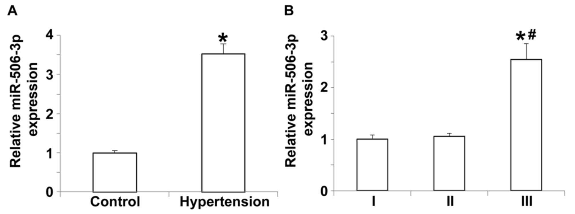
Expression of miR-506-3p in the
peripheral blood of hypertension patients is upregulated and
dependent on hypertension staging
To measure the expression of miR-506-3p in the
peripheral blood, RT-qPCR was performed. The data indicated that
miR-506-3p expression in patients with hypertension was
significantly higher compared with the control group (P<0.05;
Fig. 2A). Further analysis indicated
that the expression of miR-506-3p in patients with grade III
hypertension was significantly higher than in patients with grade I
or II hypertension (P<0.05; Fig.
2B), whereas miR-506-3p expression did not differ between
patients with grade I and II hypertension (P>0.05; Fig. 2B). These results demonstrate that
miR-506-3p expression in the peripheral blood of patients with
hypertension is upregulated and that this upregulation is dependent
on hypertension staging.
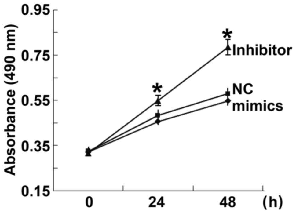
Overexpression of miR-506-3p inhibits
the proliferation of HUVECs
To determine the effect of miR-506-3p on the
proliferation of HUVECs, a CCK-8 assay was performed. The data
indicated that the absorbance of HUVECs transfected with miR-506-3p
inhibitor was significantly higher than that of HUVECs in the NC
group 24 and 48 h after transfection (P<0.05; Fig. 3). By contrast, the absorbance of
HUVECs transfected with miR-506-3p mimics did not differ from that
of HUVECs in the NC group at 24 and 48 h (P>0.05; Fig. 3). These results indicate that the
overexpression of miR-506-3p inhibits the proliferation of
HUVECs.
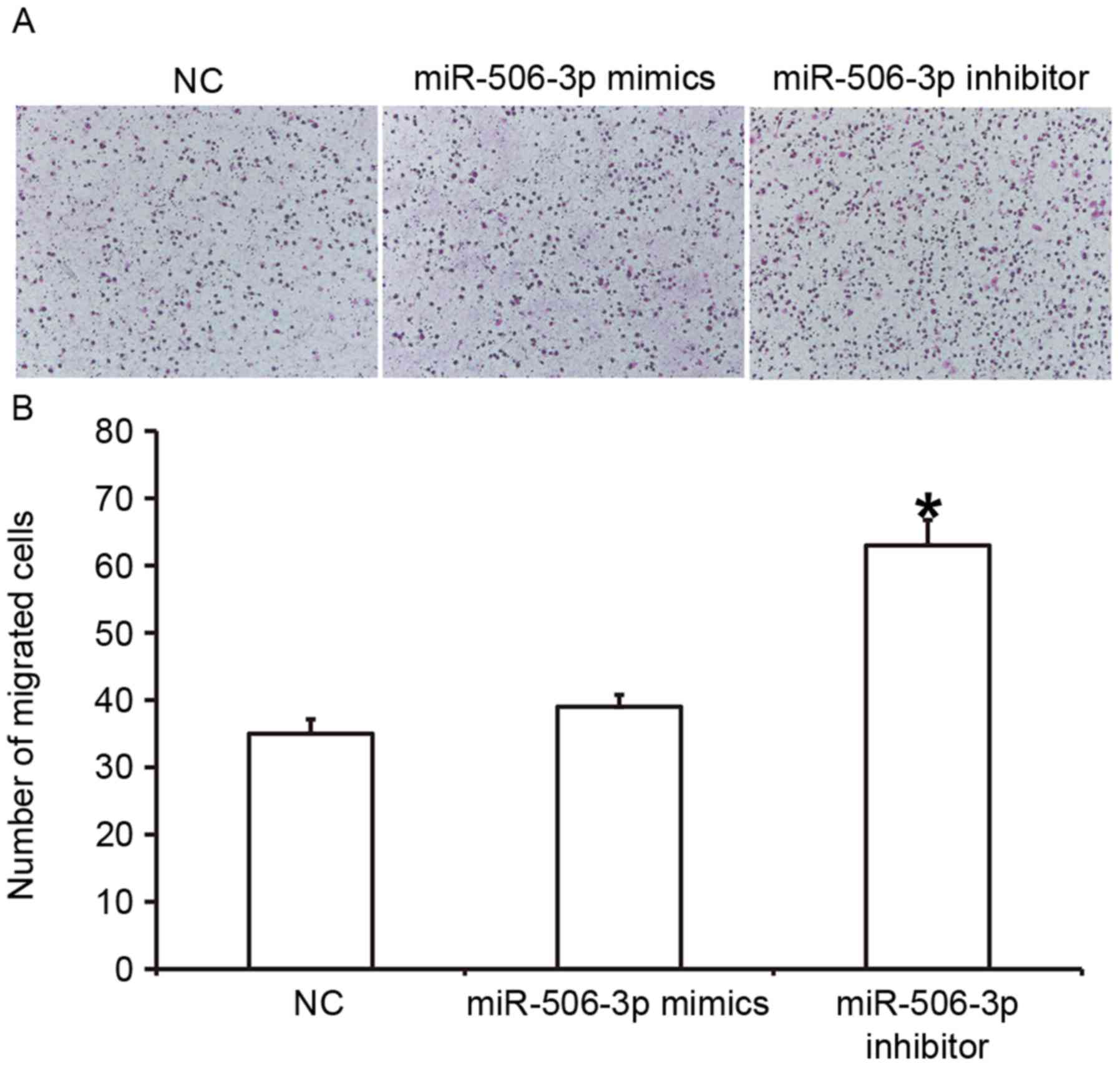
Overexpression of miR-506-3p inhibits
the migration ability of HUVECs
To determine the effect of miR-506-3p on the
migration ability of HUVECs, a Transwell assay was conducted
(Fig. 4). The data indicated that
the number of HUVECs that passed through the chamber membrane in
the miR-506-3p inhibitor group was significantly higher than in the
NC group (P<0.05; Fig. 4B).
However, the number of HUVECs did not differ significantly between
the miR-506-3p mimics group and the NC group (P>0.05; Fig. 4B). These results suggest that the
overexpression of miR-506-3p inhibits the migration ability of
HUVECs.
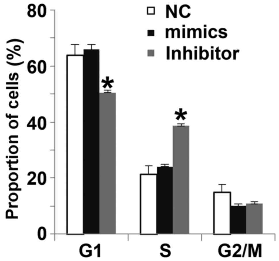
miR-506-3p regulates the G1/S phases
of HUVECs
To determine how miR-506-3p regulates the cell cycle
of HUVECs, flow cytometry was used. The data indicated that the
proportion of cells in G1 in the miR-506-3p inhibitor group
(49.5±2.1%) was significantly lower than in the NC group
(63.7±3.8%; P<0.05), whereas the proportion of cells in the
S-phase in miR-506-3p inhibitor group (38.2±1.8%) was significantly
higher than in the NC group (21.5±3.9%; P<0.05; Fig. 5). There were no differences in the
proportion of cells in each phase of the cell cycle between the
miR-506-3p mimics group and the NC group. These results indicate
that miR-506-3p may suppresses the proliferation of HUVECs by
regulating the G1/S phase transition.
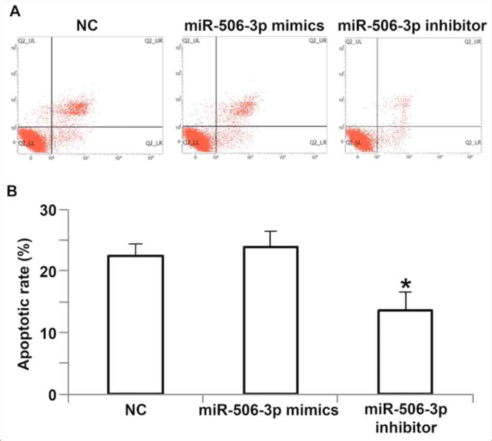
Overexpression of miR-506-3p promotes
the apoptosis of HUVECs
To determine the effect of miR-506-3p on the
apoptosis of HUVECs, flow cytometry was used (Fig. 6). The results demonstrated that the
apoptotic rate of HUVECs transfected with miR-506-3p inhibitor was
significantly lower than that of the NC group (P<0.05; Fig. 6B), whereas the apoptotic rate of
HUVECs transfected with miR-506-3p mimics did not differ from that
in the NC group (P>0.05; Fig.
6B). These results suggest that the overexpression of
miR-506-3p promotes the apoptosis of HUVECs.
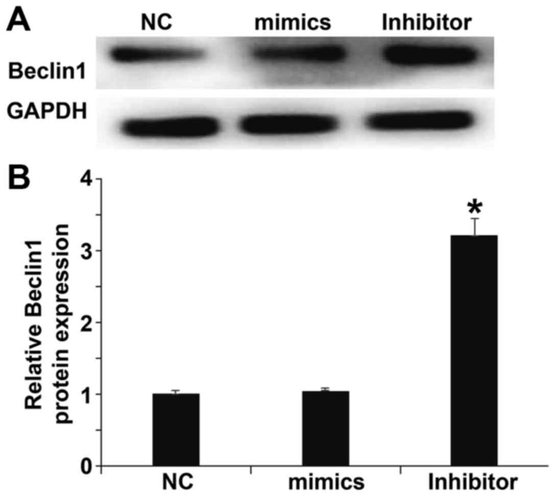
miR-506-3p exerts its biological role
by regulating the expression of BECN1 protein
To determine the expression of BECN1, which is a
target protein of miR-506-3p, western blotting was performed. The
results demonstrated that levels of BECN1 in HUVECs transfected
with miR-506-3p inhibitor were significantly higher than in the NC
group (P<0.05), however the expression of BECN1 in HUVECs
transfected with miR-506-3p mimics did not differ from that in the
NC group (P>0.05; Fig. 7). The
result indicates that miR-506-3p may exert its biological role by
regulating the expression of BECN1.
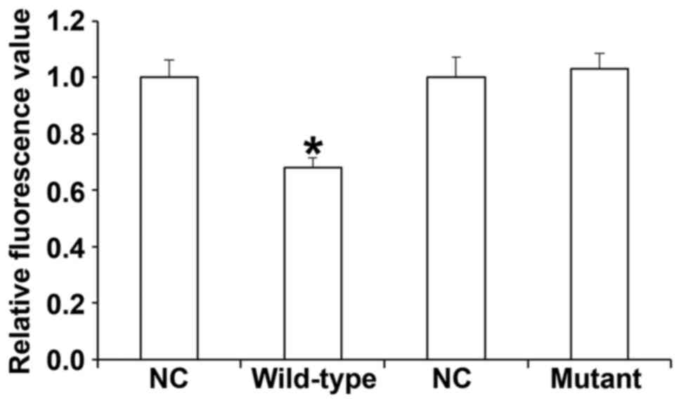
miR-506-3p regulates the expression of
BECN1 by binding to the 3′-UTR of BECN1
To identify the interaction between miR-506-3p and
BECN1, a dual-luciferase reporter assay was performed. The data
demonstrated that transfection with miR-506-3p mimics and
pMIR-REPORT in the WT group led to significantly reduced
fluorescence intensity compared with the NC (P<0.05). By
contrast, fluorescence intensity in the mutant group did not differ
significantly from that in the NC (P>0.05; Fig. 8). These results indicate that
miR-506-3p regulates the expression of BECN1 by binding to the
3′-UTR of BECN1.
Discussion
Hypertension is a complex disease caused by the
interaction between environmental and genetic factors. In recent
years, studies have demonstrated that miRNAs are closely associated
with the onset and development of hypertension (30,31). The
expression of some miRNA molecules in vascular tissues is increased
markedly. For example, miR-204 is highly expressed in vascular
tissues and helps to determine blood pressure by regulating the
proliferation and apoptosis of vascular smooth muscle cells
(32,33). In addition, miR-155 regulates the
expression of angiotensin II type 1 receptor, affects the
relaxation and contraction of vascular endothelial cells and
promotes the onset and development of hypertension (34). It has been demonstrated that miR-122
influences the expression of the amino acid transporter gene SLC7A
by changing the small nucleotide polymorphism site, disrupting
nitric oxide metabolism, regulating vascular endothelial cell
function and promoting the onset of primary hypertension (35). The results of the current study
demonstrate that miR-506-3p expression is upregulated in the
peripheral blood of patients with hypertension and is dependent on
the stage of hypertension.
It has been demonstrated that patients with
hypertension develop vascular injury in the early course of the
disease. Furthermore, vascular endothelial cell injury and
dysfunction are important signs of hypertension and may even occur
prior to the onset of hypertension (36). miRNAs serve important roles in
endothelial cell injury and dysfunction. miR-34a promotes pulmonary
arterial hypertension by regulating the proliferation and apoptosis
of vascular endothelial cells (37).
The in vitro experiments performed in the current study
demonstrate that miR-506-3p inhibits the proliferation, G1/S phase
transition and migration of HUVECs, and promotes the apoptosis of
HUVECs.
BECN1 has three structural domains,
Bcl-2-homology-3, the central coiled-coil domain and the
evolutionarily conserved domain, by which it interacts with other
molecules (38). BECN1 is essential
for the formation of autophagosomes and regulates the formation and
maturation of intracellular autophagosomes by mediating the
localization of other autophagy proteins in phagophores (39). Elevated autophagy activity is
associated with the upregulation of BECN1 expression and
downregulating BECN1 expression inhibits autophagy activity
(40). Additionally, it has been
demonstrated that BECN1 is closely associated with tumorigenesis
and organ damage (41,42).
In the current study, bioinformatics analysis
indicated that BECN1 is a potential target gene of miR-506-3p.
BECN1 is a homologous gene of autophagy-related 6 in yeast and
regulates autophagy by forming a complex with phosphoinositide
3-kinase (38). Previous studies
have demonstrated that autophagy is closely associated with
vascular endothelial injury (43,44). The
activation of autophagy contributes to the survival and
proliferation of vascular endothelial cells under stress. In the
present study, the results of the dual-luciferase reporter assay
indicated that miR-506-3p directly regulates the expression of
BECN1 and that BECN1 is a target gene of miR-506-3p.
Acknowledgements
The authors would like to thank Professor Chunting
Wang from the Department of Intensive Care Unit, Shandong
Provincial Hospital Affiliated to Shandong University for kindly
providing helpful advice in the conception, design, statistical
analysis and preparation of the manuscript.
References
|
1
|
Leopold JA: Catheter-based therapies for
patients with medication-refractory pulmonary arterial
hypertension. Circ Cardiovasc Interv. 8:e0033322015. View Article : Google Scholar : PubMed/NCBI
|
|
2
|
Olatunji LA, Seok YM, Igunnu A, Kang SH
and Kim IK: Combined oral contraceptive-induced hypertension is
accompanied by endothelial dysfunction and upregulated intrarenal
angiotensin II type 1 receptor gene expression. Naunyn
Schmiedebergs Arch Pharmacol. 389:1147–1157. 2016. View Article : Google Scholar : PubMed/NCBI
|
|
3
|
Zheng Y, Poon CC, Yan BP and Lau JY: Pulse
arrival time based cuff-less and 24-H wearable blood pressure
monitoring and its diagnostic value in hypertension. J Med Syst.
40:1952016. View Article : Google Scholar : PubMed/NCBI
|
|
4
|
Gerage AM, Bededetti TR, Ritti-Dias RM,
Dos Santos ACO, de Souza BCC and Almeida FA: Effectiveness of a
behavior change program on physical activity and eating habits in
patients with hypertension: A randomized controlled trial. J Phys
Act Health. 1–30. 2017.
|
|
5
|
Ambrozova G, Fidlerova T, Verescakova H,
Koudelka A, Rudolph TK, Woodcock SR, Freeman BA, Kubala L and
Pekarova M: Nitro-oleic acid inhibits vascular endothelial
inflammatory responses and the endothelial-mesenchymal transition.
Biochim Biophys Acta. 1860:2428–2437. 2016. View Article : Google Scholar : PubMed/NCBI
|
|
6
|
Souza PS, Gonçalves ED, Pedroso GS, Farias
HR, Junqueira SC, Marcon R, Tuon T, Cola M, Silveira PCL, Santos
AR, et al: Physical exercise attenuates experimental autoimmune
encephalomyelitis by inhibiting peripheral immune response and
blood-brain barrier disruption. Mol Neurobiol. 54:4723–4737. 2017.
View Article : Google Scholar : PubMed/NCBI
|
|
7
|
Balduino Mendes AB, Giollo-Junior LT, de
Andrade DO, Gregório ML, Yugar-Toledo JC and Vilela-Martin JF: How
to investigate the vascular changes in resistant hypertension. Curr
Hypertens Rev. 12:139–147. 2016. View Article : Google Scholar : PubMed/NCBI
|
|
8
|
Vaillancourt M, Ruffenach G, Meloche J and
Bonnet S: Adaptation and remodelling of the pulmonary circulation
in pulmonary hypertension. Can J Cardiol. 31:407–415. 2015.
View Article : Google Scholar : PubMed/NCBI
|
|
9
|
Jenkins D: Pulmonary endarterectomy: The
potentially curative treatment for patients with chronic
thromboembolic pulmonary hypertension. Eur Respir Rev. 24:263–271.
2015. View Article : Google Scholar : PubMed/NCBI
|
|
10
|
Reynolds JA, Rosenberg AZ, Smith CK,
Sergeant JC, Rice GI, Briggs TA, Bruce IN and Kaplan MJ: Brief
Report: Vitamin D deficiency is associated with endothelial
dysfunction and increases type-1 interferon gene expression in a
murine model of systemic lupus erythematosus. Arthritis Rheumatol.
68:2929–2935. 2016. View Article : Google Scholar : PubMed/NCBI
|
|
11
|
Furumoto Y, Smith CK, Blanco L, Zhao W,
Brooks SR, Thacker SG, Abdalrahman Z, Sciumè G, Tsai WL, Trier AM,
et al: Tofacitinib ameliorates murine lupus and its associated
vascular dysfunction. Arthritis Rheumatol. 69:148–160. 2017.
View Article : Google Scholar : PubMed/NCBI
|
|
12
|
Wang H, Shi H, Luo J, Yi Y, Yao D, Zhang
X, Ma G and Loor JJ: MiR-145 regulates lipogenesis in goat mammary
cells via targeting INSIG1 and epigenetic regulation of
lipid-related genes. J Cell Physiol. 232:1030–1040. 2017.
View Article : Google Scholar : PubMed/NCBI
|
|
13
|
Lu YJ, Liu RY, Hu K and Wang Y: MiR-541-3p
reverses cancer progression by directly targeting TGIF2 in
non-small cell lung cancer. Tumour Biol. 37:12685–12695. 2016.
View Article : Google Scholar : PubMed/NCBI
|
|
14
|
Wang X, Guo Y, Wang C and Yu H, Yu X and
Yu H: MicroRNA-142-3p inhibits chondrocyte apoptosis and
inflammation in osteoarthritis by targeting HMGB1. Inflammation.
39:1718–1728. 2016. View Article : Google Scholar : PubMed/NCBI
|
|
15
|
Liu B, Ding JF, Luo J, Lu L, Yang F and
Tan XD: Seven protective miRNA signatures for prognosis of cervical
cancer. Oncotarget. 7:56690–56698. 2016.PubMed/NCBI
|
|
16
|
Chang TY, Tsai WC, Huang TS, Su SH, Chang
CY, Ma HY, Wu CH, Yang CY, Lin CH, Huang PH, et al: Dysregulation
of endothelial colony-forming cell function by a negative feedback
loop of circulating miR-146a and −146b in cardiovascular disease
patients. PLoS One. 12:e01815622017. View Article : Google Scholar : PubMed/NCBI
|
|
17
|
Carr G, Barrese V, Stott JB, Povstyan OV,
Jepps TA, Figueiredo HB, Zheng D, Jamshidi Y and Greenwood IA:
MicroRNA-153 targeting of KCNQ4 contributes to vascular dysfunction
in hypertension. Cardiovasc Res. pii:cvw1772016.
|
|
18
|
Oury C, Servais L, Bouznad N, Hego A,
Nchimi A and Lancellotti P: MicroRNAs in valvular heart diseases:
Potential role as markers and actors of valvular and cardiac
remodeling. Int J Mol Sci. 17:pii: E11202016. View Article : Google Scholar
|
|
19
|
Elia L and Condorelli G: MicroRNAs and
pulmonary hypertension: A tight link. Cardiovasc Res. 111:163–164.
2016. View Article : Google Scholar : PubMed/NCBI
|
|
20
|
Yu L, Yu H, Li X, Jin C, Zhao Y, Xu S and
Sheng X: P38 MAPK/miR-1 are involved in the protective effect of
EGCG in high glucose-induced Cx43 downregulation in neonatal rat
cardiomyocytes. Cell Biol Int. 40:934–942. 2016. View Article : Google Scholar : PubMed/NCBI
|
|
21
|
Tong JL, Zhang CP, Nie F, Xu XT, Zhu MM,
Xiao SD and Ran ZH: MicroRNA 506 regulates expression of PPAR alpha
in hydroxycamptothecin-resistant human colon cancer cells. FEBS
Lett. 585:3560–3568. 2011. View Article : Google Scholar : PubMed/NCBI
|
|
22
|
Peng T, Zhou L, Zuo L and Luan Y: MiR-506
functions as a tumor suppressor in glioma by targeting STAT3. Oncol
Rep. 35:1057–1064. 2016. View Article : Google Scholar : PubMed/NCBI
|
|
23
|
Li Z, Liu Z, Dong S, Zhang J, Tan J, Wang
Y, Ge C, Li R, Xue Y, Li M, et al: miR-506 inhibits
epithelial-to-mesenchymal transition and angiogenesis in gastric
cancer. Am J Pathol. 185:2412–2420. 2015. View Article : Google Scholar : PubMed/NCBI
|
|
24
|
Chen C, Yang S, Li H, Yin Z, Fan J, Zhao
Y, Gong W, Yan M and Wang DW: Mir30c is involved in diabetic
cardiomyopathy through regulation of cardiac autophagy via BECN1.
Mol Ther Nucleic Acids. 7:127–139. 2017. View Article : Google Scholar : PubMed/NCBI
|
|
25
|
Al-Shenawy HA: Expression of Beclin-1, an
autophagy-related marker, in chronic hepatitis and hepatocellular
carcinoma and its relation with apoptotic markers. APMIS.
124:229–237. 2016. View Article : Google Scholar : PubMed/NCBI
|
|
26
|
Wang H, Wang Y, Qian L, Wang X, Gu H, Dong
X, Huang S, Jin M, Ge H, Xu C and Zhang Y: RNF216 contributes to
proliferation and migration of colorectal cancer via suppressing
BECN1-dependent autophagy. Oncotarget. 7:51174–51183.
2016.PubMed/NCBI
|
|
27
|
Liu LS: Writing Group of 2010 Chinese
Guidelines for the Management of Hypertension: 2010 Chinese
guidelines for the management of hypertension. Zhonghua Xin Xue
Guan Bing Za Zhi. 39:579–615. 2011.(In Chinese). PubMed/NCBI
|
|
28
|
Nwokeoji AO, Kilby PM, Portwood DE and
Dickman MJ: RNASwift: A rapid, versatile RNA extraction method free
from phenol and chloroform. Anal Biochem. 512:36–46. 2016.
View Article : Google Scholar : PubMed/NCBI
|
|
29
|
Livak KJ and Schmittgen TD: Analysis of
relative gene expression data using real-time quantitative PCR and
the 2(-Delta Delta C(T)) method. Methods. 25:402–408. 2001.
View Article : Google Scholar : PubMed/NCBI
|
|
30
|
Vashukova ES, Glotov AS, Fedotov PV,
Efimova OA, Pakin VS, Mozgovaya EV, Pendina AA, Tikhonov AV,
Koltsova AS and Baranov VS: Placental microRNA expression in
pregnancies complicated by superimposed pre-eclampsia on chronic
hypertension. Mol Med Rep. 14:22–32. 2016. View Article : Google Scholar : PubMed/NCBI
|
|
31
|
Klimczak D, Jazdzewski K and Kuch M:
Regulatory mechanisms in arterial hypertension: Role of microRNA in
pathophysiology and therapy. Blood Press. 26:2–8. 2017. View Article : Google Scholar : PubMed/NCBI
|
|
32
|
Potus F, Graydon C, Provencher S and
Bonnet S: Vascular remodeling process in pulmonary arterial
hypertension, with focus on miR-204 and miR-126 (2013 Grover
Conference series). Pulm Circ. 4:175–184. 2014. View Article : Google Scholar : PubMed/NCBI
|
|
33
|
Ruffenach G, Chabot S, Tanguay VF,
Courboulin A, Boucherat O, Potus F, Meloche J, Pflieger A,
Breuils-Bonnet S, Nadeau V, et al: Role for runt-related
transcription factor 2 in proliferative and calcified vascular
lesions in pulmonary arterial hypertension. Am J Respir Crit Care
Med. 194:1273–1285. 2016. View Article : Google Scholar : PubMed/NCBI
|
|
34
|
Yang LX, Liu G, Zhu GF, Liu H, Guo RW, Qi
F and Zou JH: MicroRNA-155 inhibits angiotensin II-induced vascular
smooth muscle cell proliferation. J Renin Angiotensin Aldosterone
Syst. 15:109–116. 2014. View Article : Google Scholar : PubMed/NCBI
|
|
35
|
Kamo Y, Ichikawa T, Miyaaki H, Uchida S,
Yamaguchi T, Shibata H, Honda T, Taura N, Isomoto H, Takeshima F
and Nakao K: Significance of miRNA-122 in chronic hepatitis C
patients with serotype 1 on interferon therapy. Hepatol Res.
45:88–96. 2015. View Article : Google Scholar : PubMed/NCBI
|
|
36
|
Yoruk A, Bisognano JD and Gassler JP:
Baroreceptor stimulation for resistant hypertension. Am J
Hypertens. 29:1319–1324. 2016.PubMed/NCBI
|
|
37
|
Wang MM, Fang MX, Chen LG, Wang HQ, Liu HJ
and Tang HL: Differential expression of microRNA in endothelial
cells incubated with serum of hypertension patients with
blood-stasis syndrome. Chin J Integr Med. 21:817–822. 2015.
View Article : Google Scholar : PubMed/NCBI
|
|
38
|
Kumar A, Singh UK and Chaudhary A:
Targeting autophagy to overcome drug resistance in cancer therapy.
Future Med Chem. 7:1535–1542. 2015. View Article : Google Scholar : PubMed/NCBI
|
|
39
|
Wang X, Tao Y, Huang Y, Zhan K, Xue M,
Wang Y, Ruan D, Liang Y, Huang X, Lin J, et al: Catalase
ameliorates diabetes-induced cardiac injury through reduced
p65/RelA-mediated transcription of BECN1. J Cell Mol Med. Jun
23–2017.(Epub ahead of print).
|
|
40
|
Mizuno S, Bogaard HJ, Gomez-Arroyo J,
Alhussaini A, Kraskauskas D, Cool CD and Voelkel NF:
MicroRNA-199a-5p is associated with hypoxia-inducible factor-1a
expression in lungs from patients with COPD. Chest. 142:663–672.
2012. View Article : Google Scholar : PubMed/NCBI
|
|
41
|
Goyal A, Gubbiotti MA, Chery DR, Han L and
Iozzo RV: Endorepellin-evoked autophagy contributes to angiostasis.
J Biol Chem. 291:19245–19256. 2016. View Article : Google Scholar : PubMed/NCBI
|
|
42
|
Qi JJ, Han XF, Cai XL, Li XZ, Zhang LQ,
Feng X and Liu DY: Expression and clinical significance of
autophagy-related gene Beclin1 and P62 in nasal polyps. Zhonghua Er
Bi Yan Hou Tou Jing Wai Ke Za Zhi. 51:428–432. 2016.(In Chinese).
PubMed/NCBI
|
|
43
|
Baek JH, Jung J, Seo J, Kim JH and Kim J:
Bacterial overexpression and denaturing purification of
VPS34-binding domain of Beclin 1. J Microbiol Biotechnol.
26:1808–1816. 2016. View Article : Google Scholar : PubMed/NCBI
|
|
44
|
Li Y, Jiang W, Hu Y, Da Z, Zeng C, Tu M,
Deng Z and Xiao W: MicroRNA-199a-5p inhibits cisplatin-induced drug
resistance via inhibition of autophagy in osteosarcoma cells. Oncol
Lett. 12:4203–4208. 2016. View Article : Google Scholar : PubMed/NCBI
|