Detection of Treg/Th17 cells and related cytokines in peripheral blood of chronic hepatitis B patients combined with thrombocytopenia and the clinical significance
- Authors:
- Published online on: June 14, 2018 https://doi.org/10.3892/etm.2018.6311
- Pages: 1328-1332
Abstract
Introduction
As a common complication in patients with chronic hepatitis B, thrombocytopenia (platelet count <100×109/l) occurs in 76% of these patients (1). Effects of mild to moderate thrombocytopenia on chronic hepatitis B treatment is generally light without causing spontaneous bleeding, but severe thrombocytopenia can significantly increase the risk of spontaneous bleeding in clinical practice, such as cerebral hemorrhage or gastrointestinal bleeding (2). The incidence of thrombocytopenia is affected by a variety of factors, including inhibition of the production of platelets by bone marrow and decreased activity of thrombopoietin (TPO) (3). In addition, increased intracarotid platelet damage, autoantibodies produced in spleen and blood dilution can also cause thrombocytopenia (4,5).
Autoimmunity is a major immunological factor leading to thrombocytopenia, and studies have reported that T-cell immunity plays an important role in autoimmunity (6). As CD4+ and CD25+ T cells, regulatory T (Treg) cells can inhibit T cell proliferation and effector function (7,8). Treg can inhibit antigen presenting cells to present antigens to T cells by secreting IL-10, which in turn inhibit T cell immune response (9). Treg can also secrete TGF-β to inhibit the function of T cells and the production of interferon γ (IFN-γ), thus maintaining a chronic persistent infection of hepatitis B virus (10–12). IL-6 and TGF-β can induce the production of Th17 cells, which can promote the development of inflammatory responses by secreting IL-17, IL-21 and IL-22, so as to promote the development of inflammatory responses (13). Percentage of Th17 cells and serum IL-17 levels were significantly elevated in patients with autoimmune diseases such as rheumatoid arthritis, asthma, and systemic lupus erythematosus (14).
This study showed that Treg cells were closely related to the differentiation process of Th17 cells, and these factors antagonize each other in immune response. Thus, the balance of Treg/Th17 is the key in maintaining immune homeostasis, and the imbalance of Treg/Th17 is associated with a variety of autoimmune diseases (15–17). TGF-β is the most important cytokine that affects the differentiation of Treg cells and Th17 cells. Low levels of TGF-β and IL-10 can induce the expression of transcription factor RORγ, so as to induce the differentiation of T cells to Th17, and high levels of expression of TGF-β and IL-10 can induce expression of transcription factor Foxp3, which in turn induce the differentiation of T cells to Treg (18). Previous studies have reported abnormalities in T cell function in patients with thrombocytopenia (3,4), suggesting that Treg/Th17 cell imbalance may be involved in the pathogenesis of thrombocytopenia. To this end, we investigated the relative levels of Treg cells and Th17 cells in the blood of patients with chronic hepatitis B with thrombocytopenia before and after treatment. In addition, serum levels of Treg cell function related factors IL-10, TGF-β, and Th17 cell function related factors IL-17, IL-21 and IL-22 were also measured to test whether treatment can restore the balance of Treg/Th17 and provide a reference for evaluating the therapeutic effect.
Patients and methods
General information
In this study, 45 patients with chronic hepatitis B combined with thrombocytopenia (26 males and 19 females, mean age 44.1±13.5 years, mean duration of chronic hepatitis B 12.7±8.3 years, all HBsAg+, total bilirubin >17.1 µmol/l, HBV DNA 4.8-12.1×10⁷ copies/ml) were selected in Heilongjiang Provincial Hospital (Harbin, China) from June 2015 to December 2016. This study was approved by the Ethics Committee of Heilongjiang Provincial Hospital. Signed informed consents were obtained from all participants before the study. All patients were treated with prednisone acetate tablets (SFDA approval no. H12020689; Tianjin Tianyao Pharmaceuticals Co., Ltd., Tianjin, China) and intravenous injection of immunoglobulin (SFDA approval no. S19994004; Shanxi Kangbao Biological Products Co., Ltd., Changzhi, China) for 60 days.
Sample collection
Fasting venous blood (5 ml) was extracted from each patient before and after treatment. The non-anticoagulated blood was kept at 4°C until coagulation. Centrifugation at 8,000 × g for 15 min was performed to collect serum to detect cytokines. In addition, heparin anticoagulant blood (5 ml) was also collected and gradient centrifugation was performed to isolate peripheral blood mononuclear cells (PBMC). Cell density was adjusted to 2×10⁶/ml for the detection of Treg and Th17 cells.
Routine blood examination and liver function tests
Fasting peripheral venous blood was extracted from each patient on the day of admission for routine blood examination and liver function tests including alanine aminotransferase (ALT), aspartate aminotransferase (AST) and prothrombin activity (PTA). After treatment, platelet count returned to normal levels (100-300×109/l) was defined as complete response. Platelet count lower than normal level was defined as non-response.
Detection of Treg cell ratio in peripheral blood
Anticoagulant blood (0.1 ml) was transferred to centrifuge tube, and Ficoll lymphocyte separation solution was used to separate lymphocytes. Mouse anti-human CD4-FITC and CD25-PE antibodies (1:1,000; cat. nos. sc-1176 and sc-19628; Santa Cruz Biotechnology, Inc., Dallas, TX, USA) were added, followed by incubation at room temperature for 30 min. After washing with 0.05% PBST 3 times, 0.1 ml of 0.1% Triton X-100 was added and incubated for 15 min. Then mouse anti-human Foxp3-PEcy5 monoclonal antibody (1:800; cat. no. sc-53876; Santa Cruz Biotechnology) was added and incubated at room temperature for 30 min. PEcy5-IgG1 was used as control. The percentage of CD4+, CD25+ and Foxp3+ cells in CD4+ cells was detected by flow cytometry (BD FACSCanto II; BD Biosciences, San Jose, CA, USA). FlowJo 7.0 software was used to analyze flow cytometry data.
Detection of Th17 cell ratio in peripheral blood
Anticoagulant blood (0.1 ml) was transferred to centrifuge tube, and then Ficoll lymphocyte separation solution was added to separate lymphocytes. Mouse anti-human CD4-FITC antibody was added, followed by incubation at room temperature for 30 min. After washing with 0.05% PBST 3 times, 0.1 ml of 0.1% Triton X-100 was added and incubated for 15 min. Then mouse anti-human IL17-PE antibody (1:900; cat. no. sc-376374; Santa Cruz Biotechnology) was added and incubated at room temperature for 30 min. PE-IgG1 was used as control. The percentage of CD4+ and IL-17+ cells in CD4+ cells was detected by flow cytometry (BD FACSCanto II; BD Biosciences). FlowJo 7.0 software was used to analyze flow cytometry data.
Detection of cytokines
Levels of IL-10 (pg/ml), TGF-β (pg/ml), IL-17 (pg/ml), IL-21 (pg/ml) and IL-22 (pg/ml) in serum of patients were measured before and after treatment using a kit (Invitrogen; Thermo Fisher Scientific, Inc., Waltham, MA, USA). Double antibody sandwich ELISA was used. ELISA plate was coated with corresponding mouse anti-human IL-10, TGF-β, IL-17, IL-21 and IL-22 IgG monoclonal antibodies and mouse IgGl (1:500; cat. nos. sc-365858, sc-65378, sc-53937, sc-137120, sc-134366 and sc-2025; Santa Cruz Biotechnology) was used as a control. A total of 0.1 ml serum was added and incubated at room temperature for 30 min. After washing with 0.05% PBST 3 times, horse anti-mouse secondary polyclonal antibody IgG-HRP (1:1,000; cat. no. 7076; Cell Signaling Technology, Inc., Danvers, MA, USA) was added and incubated at room temperature for 30 min. After washing with 0.05% PBST 3 times, enzyme reaction substrate TMB (0.1 ml) was added and incubated for 10 min. Duplicate wells were set for each sample. OD value at 490 nm was measured by a microplate reader (Multiskan FC; Thermo Fisher Scientific, Inc.) and results were expressed as the mean values of two duplicate wells.
Statistical analysis
Statistical analysis was performed using SPSS 19.0 software (IBM Corp., Armonk, NY, USA). The measurement data were expressed as mean ± standard deviation, and comparisons between different time-points in the same group were performed using paired t-test, and comparison between groups were performed using Student's t-test. ANOVA was used for comparison between multiple groups and the post hoc test was SNK test. P<0.05 was considered to indicate a statistically significant difference.
Results
Results of routine blood examination and liver function tests
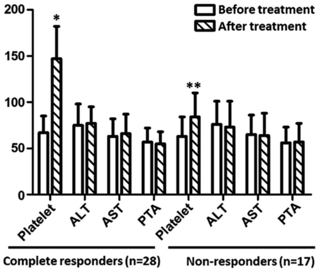
As shown in Fig. 1, after treatment, platelet count returned to normal level in complete responders (n=28). Although platelet count was significantly increased in non-responders (n=17) (P<0.05), the level is still lower than normal level (100×109/l). After treatment, no significant changes in ALT, AST and PTA were observed in complete responders or non-responders (P>0.05).
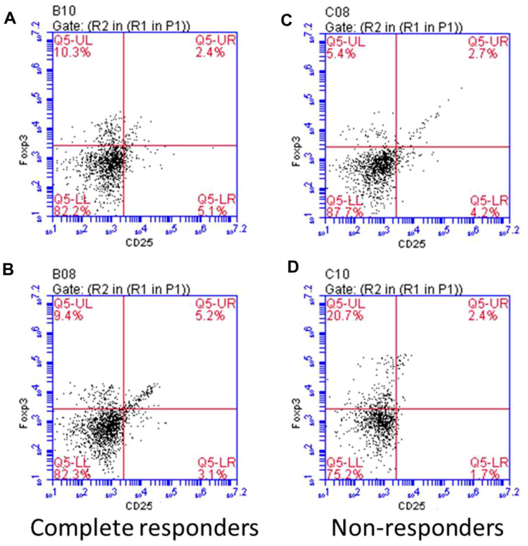
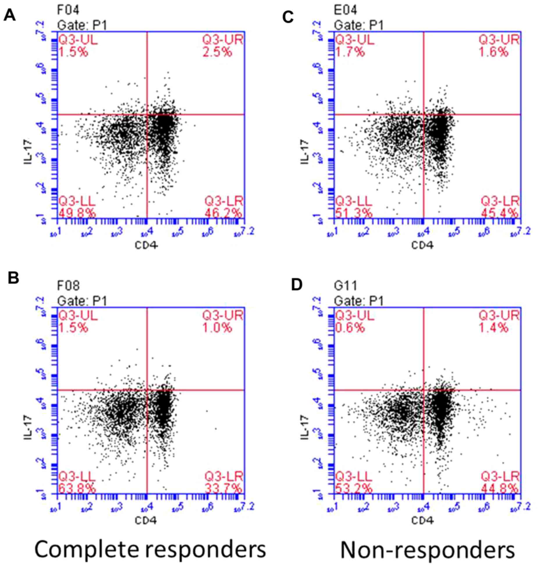
Percentages of Treg and Th17 cells in CD4+ T cells. As shown in Fig. 2, before treatment, the percentage of Treg cells was higher in complete responders than that in non-responders, but the difference was not significant, but the percentage of Treg cells was significantly higher in complete responders than that in non-responders (P<0.05). After treatment, the percentage of Treg cells was significantly increased and the percentage of Th17 cells was significantly decreased in complete responders (P<0.01). No significant changes in the percentage of Treg cells and Th17 cells were found in non-responders after treatment (P>0.05). Compared with non-responders, the percentage of Treg cells was significantly increased and percentage of Th17 cells was significantly decreased in complete responders after treatment (P<0.01). Repsresentative flow cytomerty is shown in Figs. 3 and 4.
Serum levels of cytokines in patients before and after treatment. As shown in Fig. 5, before treatment, levels of IL-10 and TGF-β were significantly lower, and levels of IL-17, IL-21 and IL-22 were significantly higher in complete responders than those in non-responders (P<0.05). After treatment, levels of IL-10 and TGF-β were significantly increased, and levels of IL-17, IL-21 and IL-22 were significantly decreased in complete responders (P<0.05). No significant differences in levels of IL-10, TGF-β, IL-17, IL-21 and IL-22 were found in non-responders after treatment. Thus, after treatment, levels of IL-10 and TGF-β were significantly higher, and levels of IL-17, IL-21 and IL-22 were significantly lower in complete responders than those in non-responders (P<0.05).
Discussion
Treg cells can maintain the tolerance of autoimmune and anti-inflammatory response. On the contrary, Th17 can mediate autoimmune diseases and inflammatory response. The balance between these two factors can maintain the steady state of immune response, and the imbalance will lead to the occurrence of autoimmune disease. Treg/Th17 cell imbalance is an important mechanism leading to persistent viral infection in patients with chronic hepatitis B. Increase in number of Tregs and decrease in the number of Th17 can lead to inhibition of T cell immunity in patients, and cellular immune response against hepatitis B virus will be inhibited (19,20). Studies have shown that platelet-related antibodies are one of the major causes of thrombocytopenia (21), suggesting that Treg/Th17 cell imbalance may be involved in the production of autoimmune platelet-associated antibodies (22).
This study showed that the percentage of Treg cells was increased, and percentage of Th17 cells was decreased, and platelet count returned to normal levels in patients with complete response. Correspondingly, levels of IL-10 and TGF-β were significantly increased, and levels of IL-17, IL-21 and IL-22 were significantly decreased after treatment, which reflects that Treg cells can inhibit inflammatory response and reduce the number of Th17, which in turn improves thrombocytopenia. No significant changes in Treg, Th17 and related cytokines were observed after treatment, indicating that treatment failed to reverse the imbalance of Treg/Th17. Therefore, Treg, Th17 and related cytokines can be used to predict prognosis. It is noteworthy that before treatment, numbers of both Treg cells and Th17 cells were higher in complete responders than those in non-responders, suggesting that the higher percentage of two cells may indicate good treatment outcomes.
Consistent with previous studies (19,23,24), this study showed that recovery of Treg/Th17 cell balance is beneficial for the improvement of thrombocytopenia in patients with chronic hepatitis B. Our study provides a theoretical basis for clinical treatment of thrombocytopenia in patients with hepatitis B.
Acknowledgements
Not applicable.
Funding
No funding was received.
Availability of data and materials
The datasets used and/or analyzed during the current study are available from the corresponding author on reasonable request.
Authors' contributions
YW and LW collected and analyzed the general information of patients. WG collected the blood and tissue samples. XC performed ELISA. YS helped with flow cytometry. All authors read and approved the final manuscript.
Ethics approval and consent to participate
This study was approved by the Ethics Committee of Heilongjiang Provincial Hospital (Harbin, China). Signed informed consents were obtained from all participants before the study.
Patient consent for publication
Not applicable.
Competing interests
The authors declare that they have no competing interests.
References
|
Peck-Radosavljevic M: Thrombocytopenia in liver disease. Can J Gastroenterol. 14:60D–66D. 2000. View Article : Google Scholar : PubMed/NCBI | |
|
Thomopoulos KC, Labropoulou-Karatza C, Mimidis KP, Katsakoulis EC, Iconomou G and Nikolopoulou VN: Non-invasive predictors of the presence of large oesophageal varices in patients with cirrhosis. Dig Liver Dis. 35:473–478. 2003. View Article : Google Scholar : PubMed/NCBI | |
|
Aref S, Mabed M, Selim T, Goda T and Khafagy N: Thrombopoietin (TPO) levels in hepatic patients with thrombocytopenia. Hematology. 9:351–356. 2004. View Article : Google Scholar : PubMed/NCBI | |
|
Afdhal N, McHutchison J, Brown R, Jacobson I, Manns M, Poordad F, Weksler B and Esteban R: Thrombocytopenia associated with chronic liver disease. J Hepatol. 48:1000–1007. 2008. View Article : Google Scholar : PubMed/NCBI | |
|
Stornaiuolo G, Amato A and Gaeta GB: Adefovir dipivoxil-associated thrombocytopenia in a patient with chronic hepatitis B. Dig Liver Dis. 38:211–212. 2006. View Article : Google Scholar : PubMed/NCBI | |
|
Rodeghiero F, Stasi R, Gernsheimer T, Michel M, Provan D, Arnold DM, Bussel JB, Cines DB, Chong BH, Cooper N, et al: Standardization of terminology, definitions and outcome criteria in immune thrombocytopenic purpura of adults and children: Report from an international working group. Blood. 113:pp. 2386–2393. 2009, View Article : Google Scholar : PubMed/NCBI | |
|
Shevach EM: Mechanisms of foxp3+ T regulatory cell-mediated suppression. Immunity. 30:636–645. 2009. View Article : Google Scholar : PubMed/NCBI | |
|
Wing K, Onishi Y, Prieto-Martin P, Yamaguchi T, Miyara M, Fehervari Z, Nomura T and Sakaguchi S: CTLA-4 control over Foxp3+ regulatory T cell function. Science. 322:271–275. 2008. View Article : Google Scholar : PubMed/NCBI | |
|
Mittal SK and Roche PA: Suppression of antigen presentation by IL-10. Curr Opin Immunol. 34:22–27. 2015. View Article : Google Scholar : PubMed/NCBI | |
|
Keswani T, Sarkar S, Sengupta A and Bhattacharyya A: Role of TGF-β and IL-6 in dendritic cells, Treg and Th17 mediated immune response during experimental cerebral malaria. Cytokine. 88:154–166. 2016. View Article : Google Scholar : PubMed/NCBI | |
|
Liu J, Hong X, Lin D, Luo X, Zhu M and Mo H: Artesunate influences Th17/Treg lymphocyte balance by modulating Treg apoptosis and Th17 proliferation in a murine model of rheumatoid arthritis. Exp Ther Med. 13:2267–2273. 2017. View Article : Google Scholar : PubMed/NCBI | |
|
Shrivastava S, TrehanPati N, Patra S, Kottilil S, Pande C, Trivedi SS and Sarin SK: Increased regulatory T cells and impaired functions of circulating CD8 T lymphocytes is associated with viral persistence in Hepatitis B virus-positive newborns. J Viral Hepat. 20:582–591. 2013. View Article : Google Scholar : PubMed/NCBI | |
|
Feng S, Chen XM, Wang JF and Xu XQ: Th17 cells associated cytokines and cancer. Eur Rev Med Pharmacol Sci. 20:4032–4040. 2016.PubMed/NCBI | |
|
Harrington LE, Hatton RD, Mangan PR, Turner H, Murphy TL, Murphy KM and Weaver CT: Interleukin 17-producing CD4+ effector T cells develop via a lineage distinct from the T helper type 1 and 2 lineages. Nat Immunol. 6:1123–1132. 2005. View Article : Google Scholar : PubMed/NCBI | |
|
Li J, Lai X, Liao W, He Y, Liu Y and Gong J: The dynamic changes of Th17/Treg cytokines in rat liver transplant rejection and tolerance. Int Immunopharmacol. 11:962–967. 2011. View Article : Google Scholar : PubMed/NCBI | |
|
Ni K, Zhao L, Wu J, Chen W, Hongya Yang and Li X: Th17/Treg balance in children with obstructive sleep apnea syndrome and the relationship with allergic rhinitis. Int J Pediatr Otorhinolaryngol. 79:1448–1454. 2015. View Article : Google Scholar : PubMed/NCBI | |
|
Zheng Y, Wang Z, Deng L, Zhang G, Yuan X, Huang L, Xu W and Shen L: Modulation of STAT3 and STAT5 activity rectifies the imbalance of Th17 and Treg cells in patients with acute coronary syndrome. Clin Immunol. 157:65–77. 2015. View Article : Google Scholar : PubMed/NCBI | |
|
Kleinewietfeld M and Hafler DA: The plasticity of human Treg and Th17 cells and its role in autoimmunity. Semin Immunol. 25:305–312. 2013. View Article : Google Scholar : PubMed/NCBI | |
|
Su ZJ, Yu XP, Guo RY, Ming DS, Huang LY, Su ML, Deng Y and Lin ZZ: Changes in the balance between Treg and Th17 cells in patients with chronic hepatitis B. Diagn Microbiol Infect Dis. 76:437–444. 2013. View Article : Google Scholar : PubMed/NCBI | |
|
Li K, Liu H and Guo T: Th17/Treg imbalance is an indicator of liver cirrhosis process and a risk factor for HCC occurrence in HBV patients. Clin Res Hepatol Gastroenterol. 41:399–407. 2017. View Article : Google Scholar : PubMed/NCBI | |
|
Gilli SC, de Souza Medina S, de Castro V, Fernandes LG and Saad ST: Platelet associated IgG may be related with thrombocytopenia in patients with myelodysplastic syndromes. Leuk Res. 36:554–559. 2012. View Article : Google Scholar : PubMed/NCBI | |
|
Aboul-Fotoh L-M, Abdel Raheem MM, El-Deen MA and Osman AM: Role of CD4+CD25+ T cells in children with idiopathic thrombocytopenic purpura. J Pediatr Hematol Oncol. 33:81–85. 2011. View Article : Google Scholar : PubMed/NCBI | |
|
Liu B, Gao W, Zhang L, Wang J, Chen M, Peng M, Ren H and Hu P: Th17/Treg imbalance and increased interleukin-21 are associated with liver injury in patients with chronic severe hepatitis B. Int Immunopharmacol. 46:48–55. 2017. View Article : Google Scholar : PubMed/NCBI | |
|
Cao J, Chen C, Zeng L, Li L, Li X, Li Z and Xu K: Elevated plasma IL-22 levels correlated with Th1 and Th22 cells in patients with immune thrombocytopenia. Clin Immunol. 141:121–123. 2011. View Article : Google Scholar : PubMed/NCBI |


