Open Access

Spinal cord injury and its underlying mechanism in rats with temporal lobe epilepsy

  • Authors:
    • Jinjie Liu
    • Zanhua Liu
    • Guoliang Liu
    • Kai Gao
    • Hengjie Zhou
    • Yongbo Zhao
    • Hong Wang
    • Lin Zhang
    • Sibo Liu
  • View Affiliations

  • Published online on: January 15, 2020     https://doi.org/10.3892/etm.2020.8453
  • Pages: 2103-2112
  • Copyright: © Liu et al. This is an open access article distributed under the terms of Creative Commons Attribution License.

Metrics: Total Views: 0 (Spandidos Publications: | PMC Statistics: )
Total PDF Downloads: 0 (Spandidos Publications: | PMC Statistics: )


Abstract

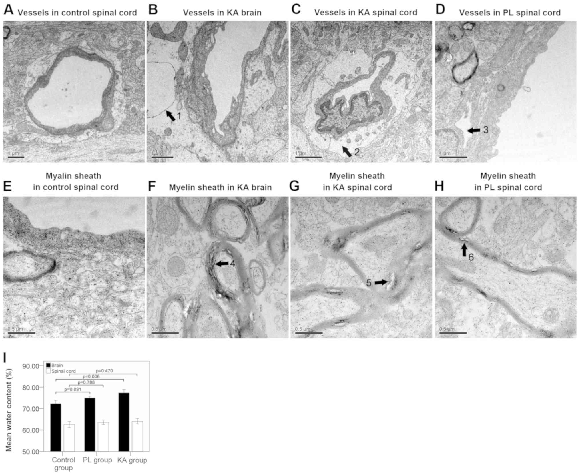
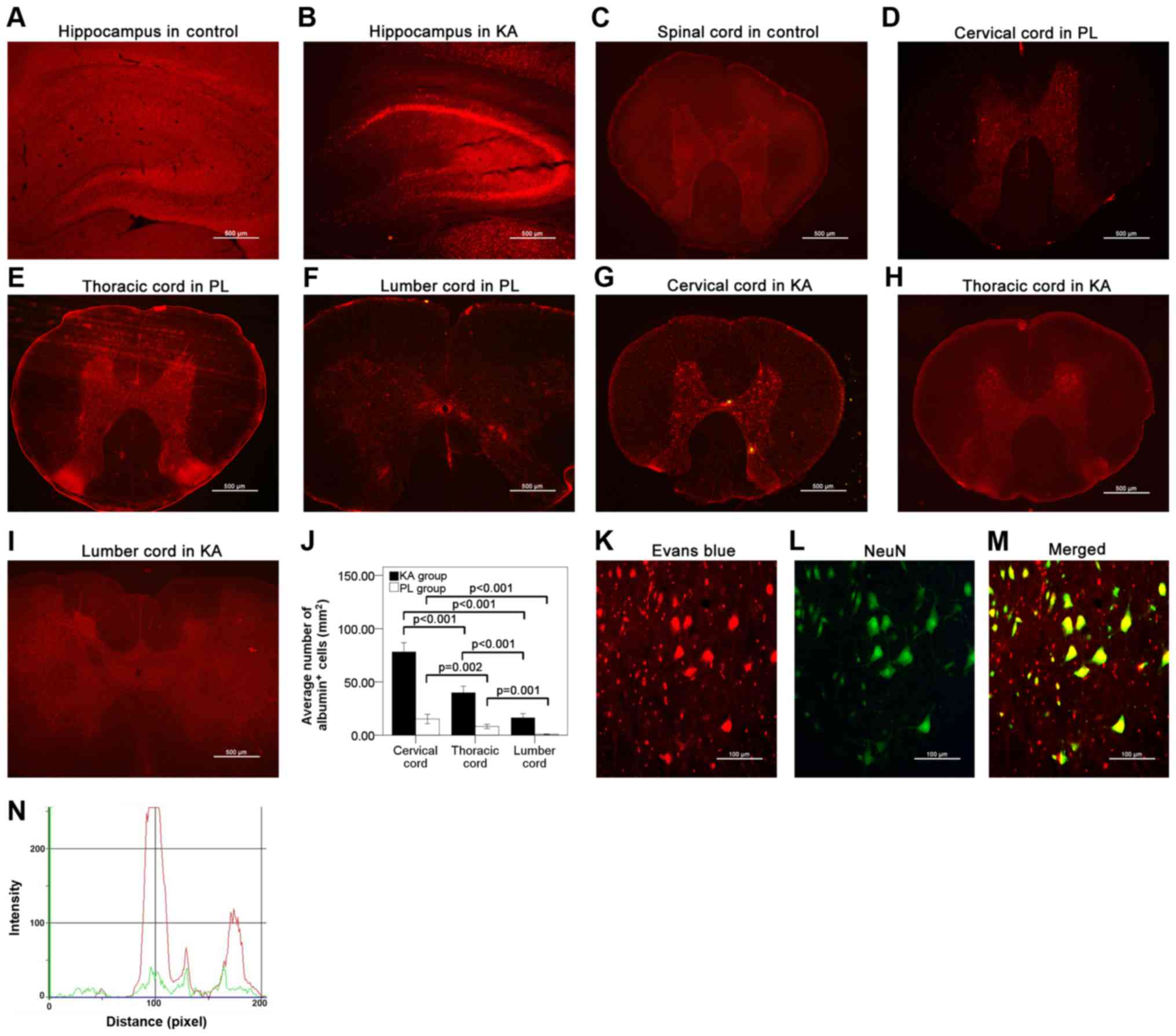
Numerous cases of spinal cord injury following seizure have been previously reported. However, whether spinal cord injury is a common occurrence after seizures and its underlying mechanisms remain unclear. The present study generated a Sprague‑Dawley rat model of temporal lobe epilepsy (TLE), and Nissl staining and transmission electron microscopy were used to detect tissue damage. In addition, Evans blue staining was used to detect damage to the blood‑brain barrier (BBB) and albumin extravasation. In addition, double‑staining was used to detect the association between neurons and extravasated albumin. Furthermore, neuronal degeneration was assessed using Fluoro‑Jade C staining, while fluorescence staining and western blotting were used to detect apoptosis and inflammation. In the present study, spinal cord injury was only observed in rats with grade IV‑V seizures, whereas Nissl staining showed structural damage and decreased neuronal cell numbers in the brain and the spinal cord. The present study identified BBB damage and albumin extravasation in rats of the TLE groups. Double‑staining for albumin and neurons showed a significant match of neurons positive for albumin. Fluoro‑Jade C staining indicated neuronal degeneration in the brain, but not the spinal cord in the TLE rats. High levels of caspase‑3 were also detected in the injured spinal cord. A small number of albumin+ neurons in the spinal cord presented caspase‑3+ signals in rats of the TLE groups. The expression levels of intercellular adhesion molecule 1, CD11b and inflammatory factors such as tumor necrosis factor‑α and interleukin‑6 were significantly elevated in the injured spinal cord. The present results suggested that spinal cord injury occurred in rats as a result of severe seizure attacks, and that BBB damage, albumin extravasation, inflammation and apoptosis contributed to the pathological changes observed during spinal cord injury.
View Figures
View References

Related Articles

Journal Cover

March-2020
Volume 19 Issue 3

Print ISSN: 1792-0981
Online ISSN:1792-1015

Sign up for eToc alerts

Recommend to Library

Copy and paste a formatted citation
x
Spandidos Publications style
Liu J, Liu Z, Liu G, Gao K, Zhou H, Zhao Y, Wang H, Zhang L and Liu S: Spinal cord injury and its underlying mechanism in rats with temporal lobe epilepsy. Exp Ther Med 19: 2103-2112, 2020.
APA
Liu, J., Liu, Z., Liu, G., Gao, K., Zhou, H., Zhao, Y. ... Liu, S. (2020). Spinal cord injury and its underlying mechanism in rats with temporal lobe epilepsy. Experimental and Therapeutic Medicine, 19, 2103-2112. https://doi.org/10.3892/etm.2020.8453
MLA
Liu, J., Liu, Z., Liu, G., Gao, K., Zhou, H., Zhao, Y., Wang, H., Zhang, L., Liu, S."Spinal cord injury and its underlying mechanism in rats with temporal lobe epilepsy". Experimental and Therapeutic Medicine 19.3 (2020): 2103-2112.
Chicago
Liu, J., Liu, Z., Liu, G., Gao, K., Zhou, H., Zhao, Y., Wang, H., Zhang, L., Liu, S."Spinal cord injury and its underlying mechanism in rats with temporal lobe epilepsy". Experimental and Therapeutic Medicine 19, no. 3 (2020): 2103-2112. https://doi.org/10.3892/etm.2020.8453