Changes in expression and distribution of attractin in the testes of rats at different developmental stages
- Authors:
- Xuejun Shang
- Juan Liu
- Qiuyu Xu
- Qi Zhang
- Bo Ma
- Yonglu Wang
- Zyuehua Zhang
- Xiaomei Cao
- Xuxin Zhan
View Affiliations
Affiliations: Department of Andrology, Jinling Hospital, Nanjing 210002, P.R. China, Shanghai Huashan Hospital North, Fudan University, Shanghai 201907, P.R. China, School of Pharmaceutical Sciences, Nanjing University of Technology, Nanjing 21009, P.R. China, Department of Andrology, School of Medicine, Southern Medical University, Guangzhou 510515, P.R. China
- Published online on: June 21, 2013 https://doi.org/10.3892/ijmm.2013.1423
-
Pages:
599-606
Metrics: Total
Views: 0 (Spandidos Publications: | PMC Statistics: )
Metrics: Total PDF Downloads: 0 (Spandidos Publications: | PMC Statistics: )
This article is mentioned in:
Abstract
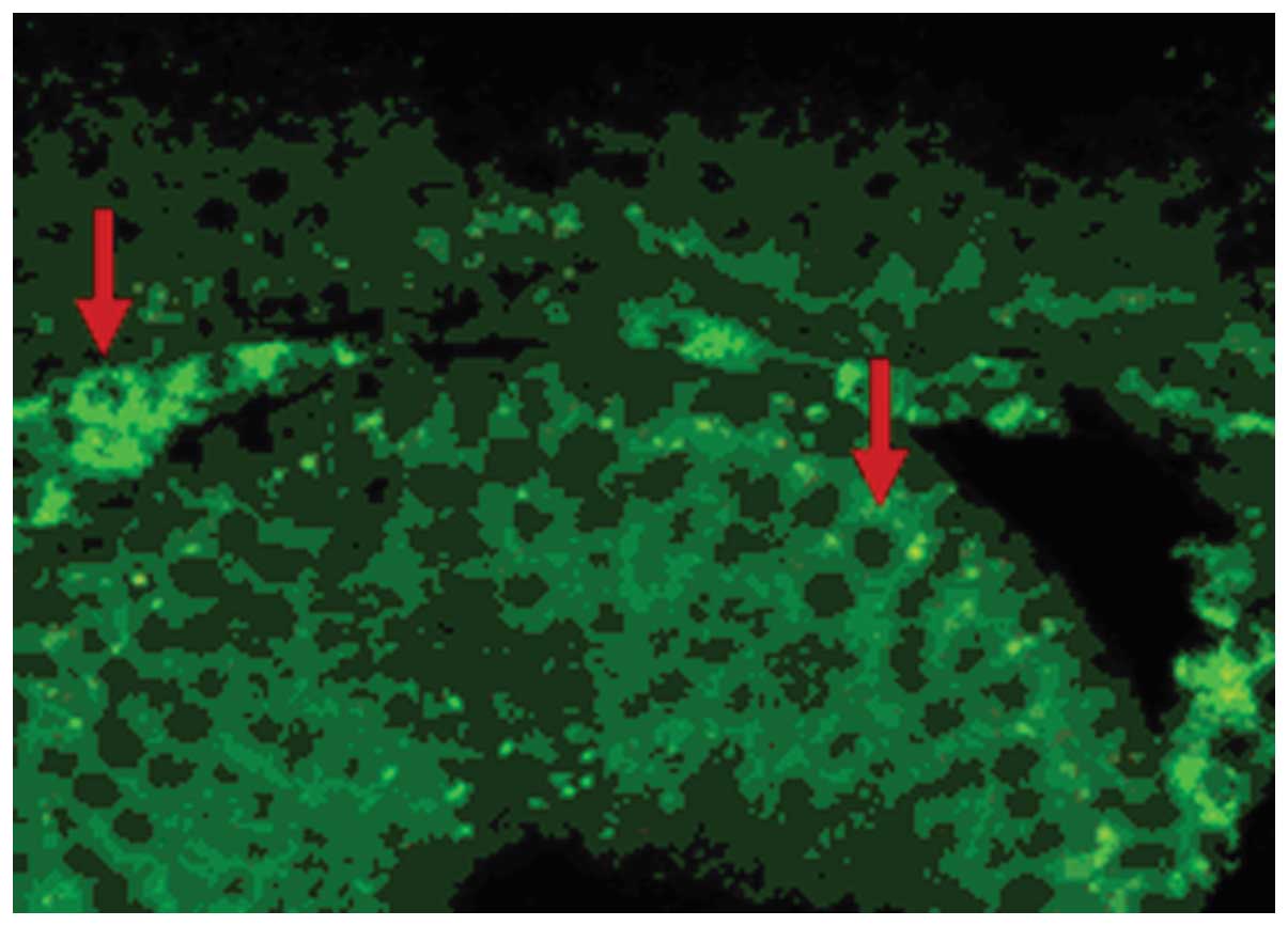
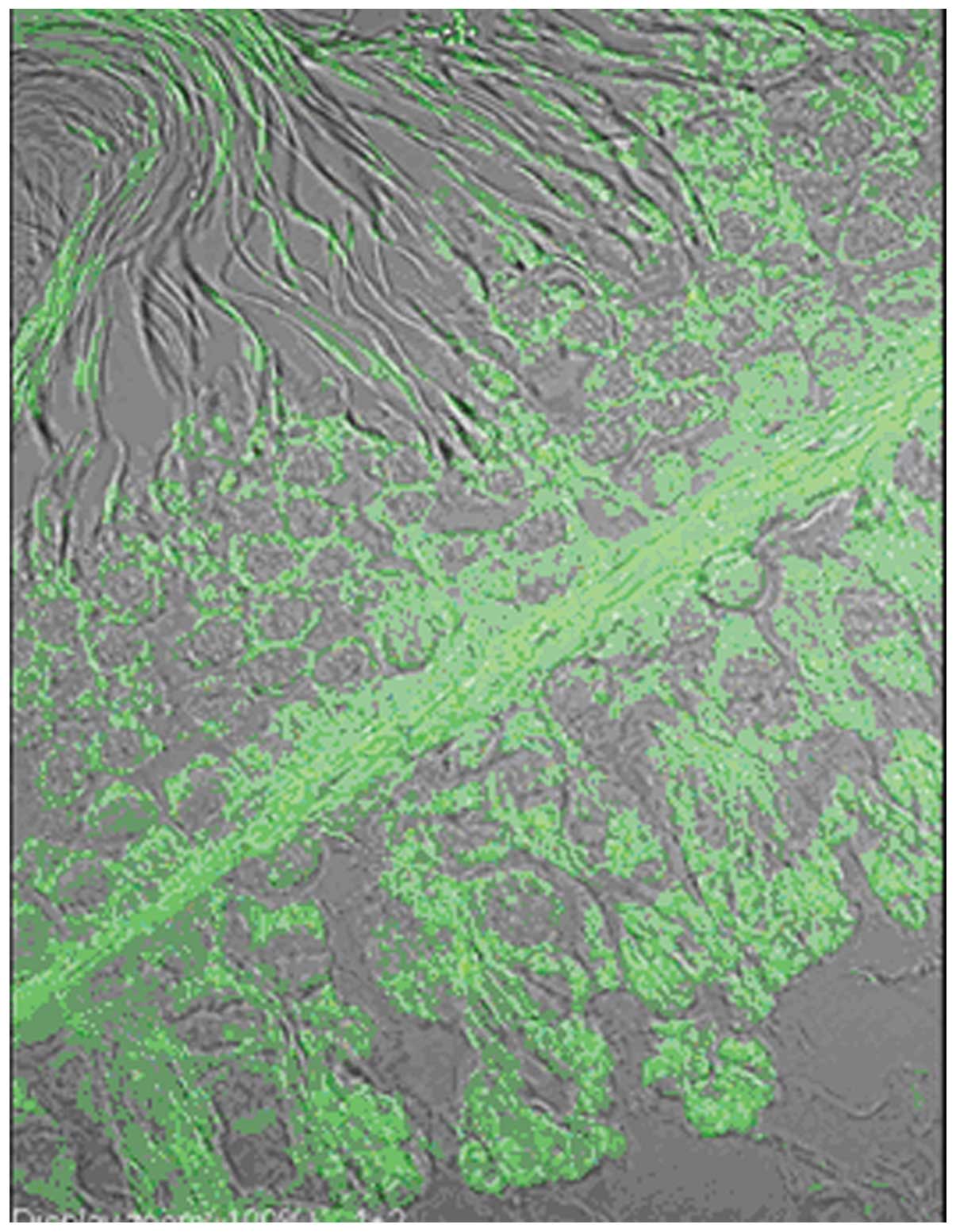
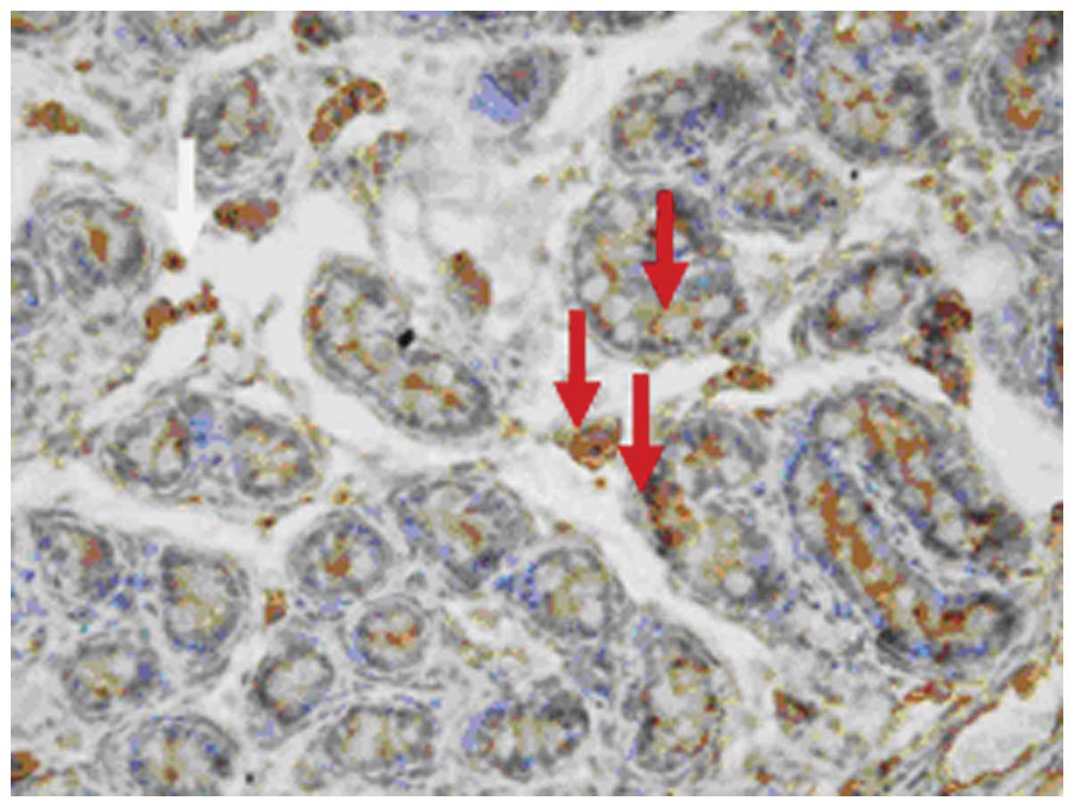
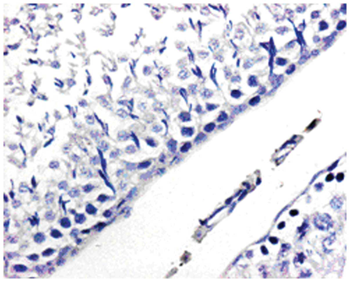
Attractin (Atrn), an autosomal recessive gene, is widely expressed in the body and displays multiple physiological and pathological functions in different types of tissues. The objective of this study was to localize Atrn protein and mRNA in the testis and epididymis of rats at different stages of maturation. Testis and epididymidis samples were obtained from the following 5 groups of Sprague Dawley (SD) rats in different developmental stages: newborn (8 h after birth), prepubertal (5 days), pubertal (20 days), postpubertal (50 days) and mature (70 days). Tissues were fixed and prepared for indirect immunofluorescence, immunohistochemistry, in situ hybridization, confocal laser scanning microscopy and western blot assays. A polyclonal antiserum against mouse Atrn and oligonucleotide riboprobes were used in the above assays. At the different stages of maturation, Atrn protein and mRNA were both widely expressed in the rat testis, including Leydig cells, primitive spermatogonia, primary spermatocytes, spermatids, Sertoli and peritubular myoid cells. Staining of the Atrn protein was mainly located on the cell membrane and in the cell cytoplasm while Atrn mRNA was distributed in both the nucleus and cytoplasm. No immunopositive staining was detected in spermatozoa and epididymides. In the epididymis, comprised of the caput, corpus and cauda, there was no definitive immunopositive staining within the efferent ductules or epididymal ducts. Taken together, Atrn protein and mRNA are both expressed widely in the rat testis at different stages of maturation, which suggests that Atrn protein is involved and plays an important role in the development of the reproductive system. In addition, the rat testis has the ability to synthesize Atrn protein throughout sexual development.
View Figures |
Figure 1
|
|
Figure 2
|
|
Figure 3
|
|
Figure 4
|
|
Figure 5
|
|
Figure 6
|
|
Figure 7
|
View References
1
|
Wrenger S, Faust J, Friedrich D, et al:
Attractin, a dipeptidyl peptidase IV/CD26-like enzyme, is expressed
on human peripheral blood monocytes and potentially influences
monocyte function. J Leukoc Biol. 80:621–629. 2006. View Article : Google Scholar
|
2
|
Gunn TM, Miller KA, He L, et al: The mouse
mahogany locus encodes a transmembrane form of human attractin.
Nature. 398:152–156. 1999. View
Article : Google Scholar : PubMed/NCBI
|
3
|
Nagle DL, McGrail SH, Vitale J, et al: The
mahogany protein is a receptor involved in suppression of obesity.
Nature. 398:148–152. 1999. View
Article : Google Scholar : PubMed/NCBI
|
4
|
Gunn TM and Barsh GS: Mahogany/attractin:
en route from phenotype to function. Trends Cardiovasc Med.
10:76–81. 2000. View Article : Google Scholar : PubMed/NCBI
|
5
|
Lu XY, Gunn TM, Shieh Kr, Barsh GS, Akil H
and Watson SJ: Distribution of Mahogany/Attractin mRNA in the rat
central nervous system. FEBS Lett. 462:101–107. 1999. View Article : Google Scholar : PubMed/NCBI
|
6
|
Malik R, Mares V, Kleibl Z, Pohlreich P,
Vlasicová K and Sedo A: Expression of attractin and its
differential enzyme activity in glioma cells. Biochem Biophys Res
Commun. 284:289–294. 2001. View Article : Google Scholar : PubMed/NCBI
|
7
|
Paz J, Yao H, Lim HS, Lu XY and Zhang W:
The neuroprotective role of attractin in neurodegeneration.
Neurobiol Aging. 28:1446–1456. 2007. View Article : Google Scholar : PubMed/NCBI
|
8
|
Duke-Cohan JS, Kim JH and Azouz A:
Attractin: cautionary tales for therapeutic intervention in
molecules with pleiotropic functionality. J Environ Pathol Toxicol
Oncol. 23:1–11. 2004. View Article : Google Scholar : PubMed/NCBI
|
9
|
Duke-Cohan JS, Gu J, McLaughlin DF, Xu Y,
Freeman GJ and Schlossman SF: Attractin (DPPT-L), a member of the
CUB family of cell adhesion and guidance proteins, is secreted by
activated human T lymphocytes and modulates immune cell
interactions. Proc Natl Acad Sci USA. 95:11336–11341. 1998.
View Article : Google Scholar : PubMed/NCBI
|
10
|
Cummins SF, Nichols AE, Schein CH and
Nagle GT: Newly identified water-borne protein pheromones interact
with attractin to stimulate mate attraction in Aplysia. Peptides.
27:597–606. 2006. View Article : Google Scholar : PubMed/NCBI
|
11
|
Kuramoto T, Kitada K, Inui T, et al:
Attractin/mahogany/zitter plays a critical role in myelination of
the central nervous system. Proc Natl Acad Sci USA. 98:559–564.
2001. View Article : Google Scholar : PubMed/NCBI
|
12
|
Tang W, Gunn TM, Mclaughlin DF, Barsh GS,
Schlossman SF and Duke-Cohan JS: Secreted and membrane attractin
result from alternative splicing of the human ATRN gene. Proc Natl
Acad Sci USA. 97:6025–6030. 2000. View Article : Google Scholar : PubMed/NCBI
|
13
|
Huckins C and Oakberg EF: Morphological
and quantitative analysis of spermatogonia in mouse testes using
whole mounted seminiferous tubules, I. The normal testes. Anat Rec.
192:519–528. 1978. View Article : Google Scholar
|
14
|
Huckins C: Spermatogonial intercellular
bridges in whole-mounted seminiferous tubules from normal and
irradiated rodent testes. Am J Anat. 153:97–121. 1978. View Article : Google Scholar : PubMed/NCBI
|